Jump to content
In this section we have spot diagnoses posted on a daily basis since June 2010, now over 4000! You can review the archived cases and read the suggested diagnoses by users and the final comment by the contributors.
Case are uploaded each week day by 10 am UK time with the correct diagnosis will generally be posted at 8 pm UK time. Why not view the most recent spot diagnosis and proffer a diagnosis?

Case Number : Case 992 - 11th April Posted By: Guest

Please read the clinical history and view the images by clicking on them before you proffer your diagnosis.
Submitted Date :
   (0 reviews)

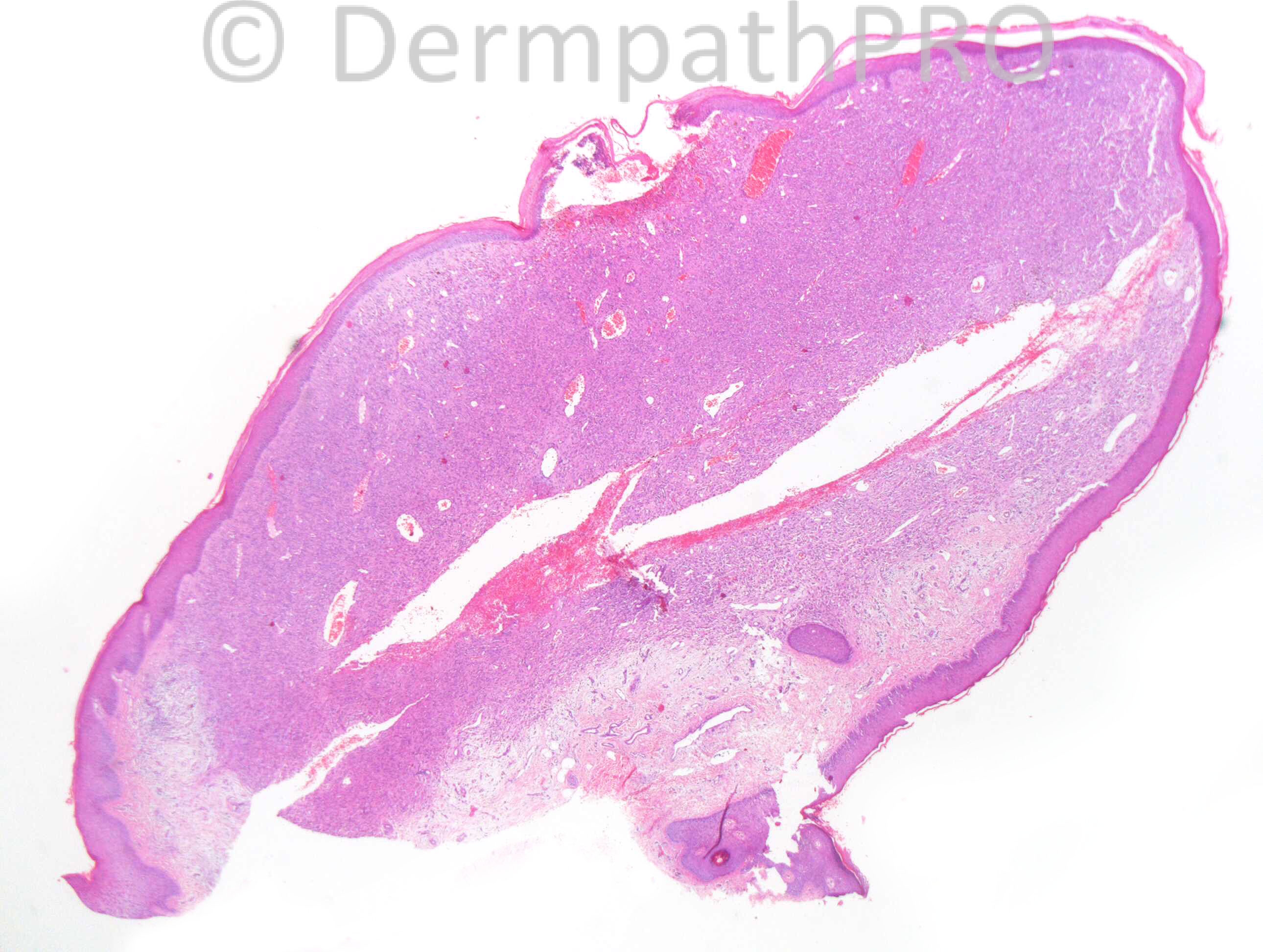
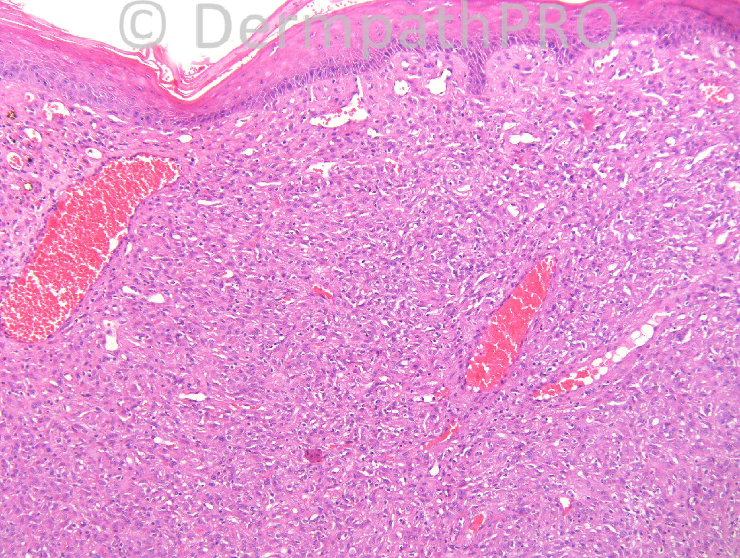
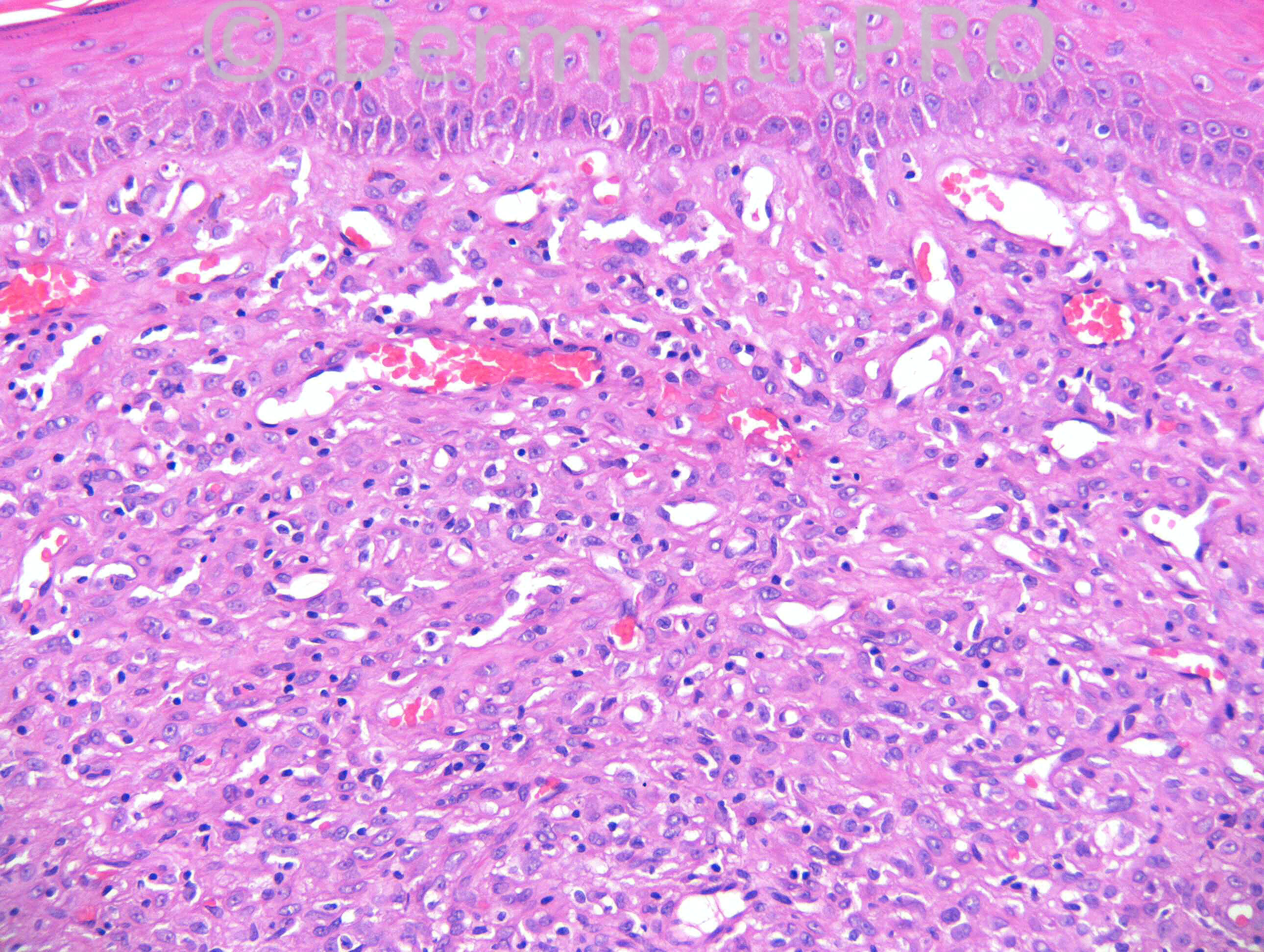
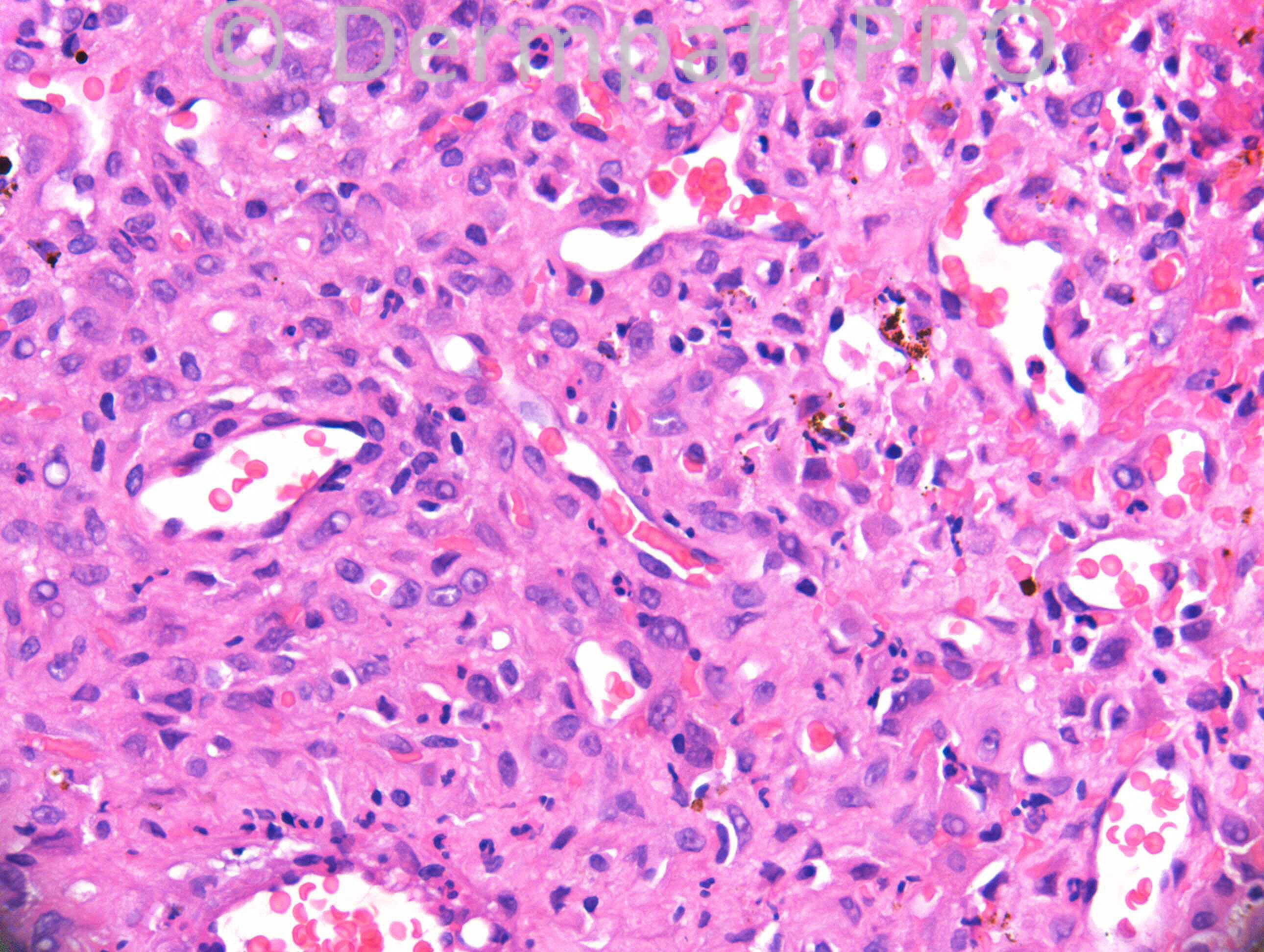
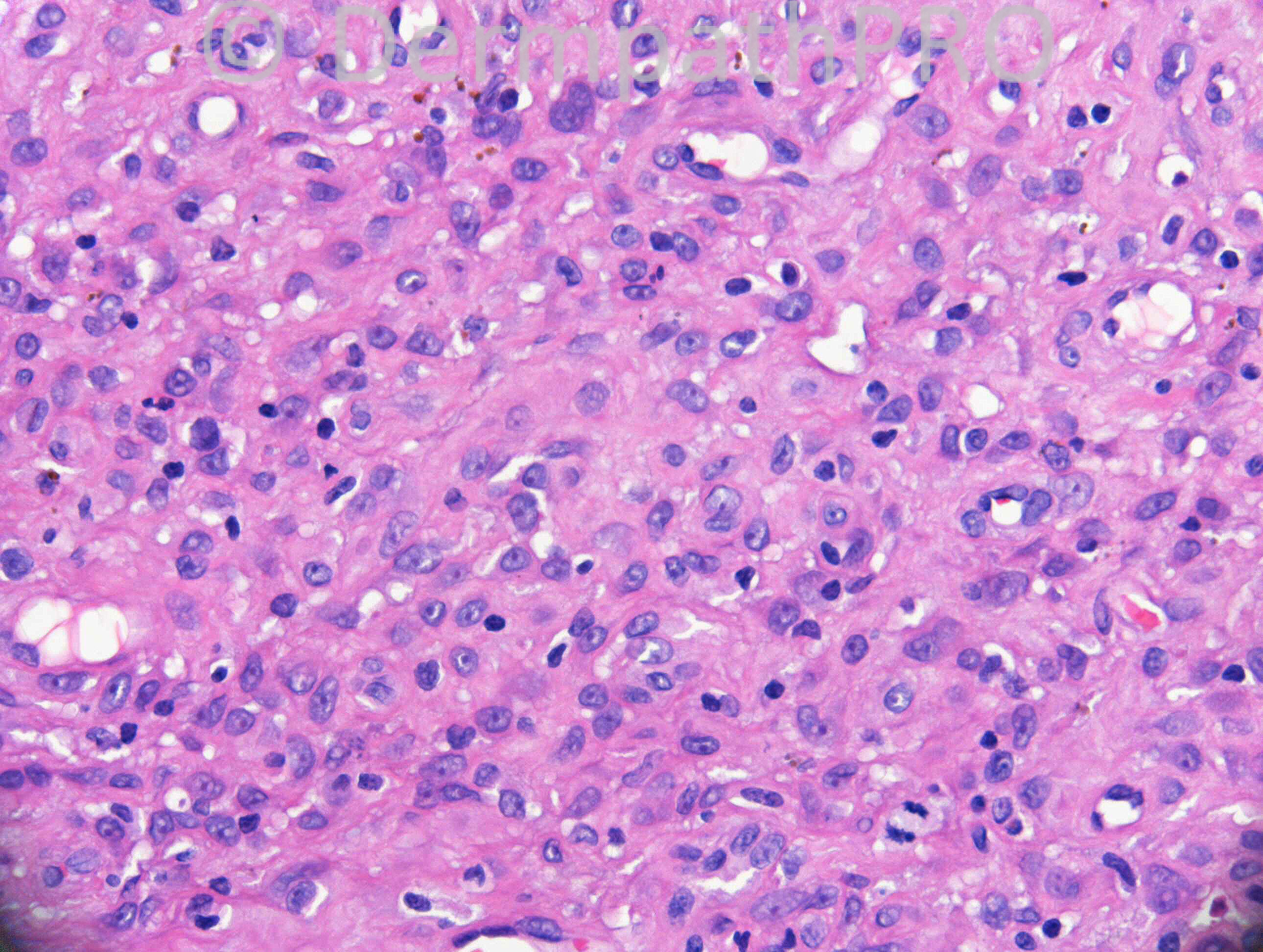
34 years old male. Skin tag on back.

Case posted by Dr. Richard Carr.

  Report Record

User Feedback


Guest Romualdo

Posted

Low-power magnification shows a polypoid vascular lesion resembling a pyogenic granuloma, except by the absence of lobularity. At higher power magnifications we can see epithelioid endothelial cells, intersticial neutrophils and nuclear dust. I hope a Warthin-Starry stain unmasks clumps of bacilli confirming my impression of bacillary angiomatosis.

Share this comment


Link to comment
share_externally

Agree with Epithelioid Hemangioma.
Once I read the entity called Cutaneous Epithelioid Angiomatous Nodule (CEAN), described in 2004 (see [url="http://www.ncbi.nlm.nih.gov/pubmed/14726818"]http://www.ncbi.nlm.nih.gov/pubmed/14726818[/url]), which differs from Epithelioid Hemangioma by some clinical (trunk and extremities) and histologic features. In 2008, other paper (see [url="http://www.ncbi.nlm.nih.gov/pubmed/18212538"]http://www.ncbi.nlm.nih.gov/pubmed/18212538[/url], with Dr. Omar Sangueza and Dr. Luiz Requena) described CEAN lesions also compromising head and neck location, and suggestes that CEAN are part of the spectrum of Epithelioid Hemangioma. I would like to know what are the opinion of the participants of this site.

Share this comment


Link to comment
share_externally

Eman El-Nabarawy

Posted

Cutaneous epithelioid angiomatous nodule.

Share this comment


Link to comment
share_externally

Robledo F. Rocha

Posted

Vascular proliferation with plump endothelial lining associated to slight microabscesses, the latter feature a clue for bacillary angiomatosis or, in the proper epidemiologic setting, verruga peruana.

Share this comment


Link to comment
share_externally

Dr. Mona Abdel-Halim

Posted

My impression is: cutaneous epithelioid angiomatous nodule.

Share this comment


Link to comment
share_externally

Dr. Richard Carr

Posted

Sorry I was at Eduardo Calonje's update today. This is not a vascular tumour as far as I remember but I am away from the office. Why don't you have another think
Regards to all from very sunny London

Share this comment


Link to comment
share_externally

Eman El-Nabarawy

Posted

So I will try the other idea that I thought as a DD: epithelioid benign fibrous histiocytoma.

Share this comment


Link to comment
share_externally

Dr. Mona Abdel-Halim

Posted

If not vascular, then as Eman said it should be epithelioid fibrous histiocytoma...

Share this comment


Link to comment
share_externally

Another differential diagnosis could be Angoimatoid Spitz nevus.

Share this comment


Link to comment
share_externally

Guest Romualdo

Posted

Agree with this new possibility: [color=#1C2837][font=arial, verdana, tahoma, sans-serif][size=4]epithelioid fibrous histiocytoma.[/size][/font][/color]

Share this comment


Link to comment
share_externally

Dr. Richard Carr

Posted

Immunohistochemistry for S100, MelanA, HMB45, EMA, MNF116 and AE1/AE3 is negative. CD31 and CD34 highlights the vessels. CD68, S100 and CD1a highlight scattered dendritic cells throughout the lesion and CD68 is weakly positive in tumour cells.

Share this comment


Link to comment
share_externally

Eman El-Nabarawy

Posted

Indeterminate dendritic cell tumor.

Share this comment


Link to comment
share_externally

Guest Tiberiu Tebeica

Posted

This could be solitary reticulohistiocytoma. I see oncocytic mononuclear histiocytes, but no multinucleate giant cells. Are there other stains available, such as CD163, factor XIIIa, lysozyme?

Share this comment


Link to comment
share_externally

Dr. Richard Carr

Posted

No sorry it is from a few years ago and sadly we don't stock those markers in any case.

Share this comment


Link to comment
share_externally

Eman El-Nabarawy

Posted

I am sorry. I think I made a mistake. Indeterminate dendritic cell tumor must be diffuse lay positive for S100 and CD1a..so it's not the case..I think I will agree with Tiberiu for solitary reticulohistiocytoma..

Share this comment


Link to comment
share_externally

Dr. Richard Carr

Posted

Yes I reported this as epithelioid fibrous histiocytoma. The polypoid nature and collarette are typical. Eduardo Calonje kindly reviewed this case and agreed. Looking forward to Mark's anecdote. Regards to all and pleased some of you are happy to contribute during your weekend.

Share this comment


Link to comment
share_externally

Dr. Richard Carr

Posted

Whoops just checked my database and this particular case was not seen by Eduardo, I was thinking of another case even older. I did call it epithelioid cell histiocytoma though but I am sure we use epithelioid fibrous histiocytoma synonymously.

Share this comment


Link to comment
share_externally


Join the conversation

You can post now and register later. If you have an account, sign in now to post with your account.

Guest
Add a comment...

×   Pasted as rich text.   Paste as plain text instead

  Only 75 emoji are allowed.

×   Your link has been automatically embedded.   Display as a link instead

×   Your previous content has been restored.   Clear editor

×   You cannot paste images directly. Upload or insert images from URL.

Loading...

×
×
  • Create New...