Jump to content
In this section we have spot diagnoses posted on a daily basis since June 2010, now over 4000! You can review the archived cases and read the suggested diagnoses by users and the final comment by the contributors.
Case are uploaded each week day by 10 am UK time with the correct diagnosis will generally be posted at 8 pm UK time. Why not view the most recent spot diagnosis and proffer a diagnosis?

Case Number : Case 996 - 17th April Posted By: Guest

Please read the clinical history and view the images by clicking on them before you proffer your diagnosis.
Submitted Date :
   (0 reviews)

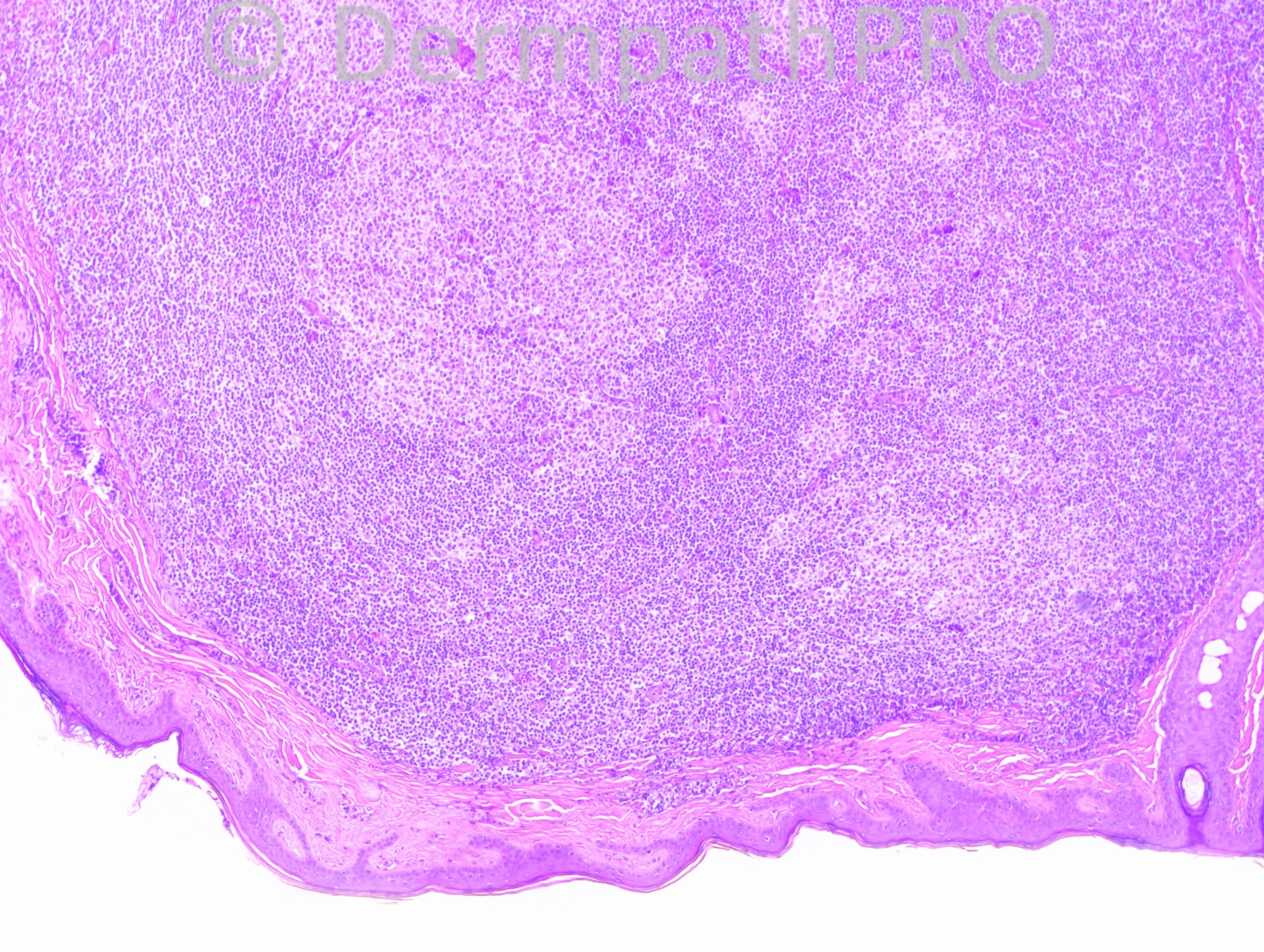
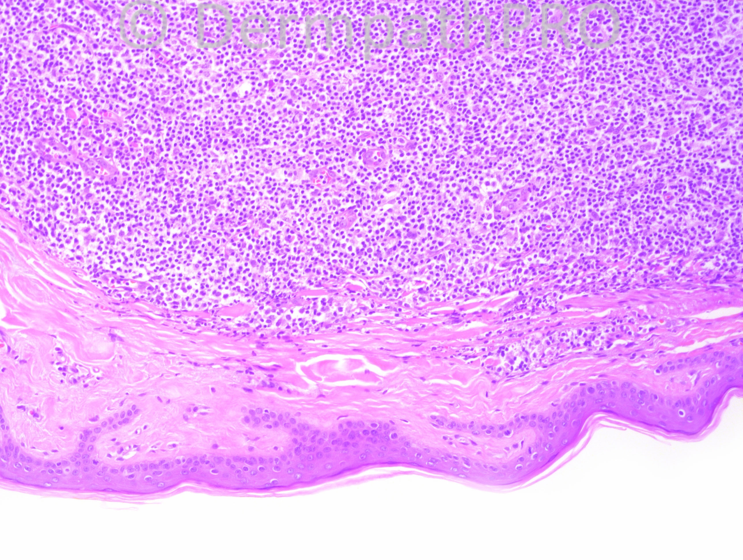
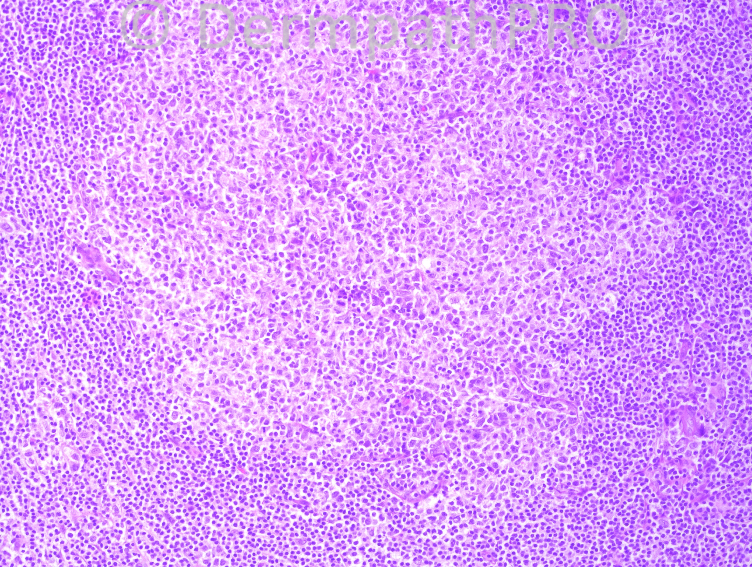
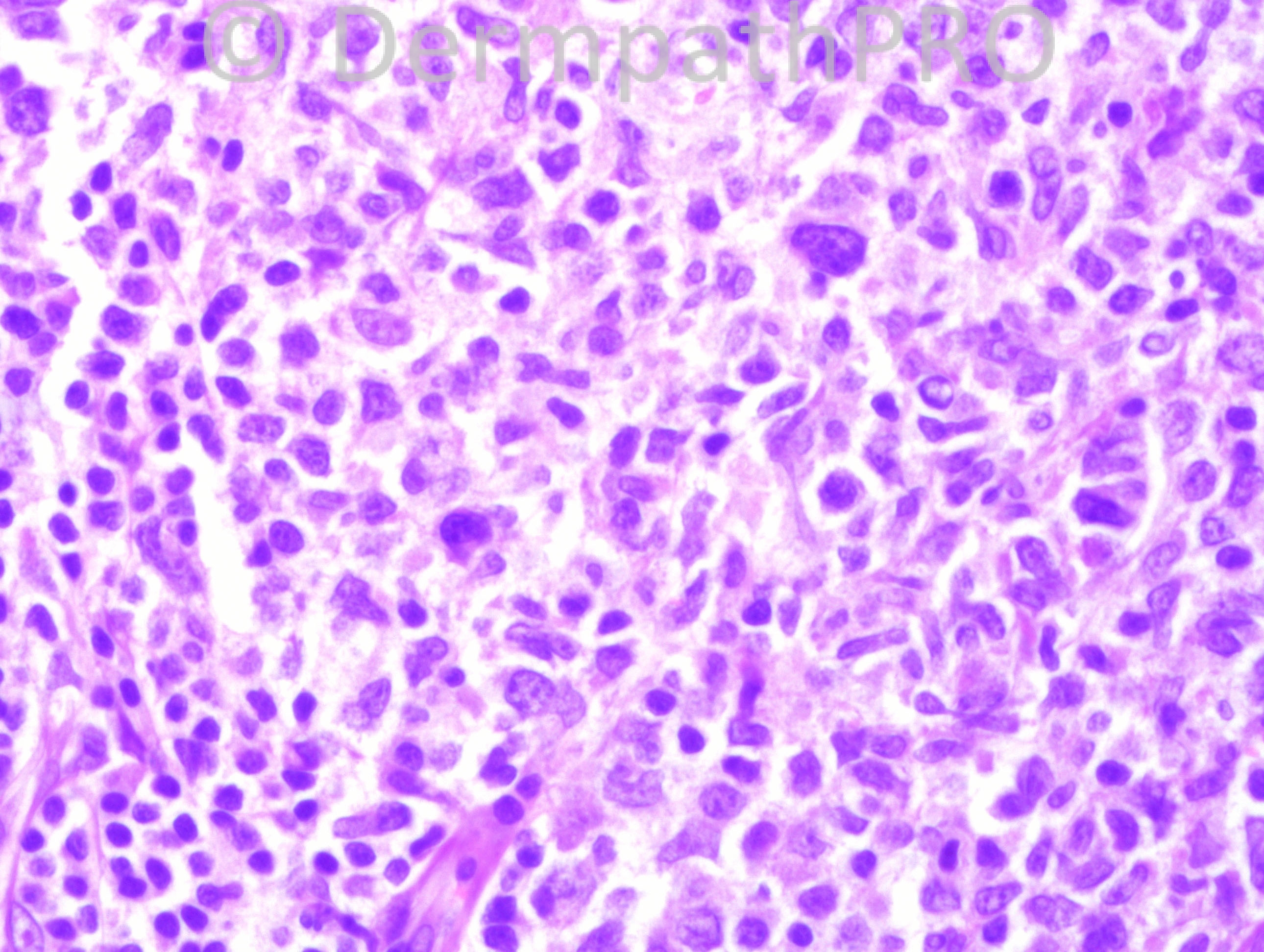
77 year-old male with scalp lesion.

Case posted by Dr. Hafeez Diwan.


  Report Record

User Feedback


Lymphomas with a nodular growth pattern are most often of B-cell origin. A nodular proliferation of lymphocites with low-grade atypia and head and neck location are most probably Follicle centre cell lymphoma. It is necessary to add bcl-2 and CD10 immunostains to exclude cutaneouos involvment of a systemic nodal lymphoma.

Share this comment


Link to comment
share_externally

Robledo F. Rocha

Posted

Primary cutaneous follicle center cell lymphoma. There’s a mixed proliferation of cleaved centrocytes and noncleaved centroblasts in a rare follicular growth pattern admixed with small lymphocytes in the interfollicular areas.
B-cell pseudolymphoma must be considered, but I didn’t find tingible body macrophages nor well-developed marginal and mantle zones for a reactive lymphoid hyperplasia.

Share this comment


Link to comment
share_externally

Dr. Mona Abdel-Halim

Posted

My impression also is follicle centre BCL for complete immunos.

Share this comment


Link to comment
share_externally

Guest Romualdo

Posted

Agree: primary cutaneous follicle centre lymphoma.

Share this comment


Link to comment
share_externally

Dr. Hafeez Diwan

Posted

Follicular lymphoma. This one was Bcl-2 and CD10 positive.

Share this comment


Link to comment
share_externally

Guest Jim Davie MD

Posted

Late to the party. For me, the absence of brisk mitotic activity in the follicular centers was an additional helpful clue in addition to the absence of tingibles, for the Dx of FCCL.

Share this comment


Link to comment
share_externally


Join the conversation

You can post now and register later. If you have an account, sign in now to post with your account.

Guest
Add a comment...

×   Pasted as rich text.   Paste as plain text instead

  Only 75 emoji are allowed.

×   Your link has been automatically embedded.   Display as a link instead

×   Your previous content has been restored.   Clear editor

×   You cannot paste images directly. Upload or insert images from URL.

Loading...

×
×
  • Create New...