Jump to content
In this section we have spot diagnoses posted on a daily basis since June 2010, now over 4000! You can review the archived cases and read the suggested diagnoses by users and the final comment by the contributors.
Case are uploaded each week day by 10 am UK time with the correct diagnosis will generally be posted at 8 pm UK time. Why not view the most recent spot diagnosis and proffer a diagnosis?

Case Number : Case 997 - 18th April Posted By: Guest

Please read the clinical history and view the images by clicking on them before you proffer your diagnosis.
Submitted Date :
   (0 reviews)

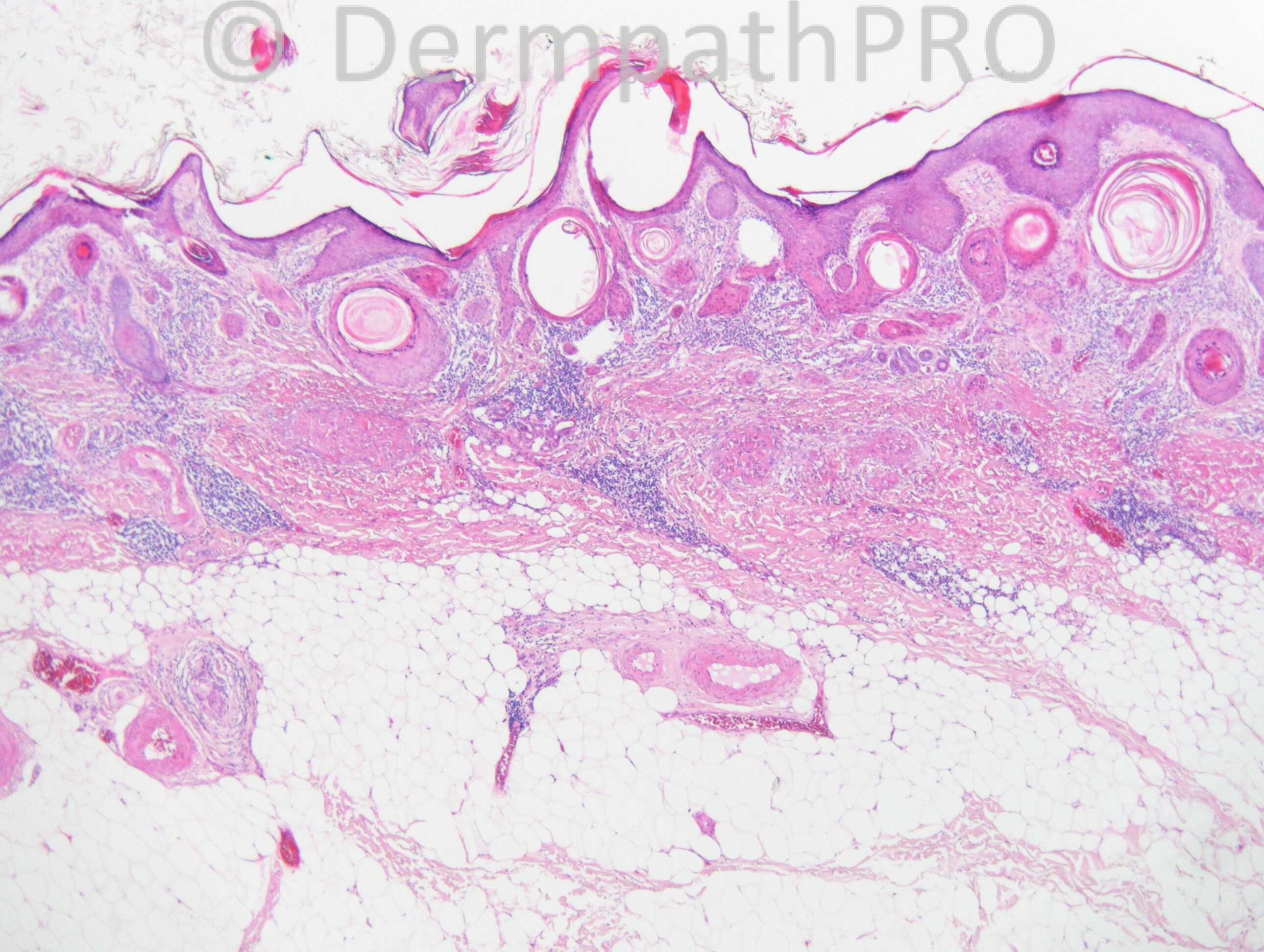
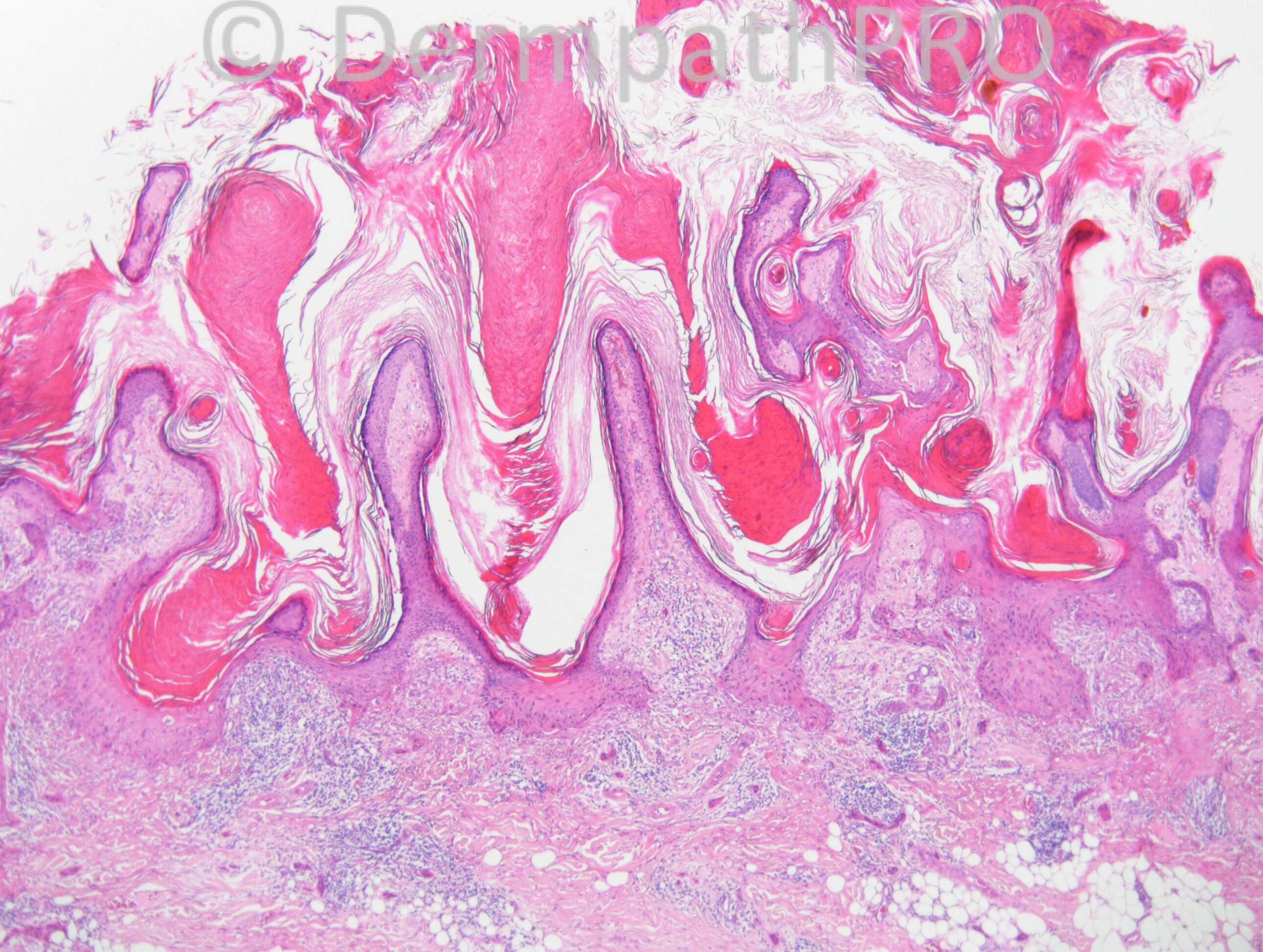
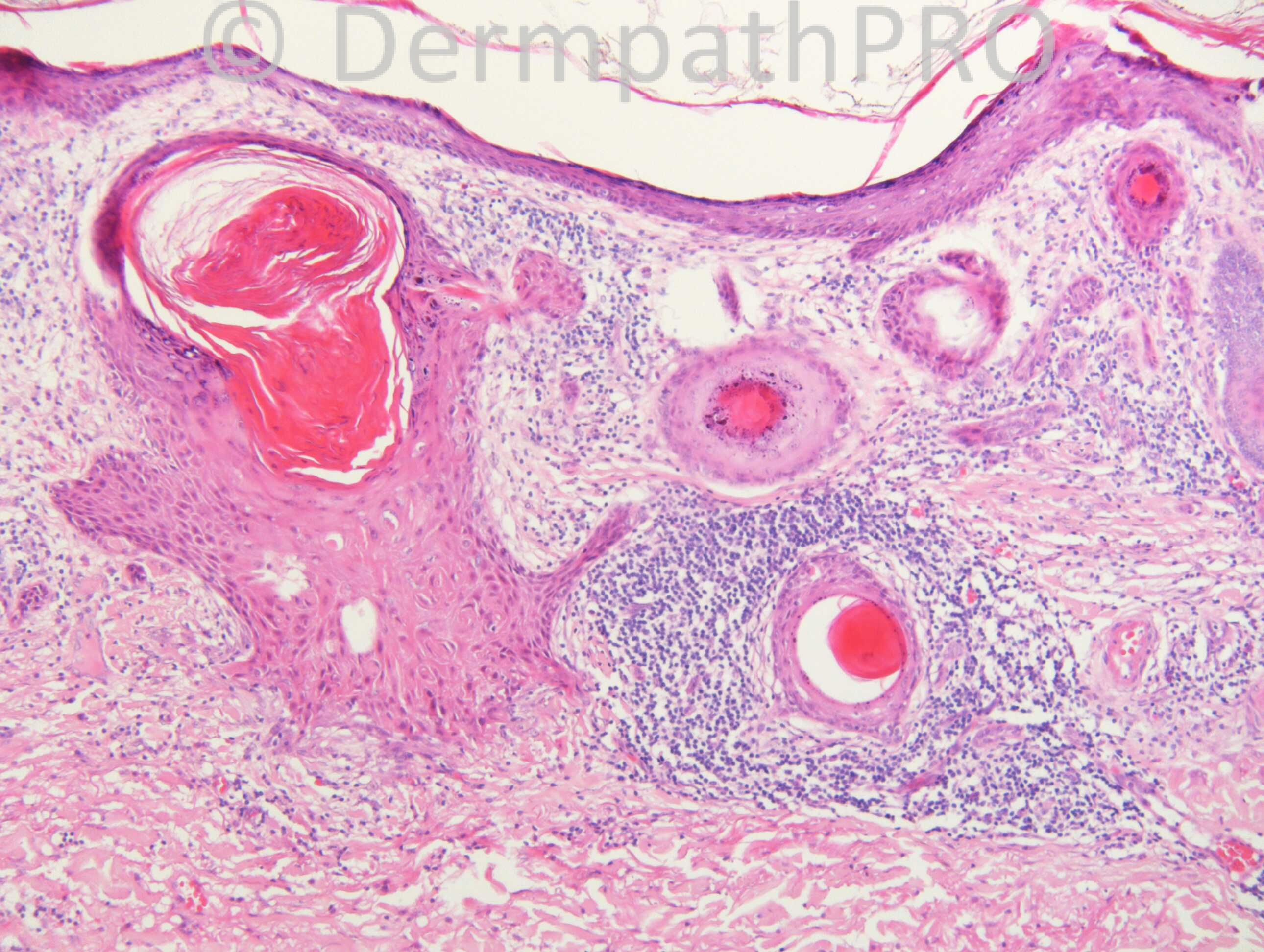
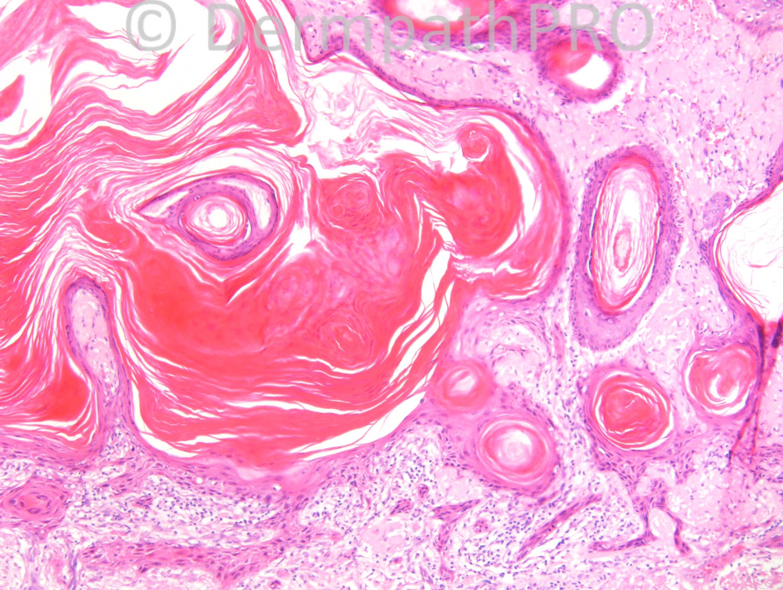
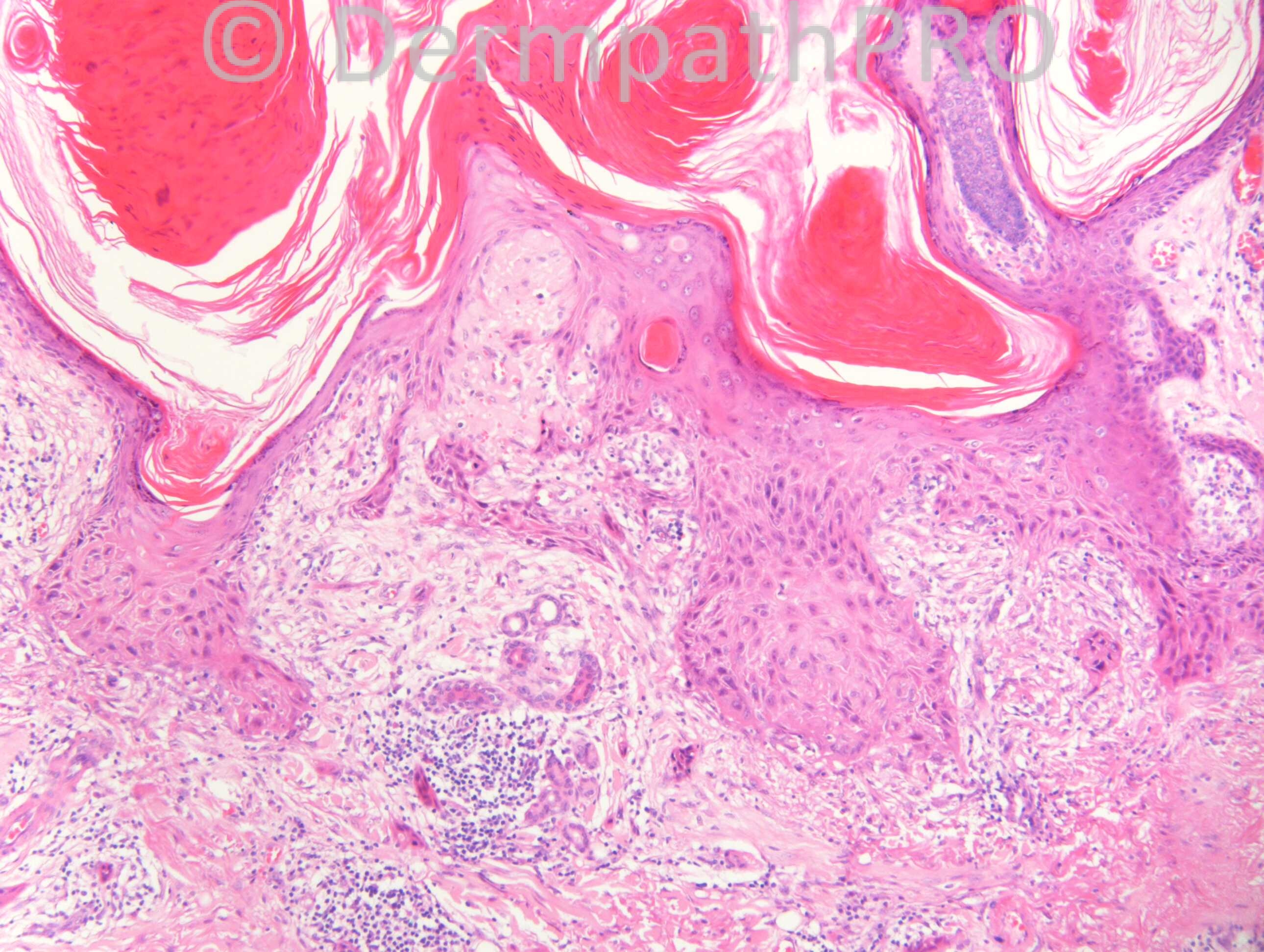
M85. Lesions 13 x 10mm ?following local trauma years ago. Previous liquid nitrogen. ?Bowen’s ?SEBK

Case posted by Dr. Richard Carr.


  Report Record

User Feedback


Dr. Mona Abdel-Halim

Posted

Follicular SCC, atypical proliferation related to follicular structures with characteristic orange red pilar keratinization.

Share this comment


Link to comment
share_externally

Eman El-Nabarawy

Posted

First impression SCC(probably follicular).

Share this comment


Link to comment
share_externally

Guest Romualdo

Posted

I completely agree with Mona: the presence of squamous nests with central tricholemmal type keratinisation, the absence of an in situ epidermal component, the absence of epidermal conections, except via dilated infundibula and the cytologic atypia are all consistent with follicular squamous cell carcinoma.

Share this comment


Link to comment
share_externally

Dr. Richard Carr

Posted

Well done. I am glad you are recognising this rather rare, predominantly infundibular-cystic variant of follicular SCC that was drawn attention to by Kossard in 2008 and Misago 2011. There is perineural invasion in the last image.

Kossard S, Tan KB, Choy C. Keratoacanthoma and [size=4]infundibulocystic squamous cell carcinoma. Am J Dermatopathol. [/size][size=4]2008 Apr;30(2):127-34. [/size]

Misago N, Inoue T, Toda S, Narisawa Y. Infundibular [size=4](follicular) and infundibulocystic squamous cell carcinoma: a [/size]clinicopathological and immunohistochemical study. Am J [size=4]Dermatopathol. 2011;33:687-94. [/size]

Share this comment


Link to comment
share_externally

Guest Jim Davie MD

Posted

The first low power image also seems to show a subcutis focus of perineural invasion with inflammatory cuffing (bottom, left of image). Thanks for sharing this interesting case!

Share this comment


Link to comment
share_externally

Dr. Richard Carr

Posted

Yes well spotted Jim, quite correct and I forgot to point that out as the image was carefully chosen for that feature!

Share this comment


Link to comment
share_externally


Join the conversation

You can post now and register later. If you have an account, sign in now to post with your account.

Guest
Add a comment...

×   Pasted as rich text.   Paste as plain text instead

  Only 75 emoji are allowed.

×   Your link has been automatically embedded.   Display as a link instead

×   Your previous content has been restored.   Clear editor

×   You cannot paste images directly. Upload or insert images from URL.

Loading...

×
×
  • Create New...