Jump to content
In this section we have spot diagnoses posted on a daily basis since June 2010, now over 4000! You can review the archived cases and read the suggested diagnoses by users and the final comment by the contributors.
Case are uploaded each week day by 10 am UK time with the correct diagnosis will generally be posted at 8 pm UK time. Why not view the most recent spot diagnosis and proffer a diagnosis?

Case Number : Case 998 - 21st April Posted By: Guest

Please read the clinical history and view the images by clicking on them before you proffer your diagnosis.
Submitted Date :
   (0 reviews)

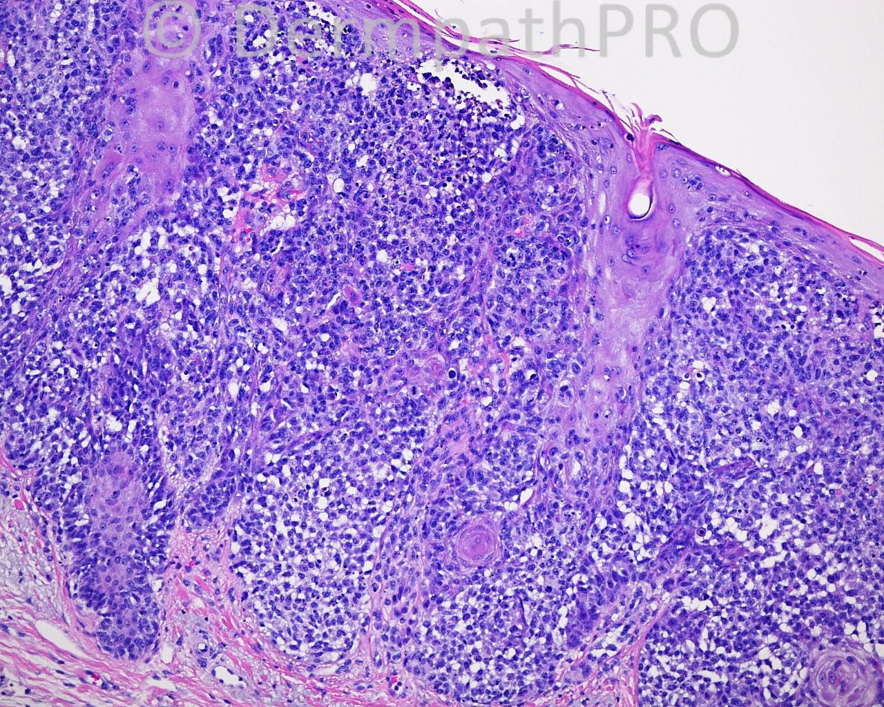
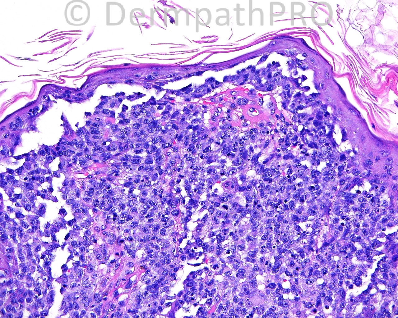
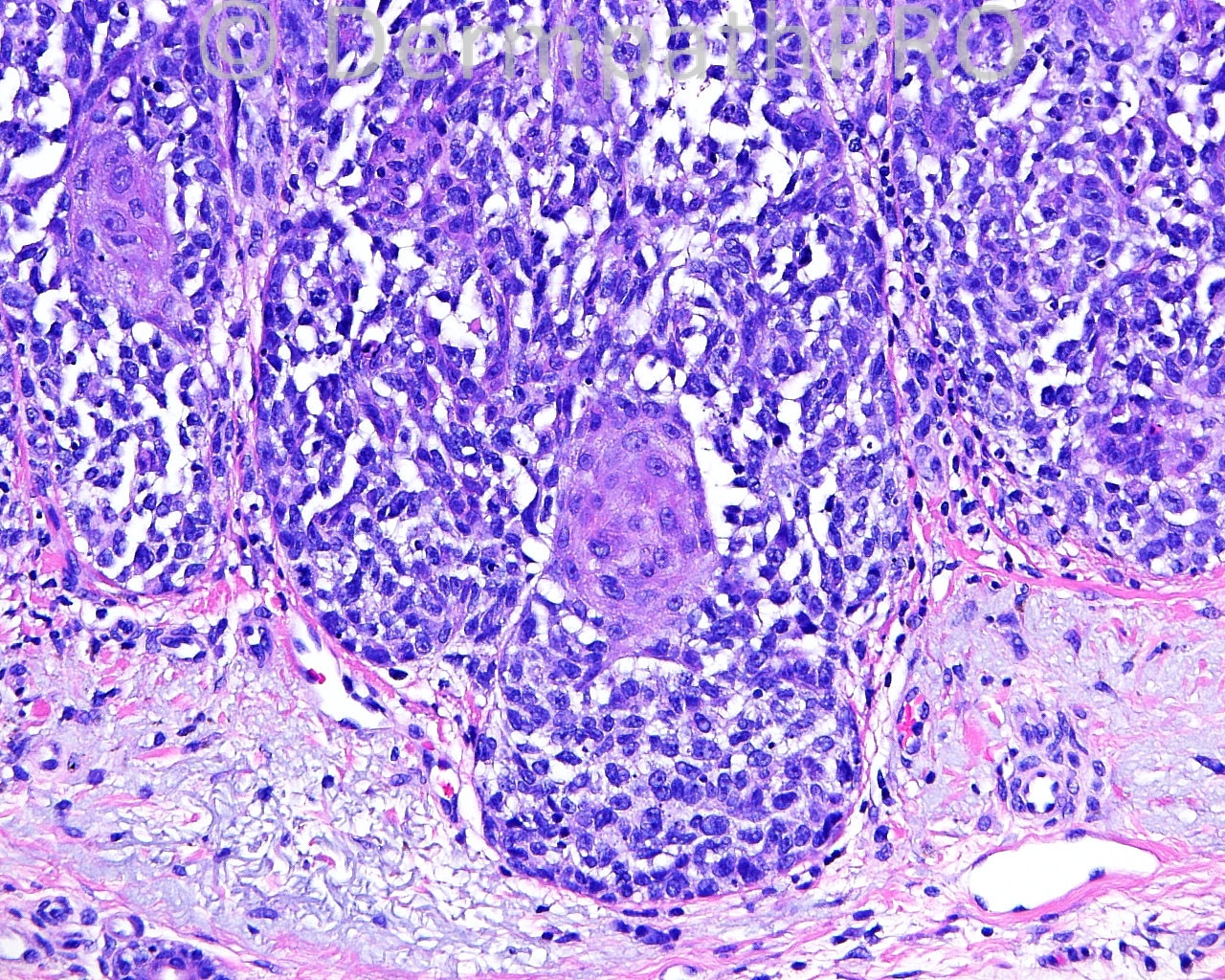
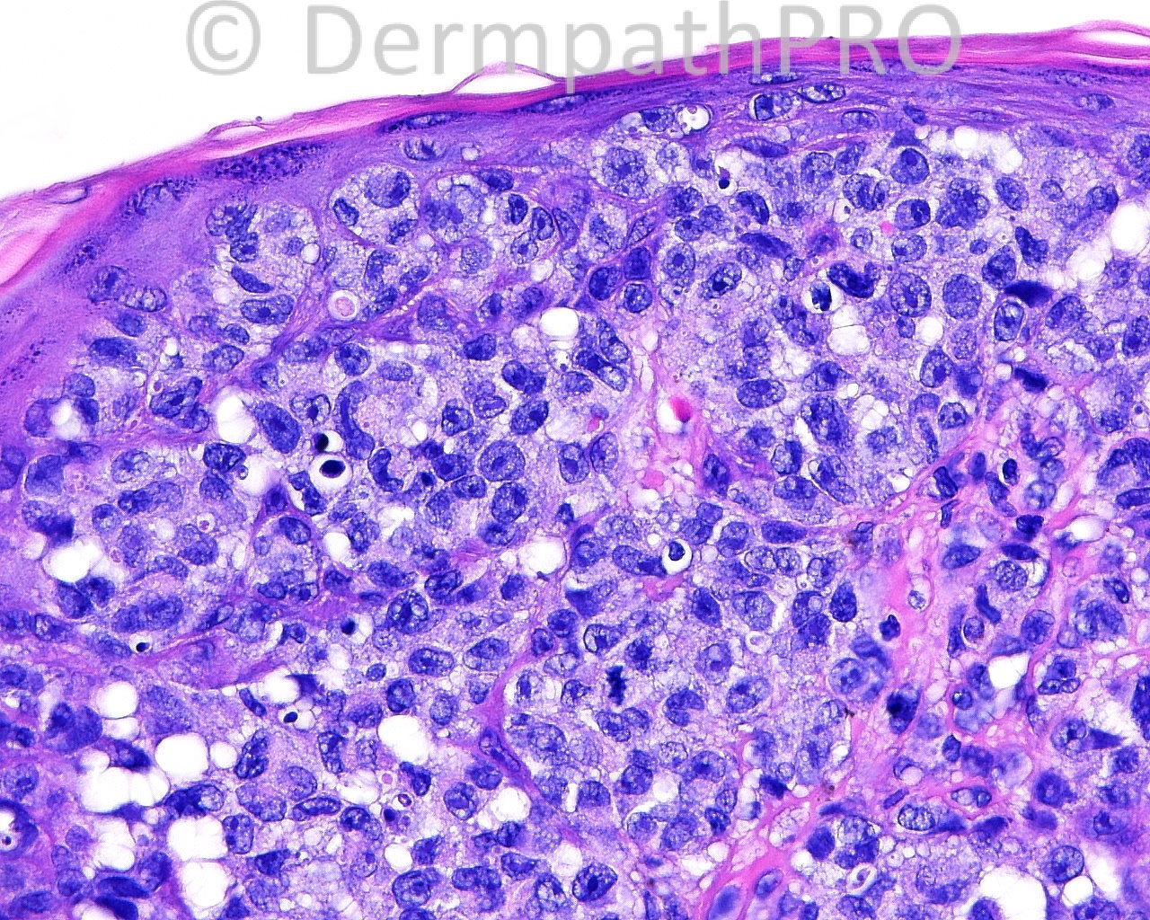
The patient is an 84 year old white woman with excisions with margin exam of enlarging lesions, present five weeks, taken from A- the left medial malar complex.

Case posted by Dr. Mark Hurt.


  Report Record

User Feedback


Guest Jim Davie MD

Posted

[size=4]In addition to melanoma, the differential would include: [/size]
SCC in situ, Merkel cell carcinoma, and sebaceous CA

This is a fast growing lesion. There is no significant melanin pigment or melanophages, equivocal squamous differentiation in some atypical cells, physaliferous vacuolation in malignant population, rapid growth, and high mitotic / apoptotic index. Stains will help differentiate.

Share this comment


Link to comment
share_externally

Dr. Mona Abdel-Halim

Posted

Wao, beautifully demonstrated,, yet scary ,, poor old lady :-((

Share this comment


Link to comment
share_externally


Join the conversation

You can post now and register later. If you have an account, sign in now to post with your account.

Guest
Add a comment...

×   Pasted as rich text.   Paste as plain text instead

  Only 75 emoji are allowed.

×   Your link has been automatically embedded.   Display as a link instead

×   Your previous content has been restored.   Clear editor

×   You cannot paste images directly. Upload or insert images from URL.

Loading...

×
×
  • Create New...