Jump to content
In this section we have spot diagnoses posted on a daily basis since June 2010, now over 4000! You can review the archived cases and read the suggested diagnoses by users and the final comment by the contributors.
Case are uploaded each week day by 10 am UK time with the correct diagnosis will generally be posted at 8 pm UK time. Why not view the most recent spot diagnosis and proffer a diagnosis?

Case Number : Case 1072 - 1st August Posted By: Guest

Please read the clinical history and view the images by clicking on them before you proffer your diagnosis.
Submitted Date :
   (0 reviews)

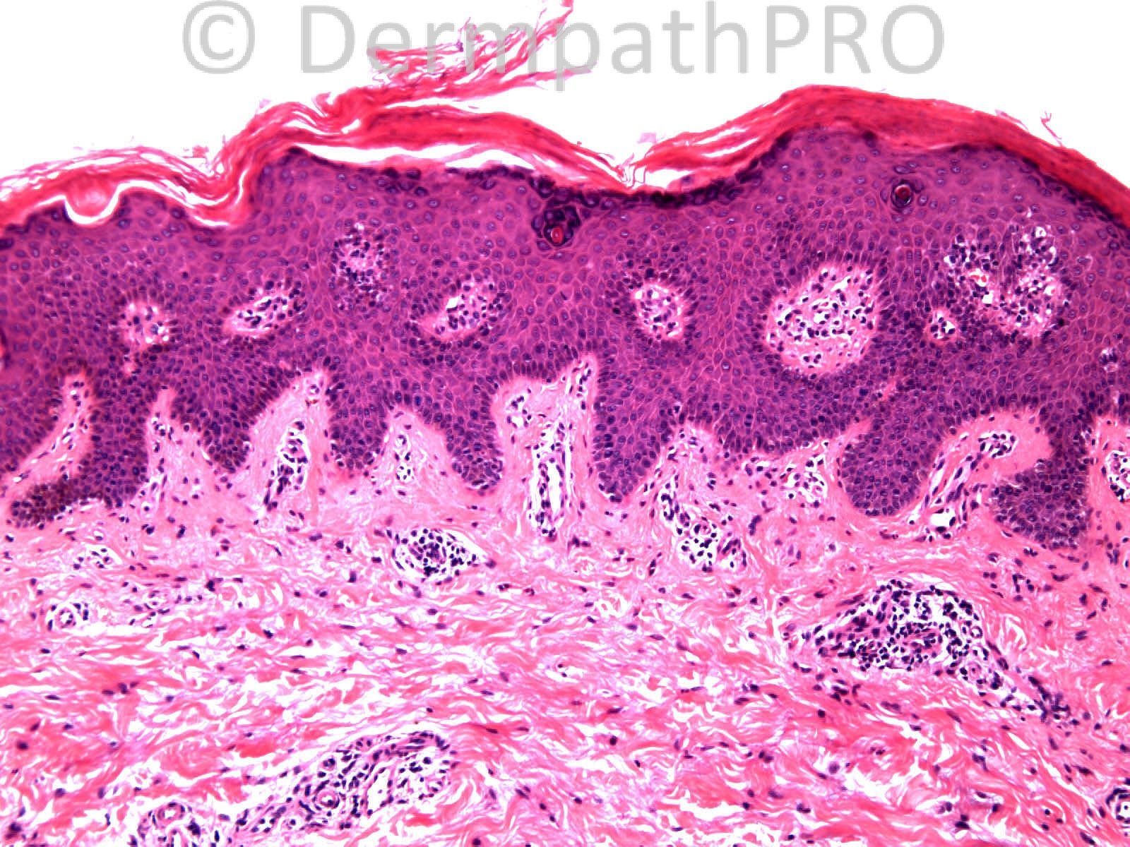
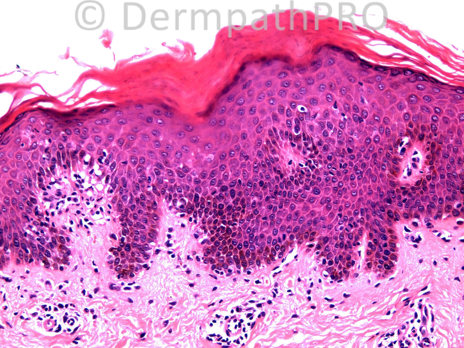
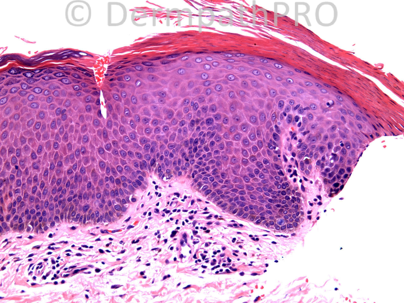
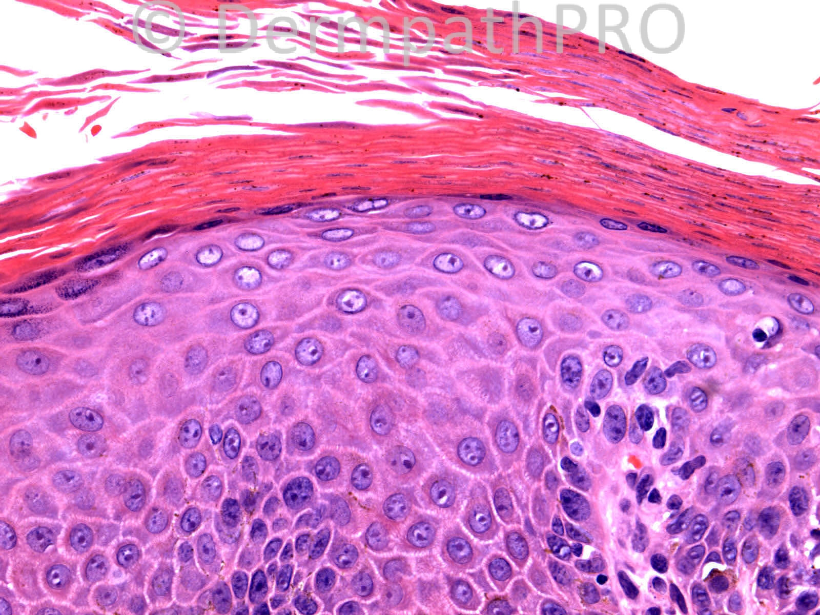
Adult female with 12 year history of extensive scaly rash. ?lichenoid.

Case Posted by Dr Richard Carr


  Report Record

User Feedback


Eman El-Nabarawy

Posted

Almost confluent parakeratosis with neutrophils, areas of hypogranulosis, psoriasiform hyperplasia, dilated capillaries in some dermal papillae, lower epidermal exocytosis. Favor psoriasis over PLC. Clinical sense is crucial. Exclusion of MF may be required.

Share this comment


Link to comment
share_externally

Guest Tiberiu Tebeica

Posted

This looks to me as a psoriasiform dermatitis, one in which the epidermal rete ridges are unevenly elongated and the suprapapillary plates are thick. Given also the suggestion of checkerboard pattern of parakeratosis depicted in pictures 4 and 6, the first diagnosis on my list is PRP.

Share this comment


Link to comment
share_externally

Robledo F. Rocha

Posted

I go with pityriasis rubra pilaris. Stratum corneum shows vertically and horizontally alternating orthokeratosis and parakeratosis. Psoriasiform epidermal hyperplasia and mild superficial perivascular lymphocytic infiltrate are also seen.

Share this comment


Link to comment
share_externally

I see neither a lichenoid nor a convincing interface pattern. There is mild, irregular psoriasiform hyperplasia and a present, if discontinuous, granular layer - which I take as being against psoriasis. Agree, PRP.

Share this comment


Link to comment
share_externally

I also go with the diagnosis of PRP, but I think picture 4 shows us more confluent parakeratosis than a checkerboard pattern.

Share this comment


Link to comment
share_externally

Dr. Richard Carr

Posted

I reported this as suggesting pityriasis rubra pilaris based on the checker-board pattern of spotty parakeratosis, supra-papillary plates not thinned and granular layer largely preserved. Certainly nothing here to suggest MF or a lichenoid dermatosis. Follow-up that after receiving the histology report the clinico-pathological correlation diagnosis also favoured PRP.
Enjoy your weekends.

Share this comment


Link to comment
share_externally


Join the conversation

You can post now and register later. If you have an account, sign in now to post with your account.

Guest
Add a comment...

×   Pasted as rich text.   Paste as plain text instead

  Only 75 emoji are allowed.

×   Your link has been automatically embedded.   Display as a link instead

×   Your previous content has been restored.   Clear editor

×   You cannot paste images directly. Upload or insert images from URL.

Loading...

×
×
  • Create New...