Jump to content
In this section we have spot diagnoses posted on a daily basis since June 2010, now over 4000! You can review the archived cases and read the suggested diagnoses by users and the final comment by the contributors.
Case are uploaded each week day by 10 am UK time with the correct diagnosis will generally be posted at 8 pm UK time. Why not view the most recent spot diagnosis and proffer a diagnosis?

Case Number : Case 1082 - 15th August Posted By: Guest

Please read the clinical history and view the images by clicking on them before you proffer your diagnosis.
Submitted Date :
   (0 reviews)

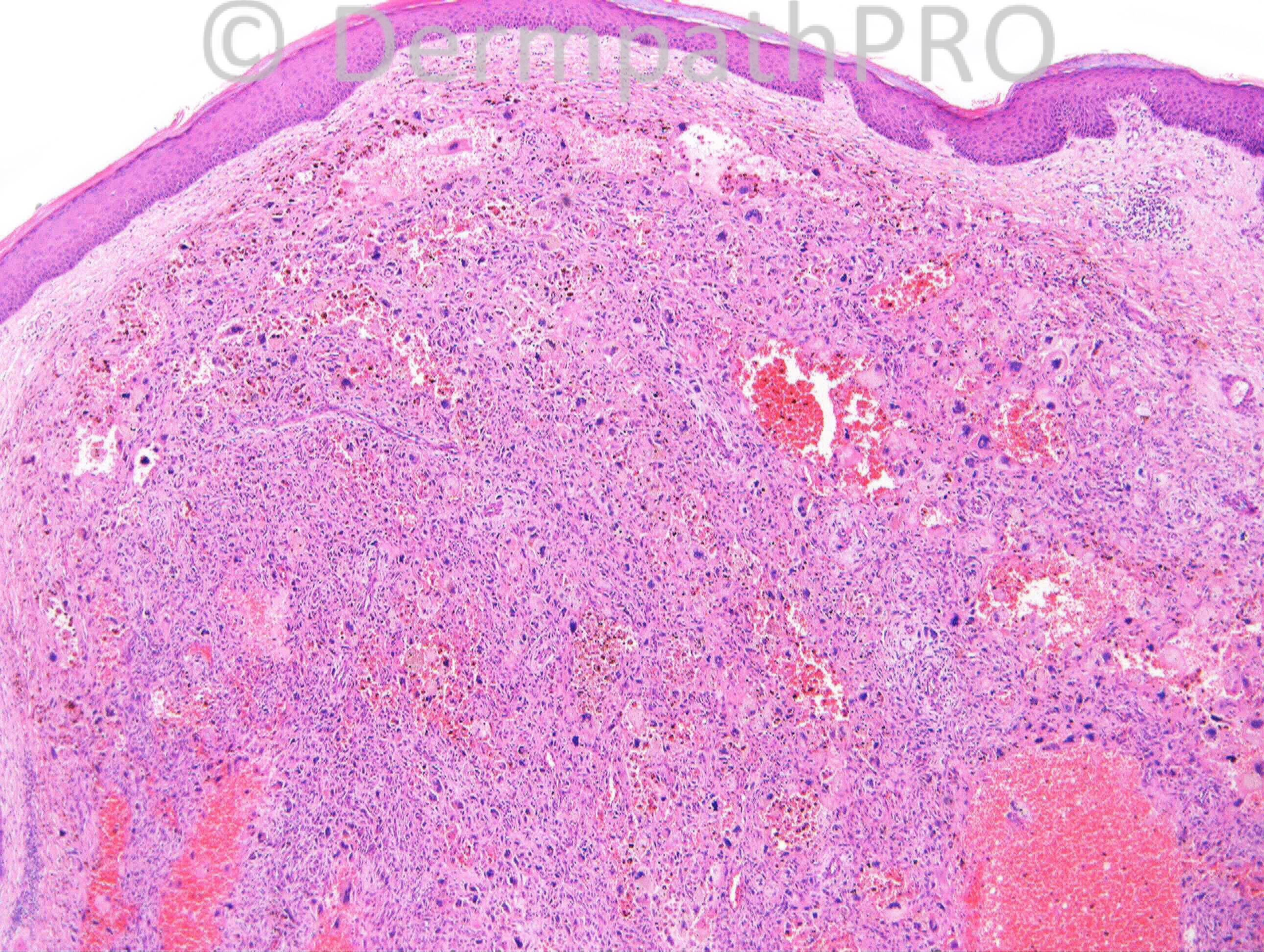
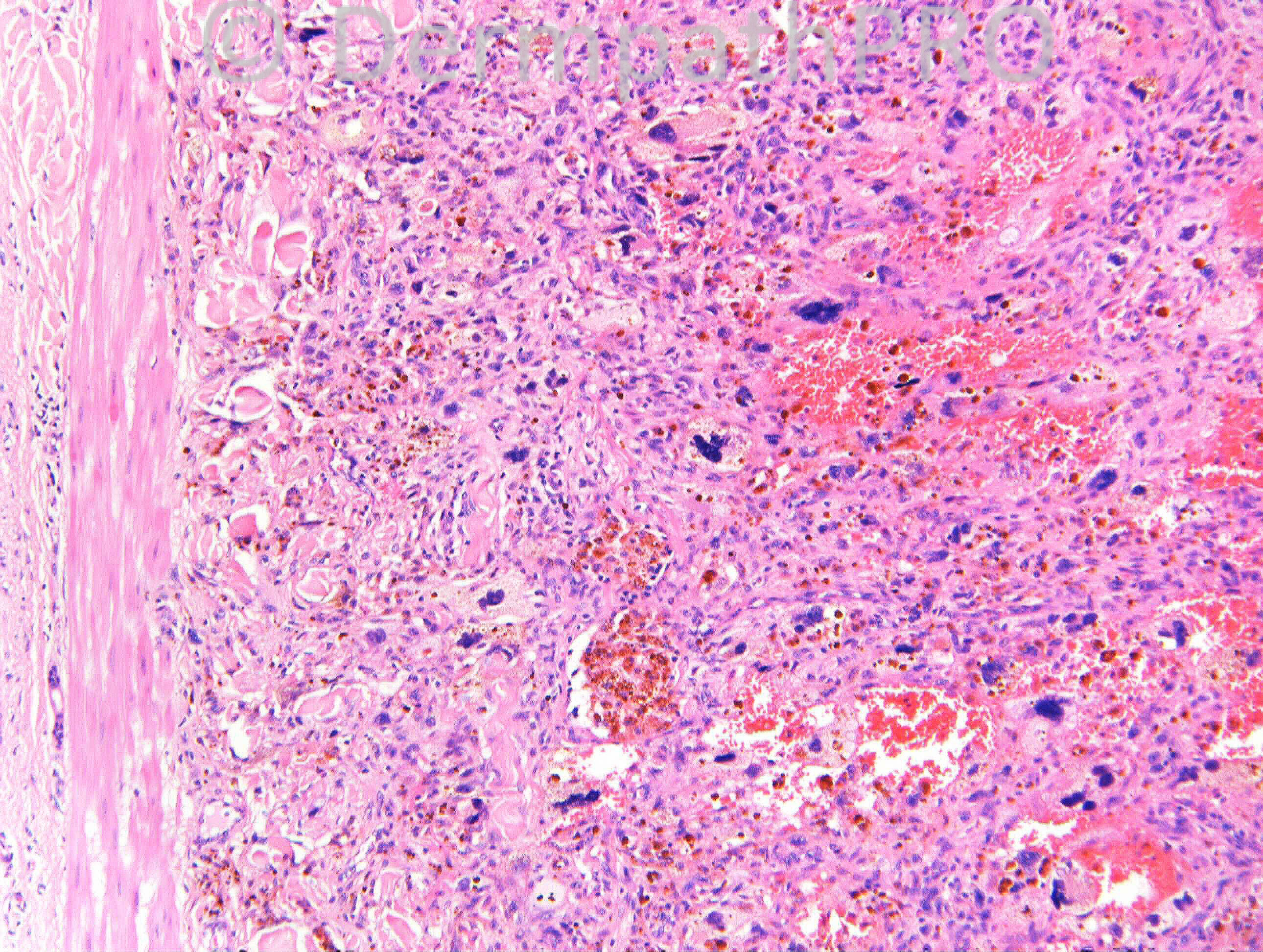
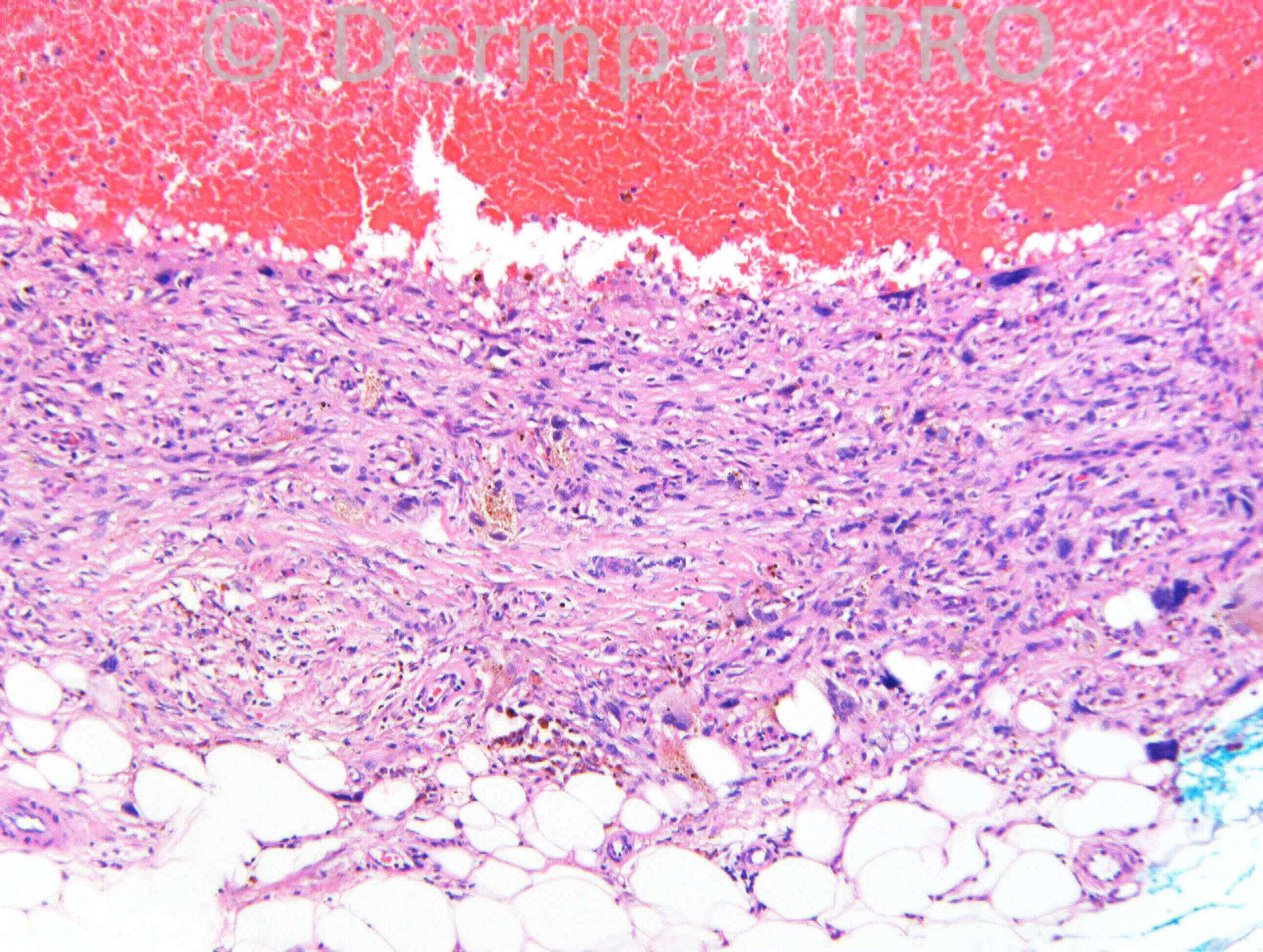
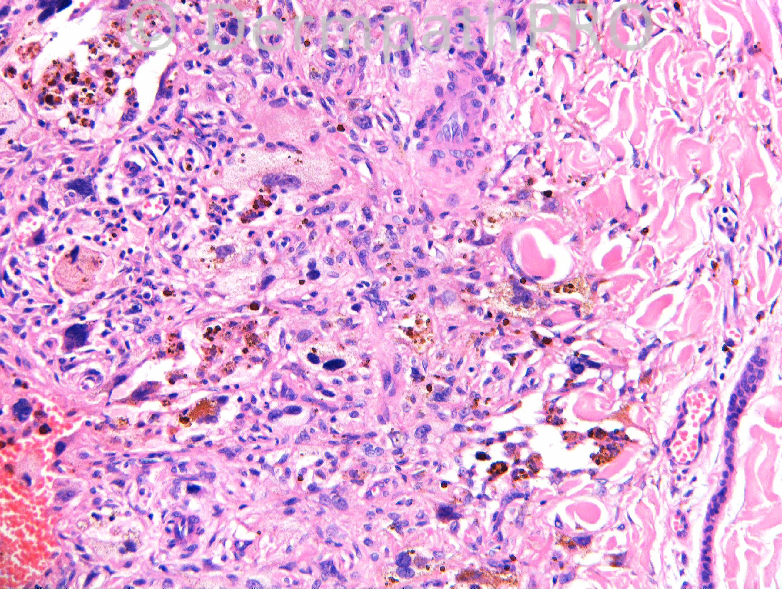
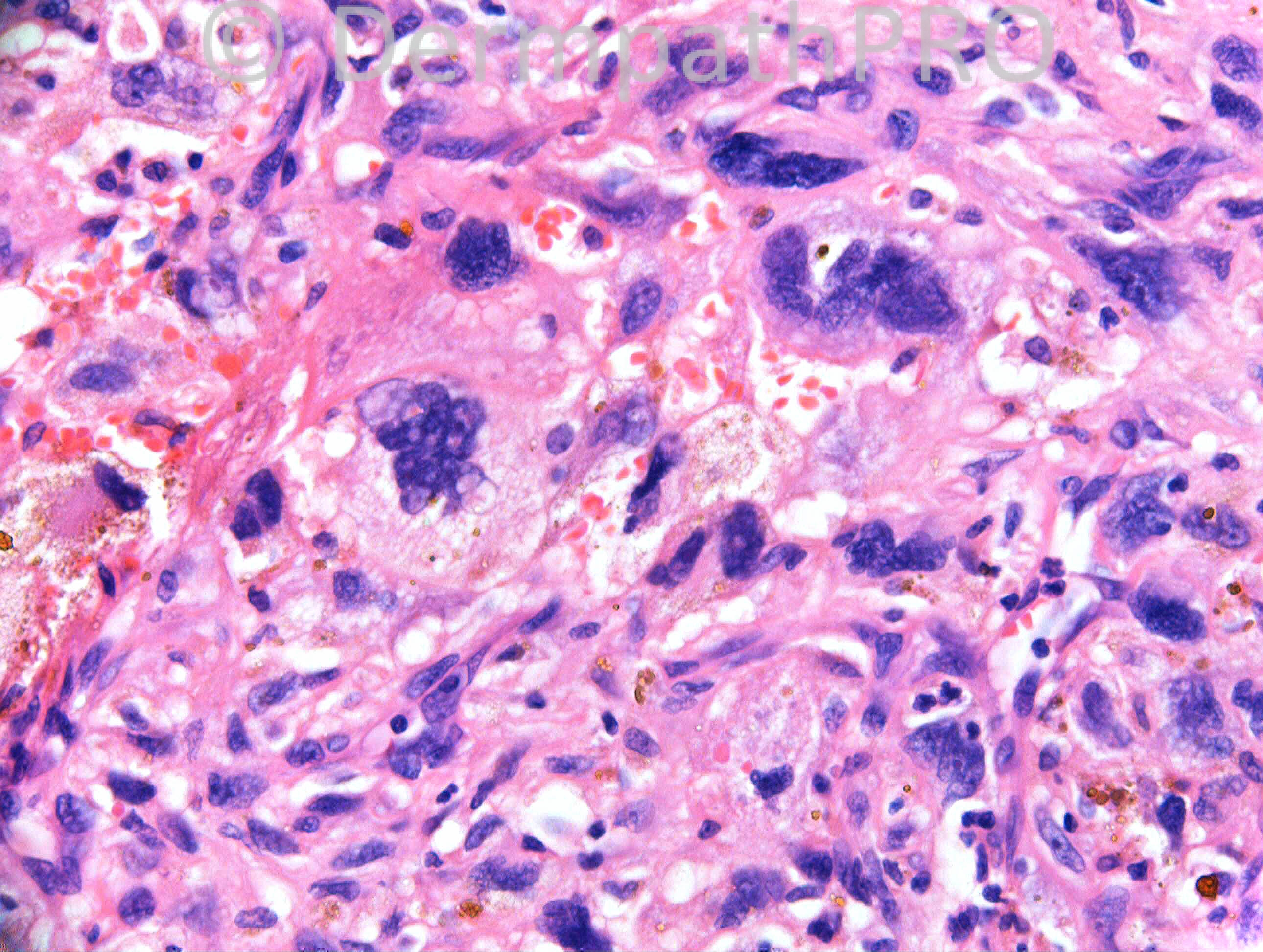
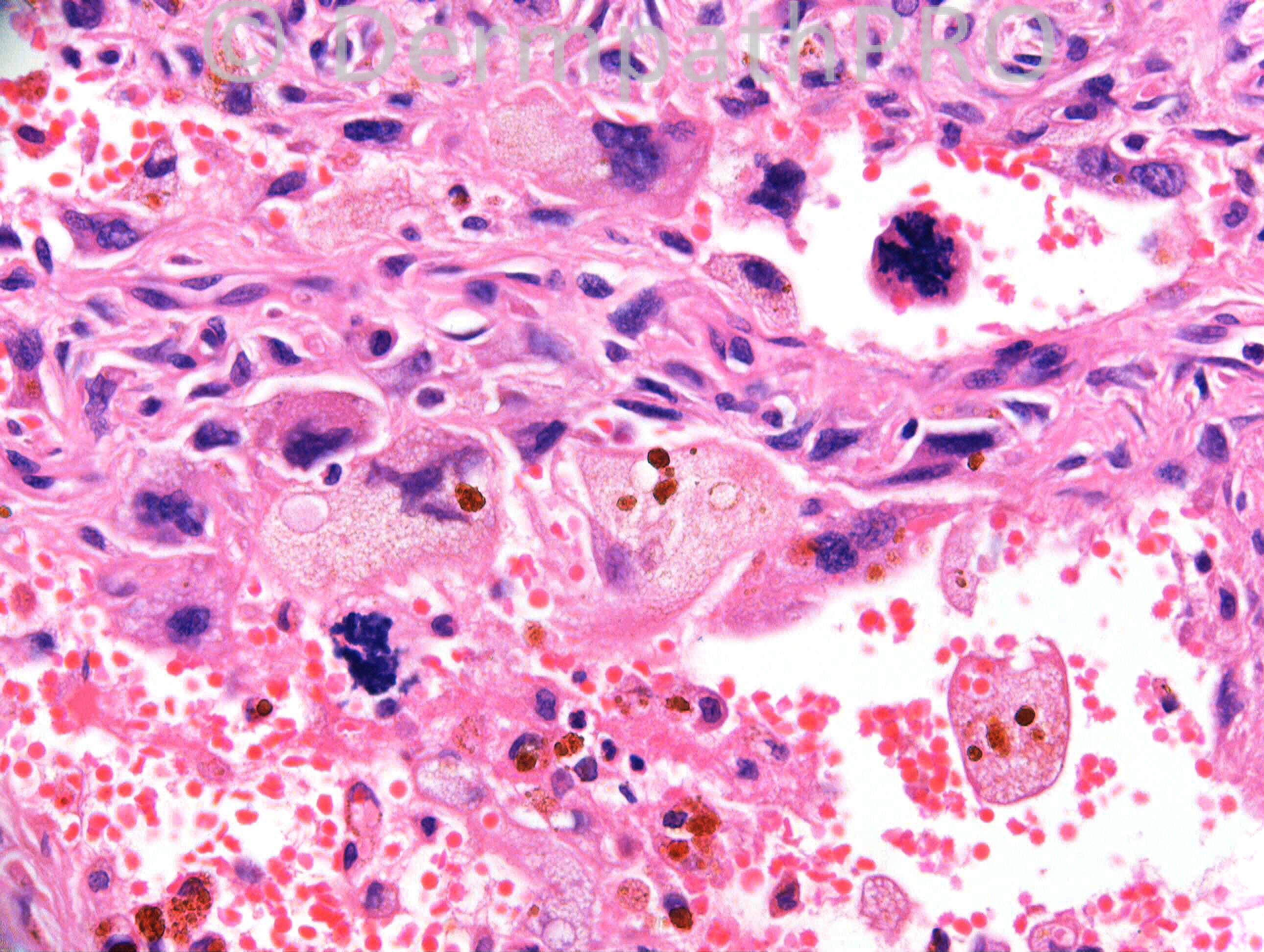
1 year, dark lump, upper thigh. h/o penile carcinoma and lymph node dissection (Case c/o Dr Kathleen Romain).

Case Posted by Dr Richard Carr

  Report Record

User Feedback


Guest Romualdo

Posted

Aneurysmal fibrous histiocytoma with monster cells.

Share this comment


Link to comment
share_externally

Atypical and aneurysmal fibrous histiocytoma. Complete excision is recommended.

Share this comment


Link to comment
share_externally

Dr. Mona Abdel-Halim

Posted

Atypical aneurysmal fibrous histiocytoma

Share this comment


Link to comment
share_externally

Arti Bakshi

Posted

Agree. The monster cells are truly monstrous in this case!!
Not sure about the atypical mitoses in the last image though...should that be concerning?

Share this comment


Link to comment
share_externally

Guest Tiberiu Tebeica

Posted

I agree with atypical aneurysmal fibrous histiocytoma. If there is history of irradiation in that anatomical region, angiosarc has to be r/o.

Share this comment


Link to comment
share_externally

Dr. Richard Carr

Posted

I called it atypical fibrous histiocytoma and cited the reference below. It is certainly scary but I think atypical mitotic figures are to be expected in tumours with monster cells (e.g. trichoepithelioma, Spitz naevus) and do not necessarily predict a malignant outcome.

Kaddu S, McMenamin ME, Fletcher CD. Atypical fibrous histiocytoma of the skin: clinicopathologic analysis of 59 cases with evidence of infrequent metastasis. Am J Surg Pathol. 2002 Jan;26(1):35-46.
Atypical fibrous histiocytoma is an uncommon, poorly documented variant of cutaneous fibrous histiocytoma. We studied 59 cases of atypical fibrous histiocytoma to better characterize the clinicopathologic spectrum. There were 33 males and 26 females (median age 38 years; range 5-79 years) with solitary lesions arising on lower (25 cases) and upper (17 cases) extremities, trunk (6 cases), head and neck (4 cases), and vulva (1 case); anatomic location was not stated in six cases. Lesions measured 0.4-8 cm in diameter (median 1.5 cm) and clinically were nodules (40 cases), polypoid tumors (18 cases), or a slightly elevated plaque (1 case). Histologically, the lesions were primarily dermal with superficial involvement of the subcutis in one third of the cases. Salient features included a proliferation of pleomorphic, plump, spindle, and/or polyhedral cells with mainly large, hyperchromatic, irregular, or bizarre nuclei, set in a background of classic features of fibrous histiocytoma, including spindle cell areas showing a storiform pattern and entrapped thickened, hyaline collagen bundles, especially at the periphery. Multinucleated giant cells, often with bizarre nuclei and foamy, sometimes hemosiderin-rich, cytoplasm were also variably present. The degree of pleomorphism varied from only focal and minimal (14 cases) or moderate (24 cases) to marked (21 cases). Mitotic activity was observed in 55 lesions, and the number of mitotic figures ranged from 1 to 15 per 10 high power fields. Atypical mitoses were noted in 20 lesions. Furthermore, some cases of atypical fibrous histiocytoma displayed other worrisome features less often observed in ordinary FH, including unusually large size (diameter >2 cm, 8 cases), involvement of the superficial subcutis (19 cases), and geographic necrosis (7 cases). Immunohistochemical studies performed in 42 cases showed only focal smooth muscle actin (10 cases) and CD34 (4 cases) positivity, whereas CD68, S-100 protein, desmin, pan-keratin, and epithelial membrane antigen were negative. Clinical follow-up data available in 21 patients (mean duration of follow-up 50.6 months, median 43 months) revealed local recurrences in three patients (one repeated); two patients developed distant metastases, one of whom died after 96 months. These two cases were not histologically distinct from the group as a whole. We conclude that atypical fibrous histiocytoma has a broader clinicopathologic spectrum than previously realized. Lesions with floridly atypical features represent potential pitfalls for overinterpretation as pleomorphic sarcoma, which would appear to be inappropriate in most cases. Provided that atypical fibrous histiocytoma is treated by complete excision, a benign outcome is to be expected in most cases. However, similar to the cellular and aneurysmal variants of fibrous histiocytoma, atypical fibrous histiocytoma shows a higher tendency to recur locally than ordinary fibrous histiocytoma and may rarely metastasize.

Share this comment


Link to comment
share_externally

Dr. Richard Carr

Posted

I don't think I can refute the argument based on our current knowledge. Cases such as this challenge our concepts tremendously and I certainly don't feel at peace at all but I have to sleep at night so must be pragmatic and explain that the diagnostic label is one thing, predicting the potential behaviour is another thing altogether. In this case the group label suggests a low risk for metastasis. Whether there are individual tumour or host factors that affected the behaviour in the lesions that did and did not metastasise remains to be determined. Perhaps some are essentially benign and do not have any inate capacity (at the time of removal) to metastasise and kill and some are malignant and do have that capacity. I don't think anyone can currently answer these questions.

Share this comment


Link to comment
share_externally


Join the conversation

You can post now and register later. If you have an account, sign in now to post with your account.

Guest
Add a comment...

×   Pasted as rich text.   Paste as plain text instead

  Only 75 emoji are allowed.

×   Your link has been automatically embedded.   Display as a link instead

×   Your previous content has been restored.   Clear editor

×   You cannot paste images directly. Upload or insert images from URL.

Loading...

×
×
  • Create New...