Jump to content
In this section we have spot diagnoses posted on a daily basis since June 2010, now over 4000! You can review the archived cases and read the suggested diagnoses by users and the final comment by the contributors.
Case are uploaded each week day by 10 am UK time with the correct diagnosis will generally be posted at 8 pm UK time. Why not view the most recent spot diagnosis and proffer a diagnosis?

Case Number : Case 1162- 5th December Posted By: Guest

Please read the clinical history and view the images by clicking on them before you proffer your diagnosis.
Submitted Date :
   (0 reviews)

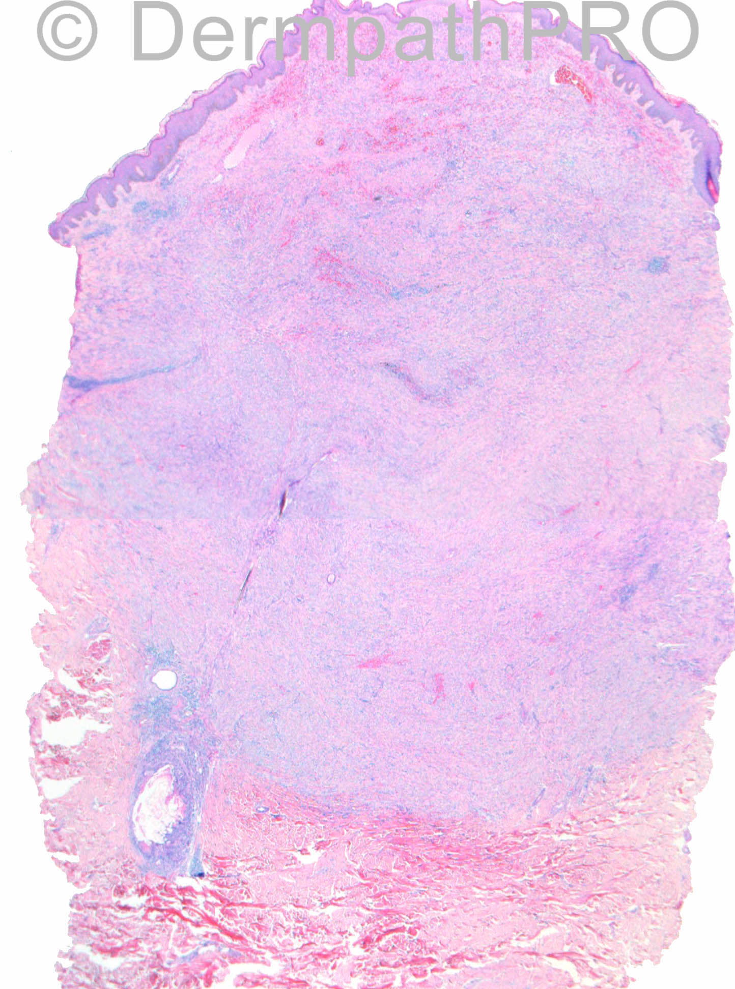
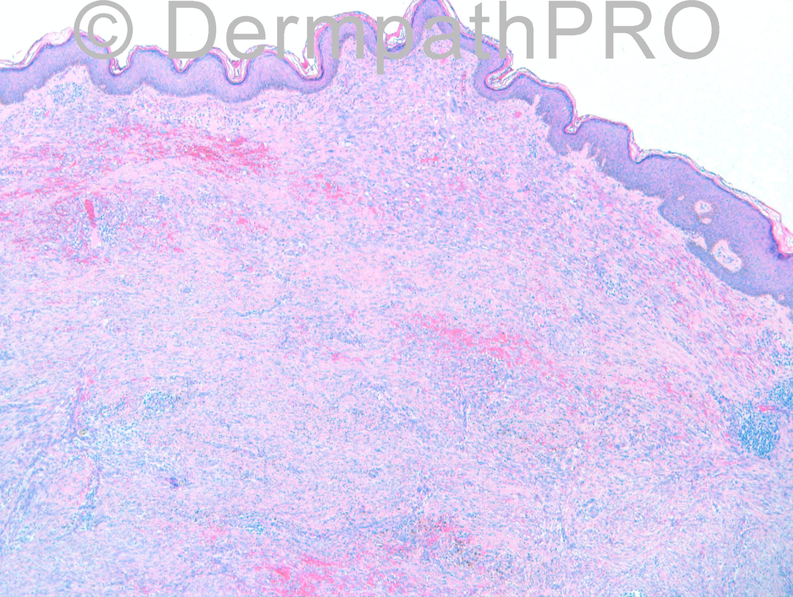
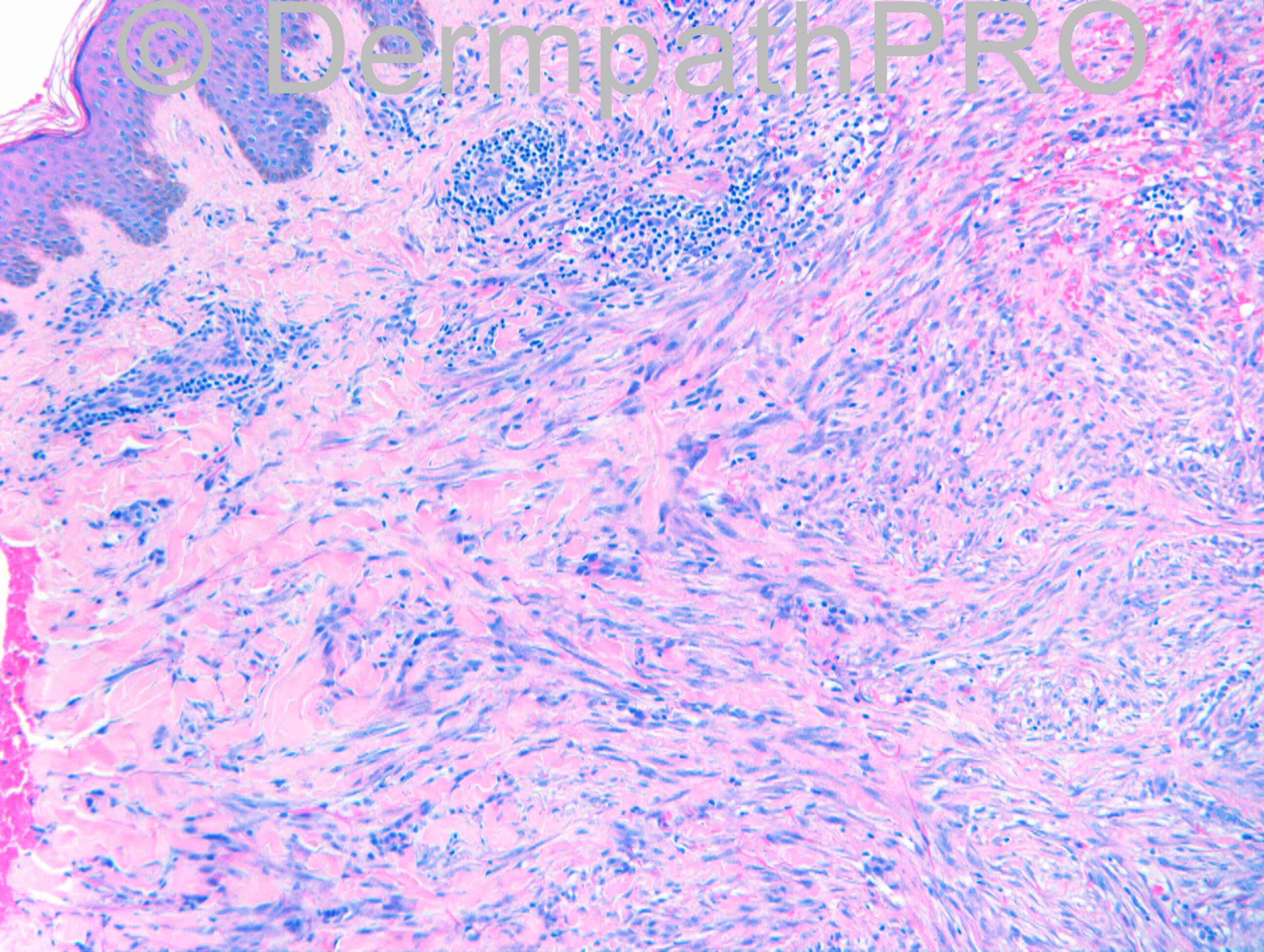
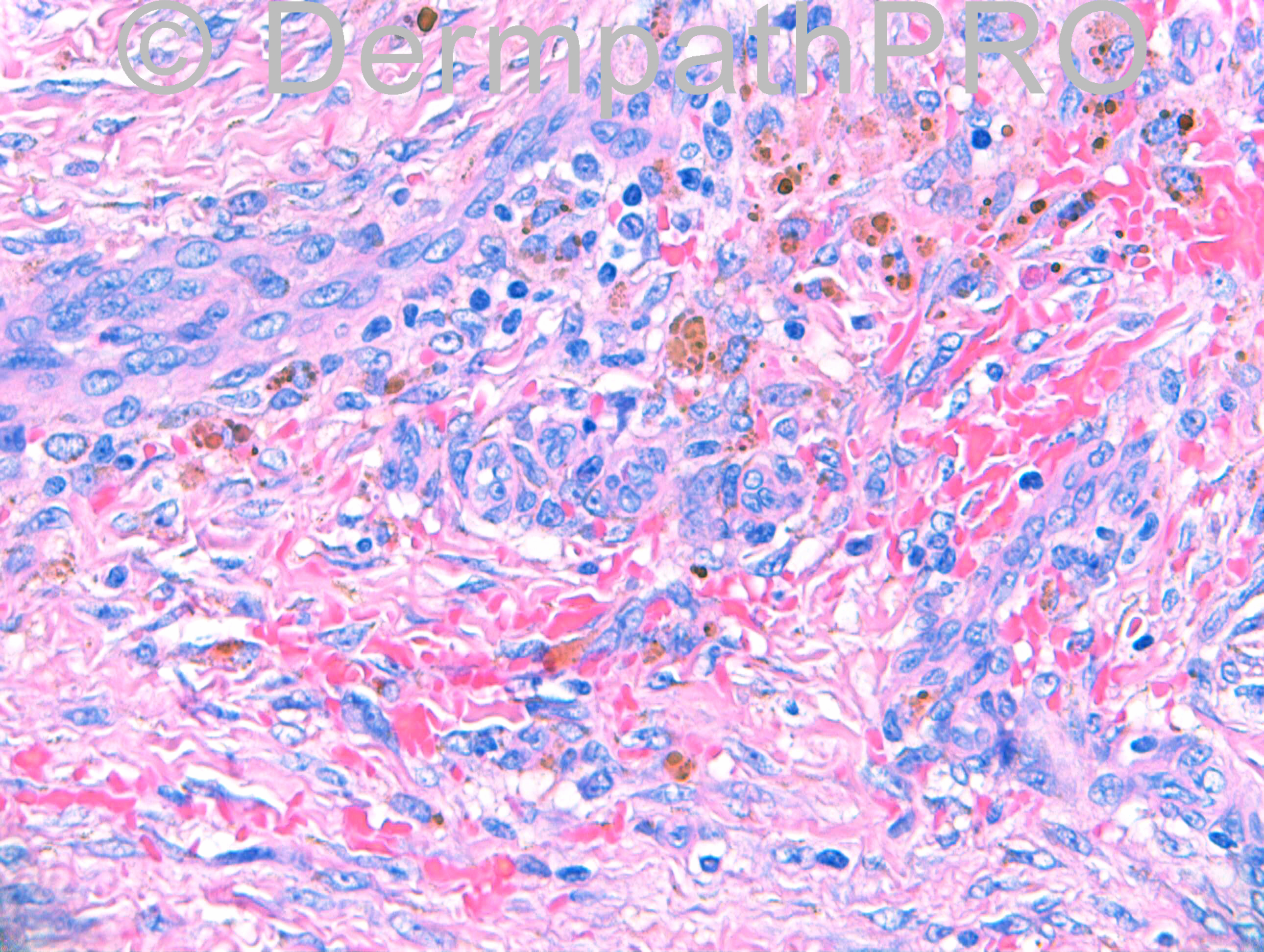
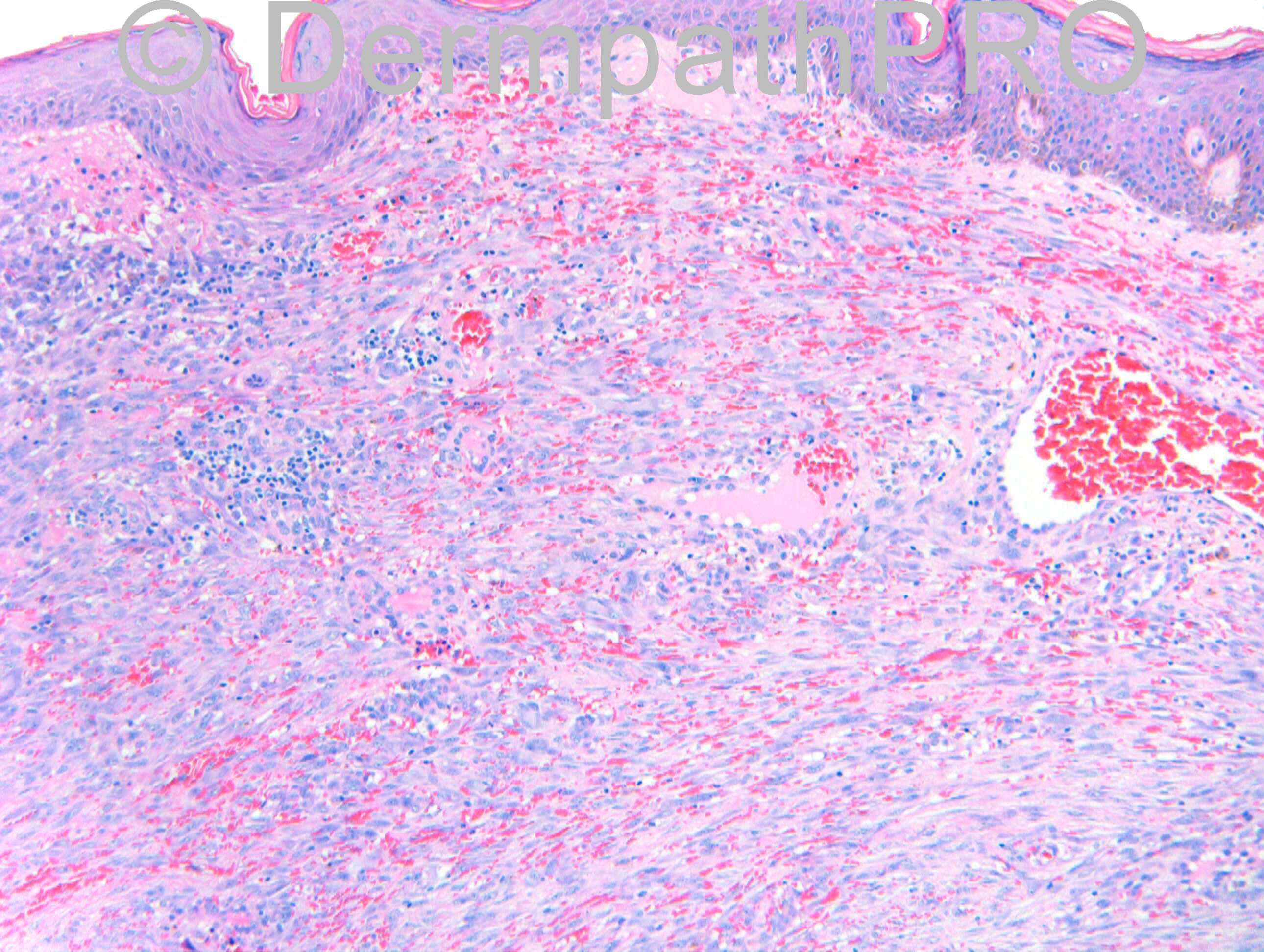
Case History:M26. Keloid-like lesion on back. No h/o trauma. ?Keloid, ?Dermatofibroma, ?Other spindle cell tumour

Case Posted By Dr.Richard Carr

  Report Record

User Feedback


Guest Romualdo

Posted

Dermatofibroma, possibly cellular and secondary to folliculitis.

Share this comment


Link to comment
share_externally

Dr. Richard Carr

Posted

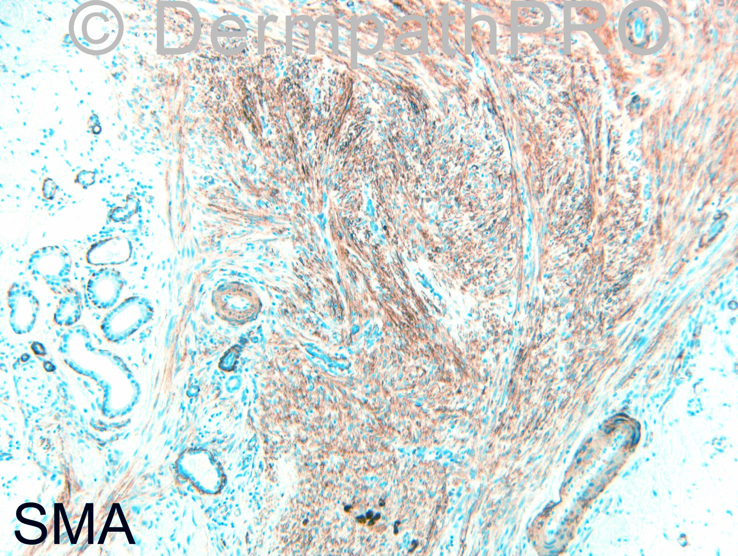
I had hoped the IHC would be posted 4pm but never mind.

Share this comment


Link to comment
share_externally

Guest Romualdo

Posted

[font=arial,helvetica,sans-serif][size=4][color=#222222]Mona, according to these articles desmin positivity is seen in about 10 % of dermatofibromas in general and 32 % of cellular benign fibrous histiocytomas.[/color]
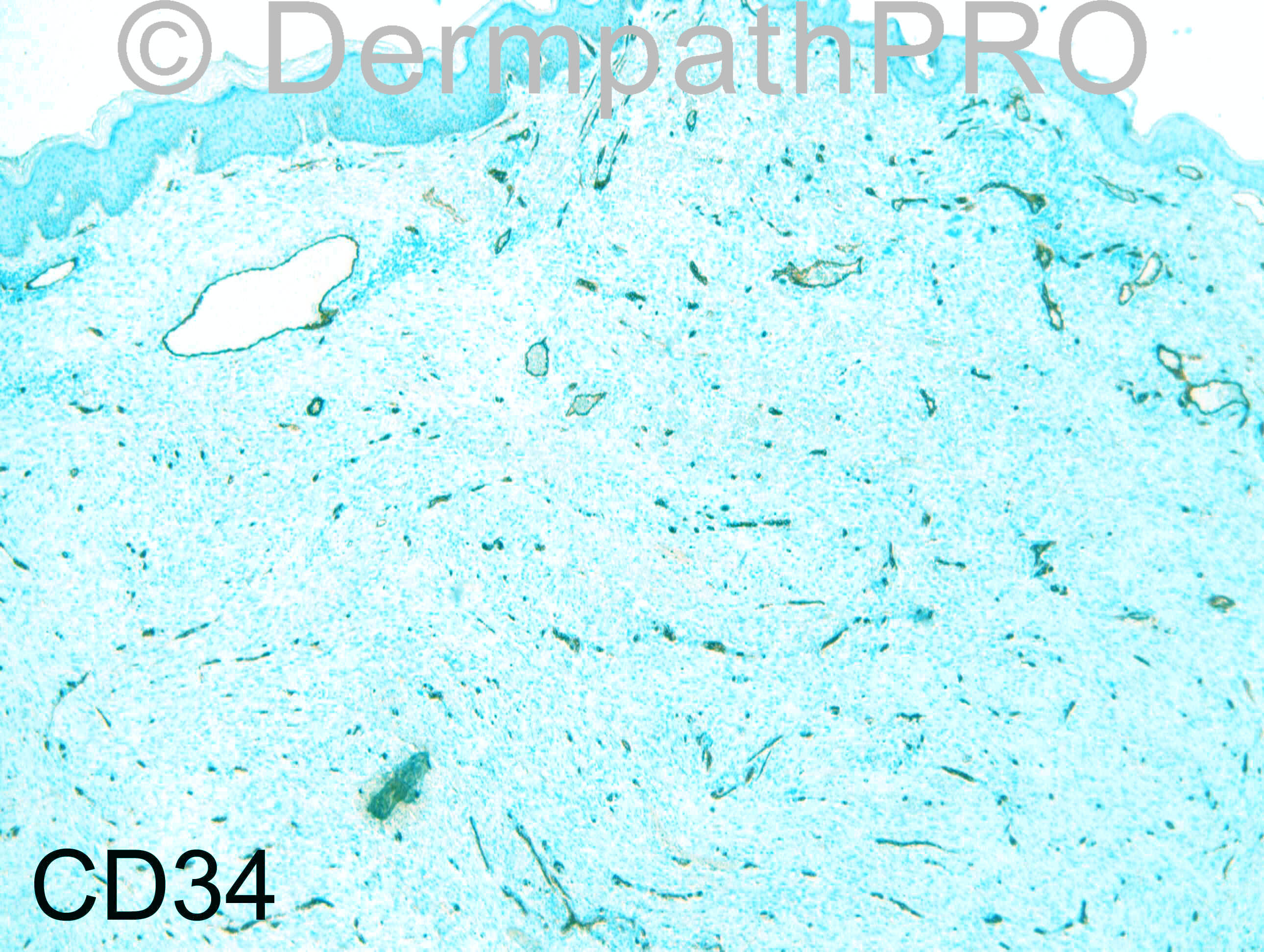
[url="http://www.ncbi.nlm.nih.gov/pubmed/22775584"][b][color=#2222CC]Desmin[/color][/b][color=#2222CC] and CD34 positivity in [b]cellular[/b] fibrous histiocytoma: an immunohistochemical analysis of 100 cases.[/color][/url]

[color=#000000]Volpicelli ER, [b]Fletcher[/b] CD.[/color]

[color=#000000]J Cutan Pathol. 2012 Aug;39(8):747-52[/color]


[url="http://www.ncbi.nlm.nih.gov/pubmed/12296754"][color=#2222CC]Expression of [b]desmin[/b] and [b]smooth muscle[/b] myosin heavy chain in dermatofibromas.[/color][/url]

[color=#000000]Bruecks AK, Trotter MJ.[/color]

[color=#000000]Arch Pathol Lab Med. 2002 Oct;126(10):1179-83 [/color][/size][/font]

Share this comment


Link to comment
share_externally

Robledo F. Rocha

Posted

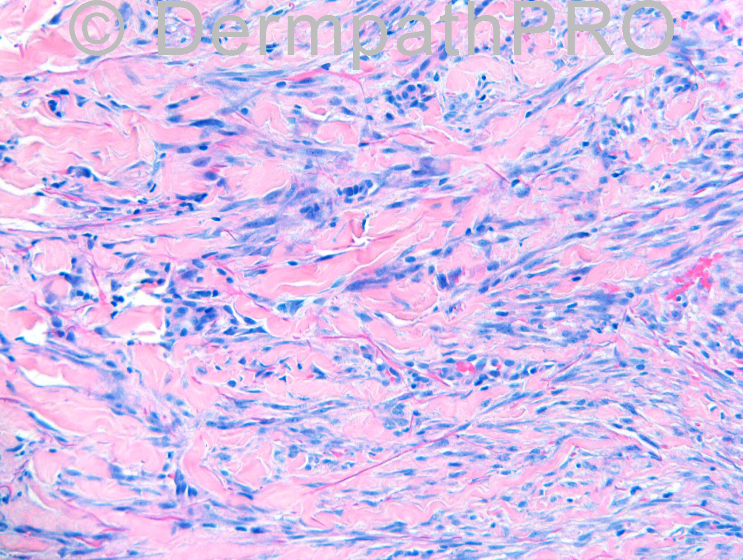
I also go with dermatofibroma. Extravasated erythrocytes, hemosiderin-laden macrophages, lymphocytic infiltrate, and peripheral collagen entrapment are some findings that point to this diagnosis. Whether follicular rupture is the cause or a consequence of dermatofibroma, well it’s another debate...

Share this comment


Link to comment
share_externally

Sasi Attili

Posted

Difficult case- Agree that this is in the spectrum of a benign cellular hemosiderotic fibrous histiocytoma

Share this comment


Link to comment
share_externally

Dr. Richard Carr

Posted

Good discussion:
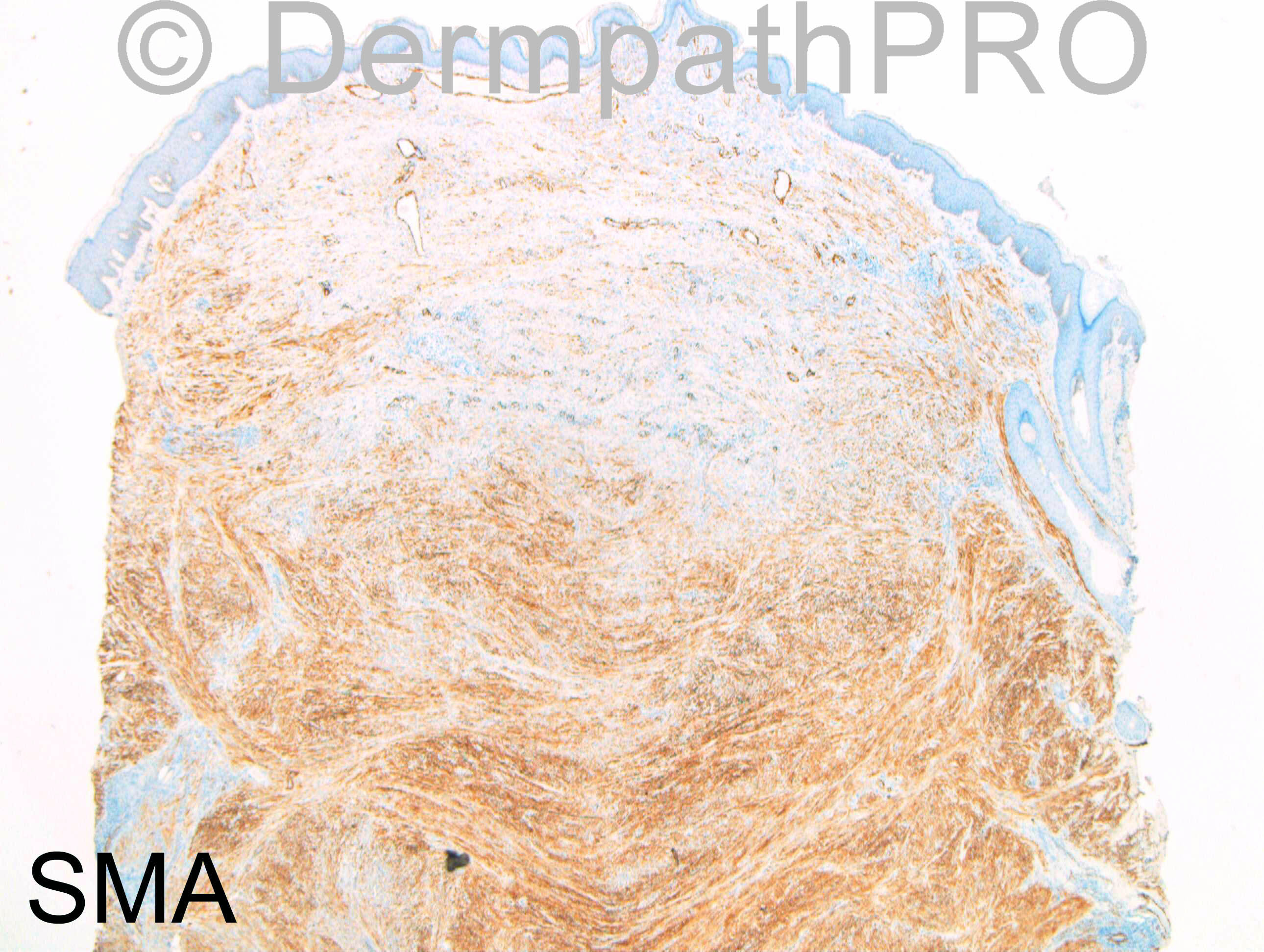
My diagnosis: Cellular fibrous histiocytoma (rather fascicular and desmin positive)

This was a referred case initially reported as favouring a leiomyosarcoma and referred to soft tissue pathologists who favoured a scar. My comments as follows:

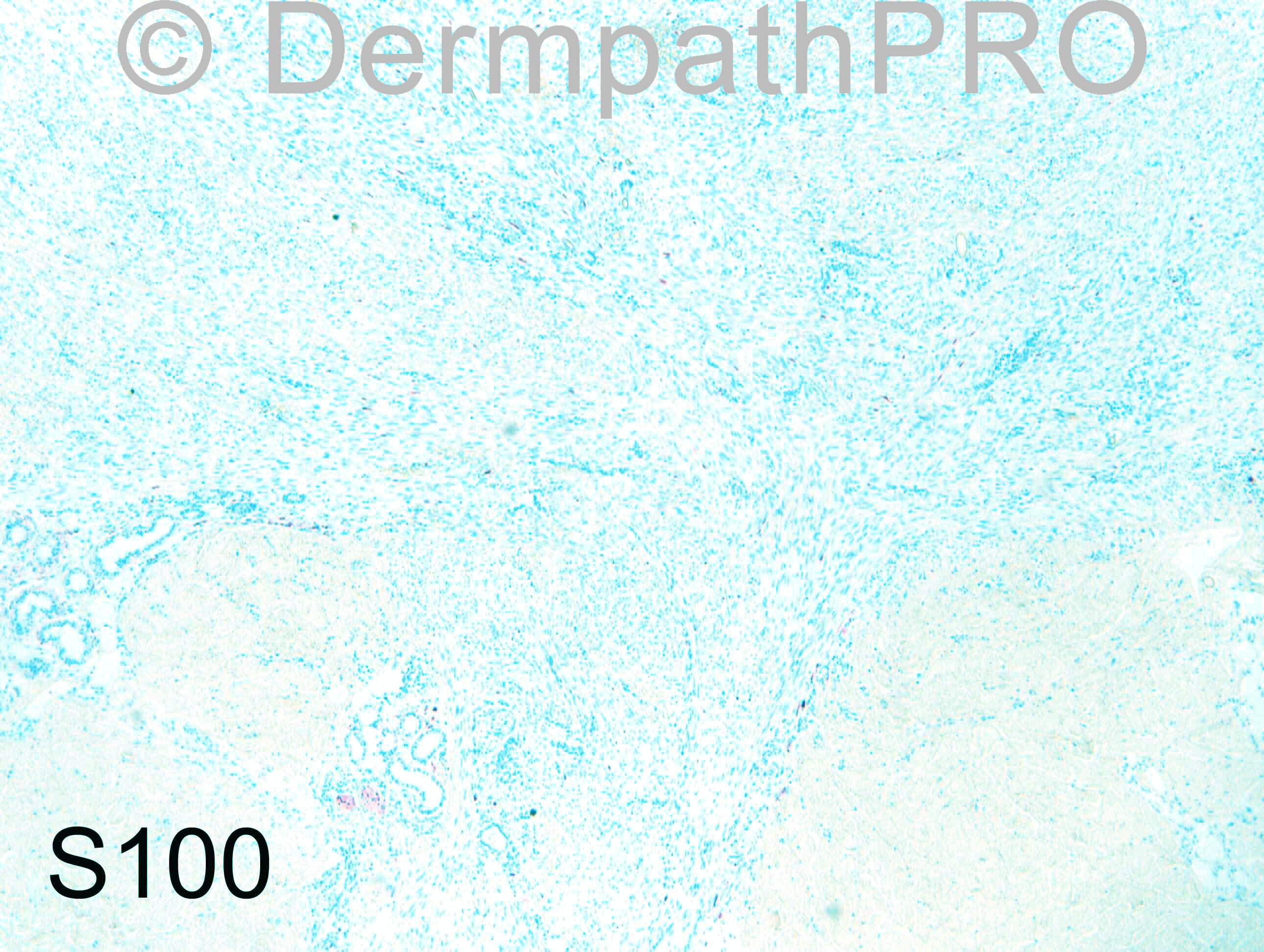
Many thanks for asking me to see this fascinating and challenging case. In the initial specimen, morphologically I interpreted this as a rather cellular monotonous and fasicular dermatofibroma/fibrous histiocytoma. Clues to the diagnosis are the more cellular superficial areas with interstitial haemorrhage and iron. The preservation of elastic fibres throughout the lesion are against scar tissue. The cells have myofibroblastic appearances lacking the typical cigar nuclei of the smooth muscle cell tumour. Widespread SMA and focal Desmin is reported in cellular fibrous histiocytomas (Volpicelli ER, Fletcher CD. J. Cutan. Pathol. 2012;39:747-52). It is common to recommend complete excision of these lesions as they can grow to large size and local recurrence is frequent. In the re-excision specimen, there is central scar tissue that merges almost imperceptibly with residual fibrous histiocytoma, but focally abuts closely to a radial margin, although the cellularity is reduced compared with that of the initial specimen. I think it would be reasonable to adopt a wait and watch policy in this case with the small potential for local recurrence noted.

Share this comment


Link to comment
share_externally


Join the conversation

You can post now and register later. If you have an account, sign in now to post with your account.

Guest
Add a comment...

×   Pasted as rich text.   Paste as plain text instead

  Only 75 emoji are allowed.

×   Your link has been automatically embedded.   Display as a link instead

×   Your previous content has been restored.   Clear editor

×   You cannot paste images directly. Upload or insert images from URL.

Loading...

×
×
  • Create New...