Jump to content
In this section we have spot diagnoses posted on a daily basis since June 2010, now over 4000! You can review the archived cases and read the suggested diagnoses by users and the final comment by the contributors.
Case are uploaded each week day by 10 am UK time with the correct diagnosis will generally be posted at 8 pm UK time. Why not view the most recent spot diagnosis and proffer a diagnosis?

Case Number : Case 1172 - 19th December Posted By: Guest

Please read the clinical history and view the images by clicking on them before you proffer your diagnosis.
Submitted Date :
   (0 reviews)

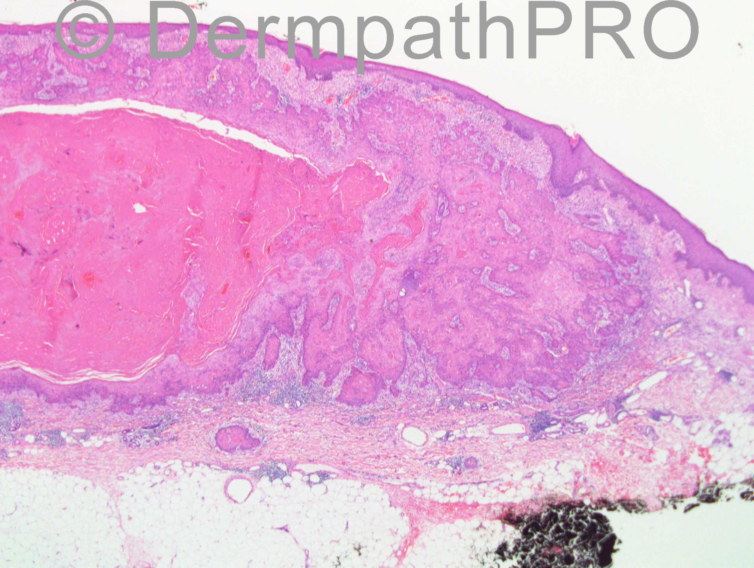
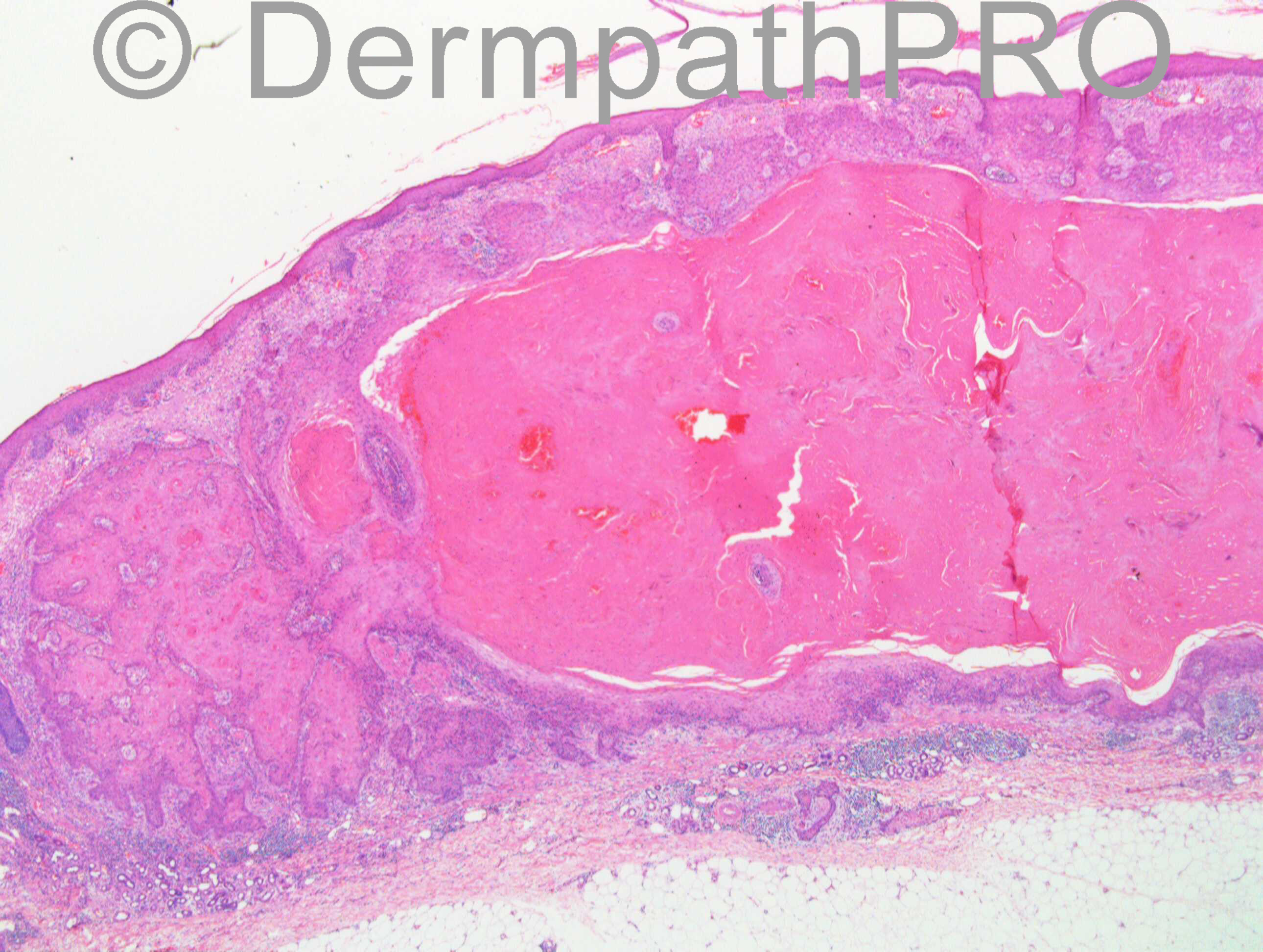
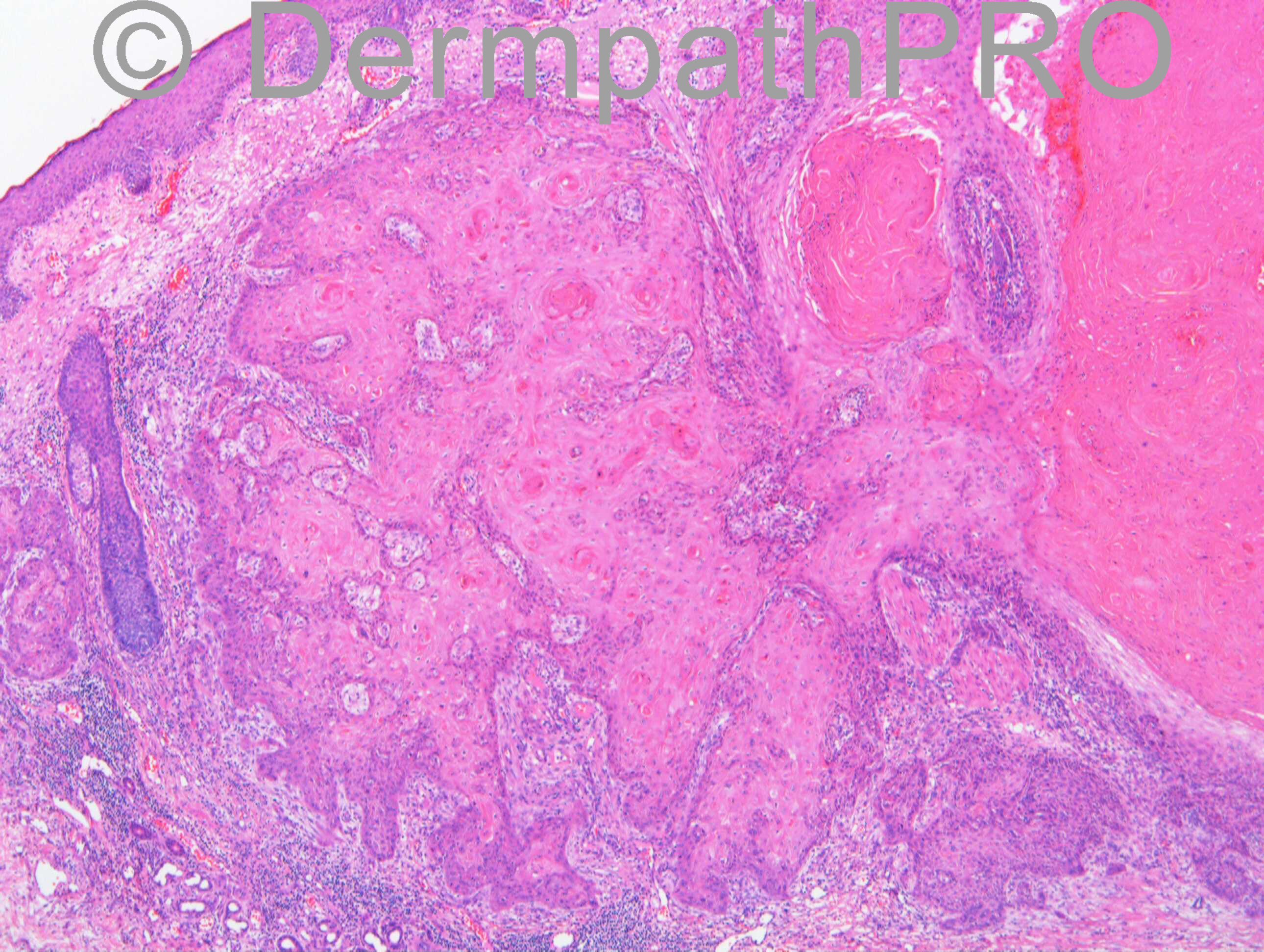
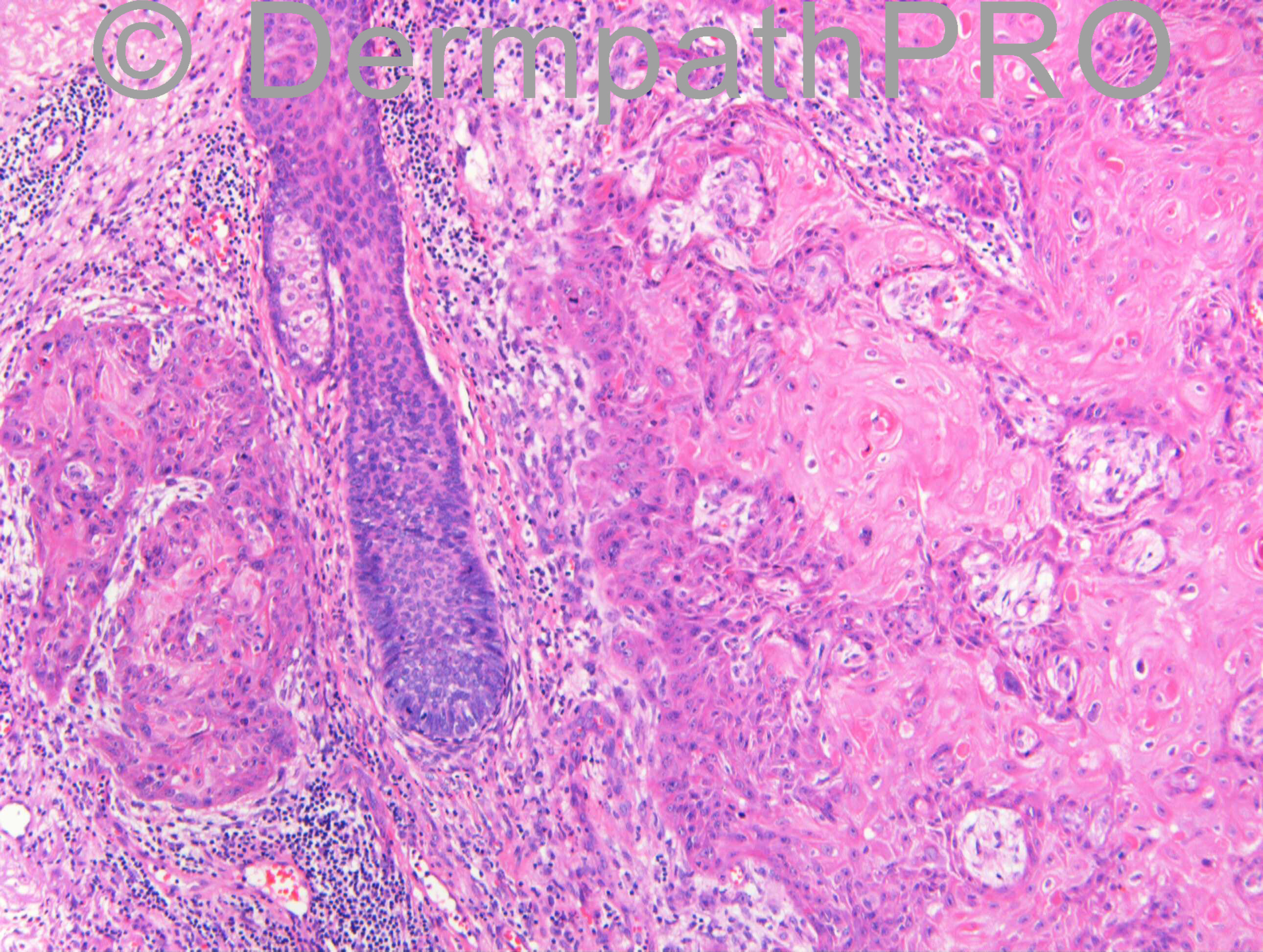
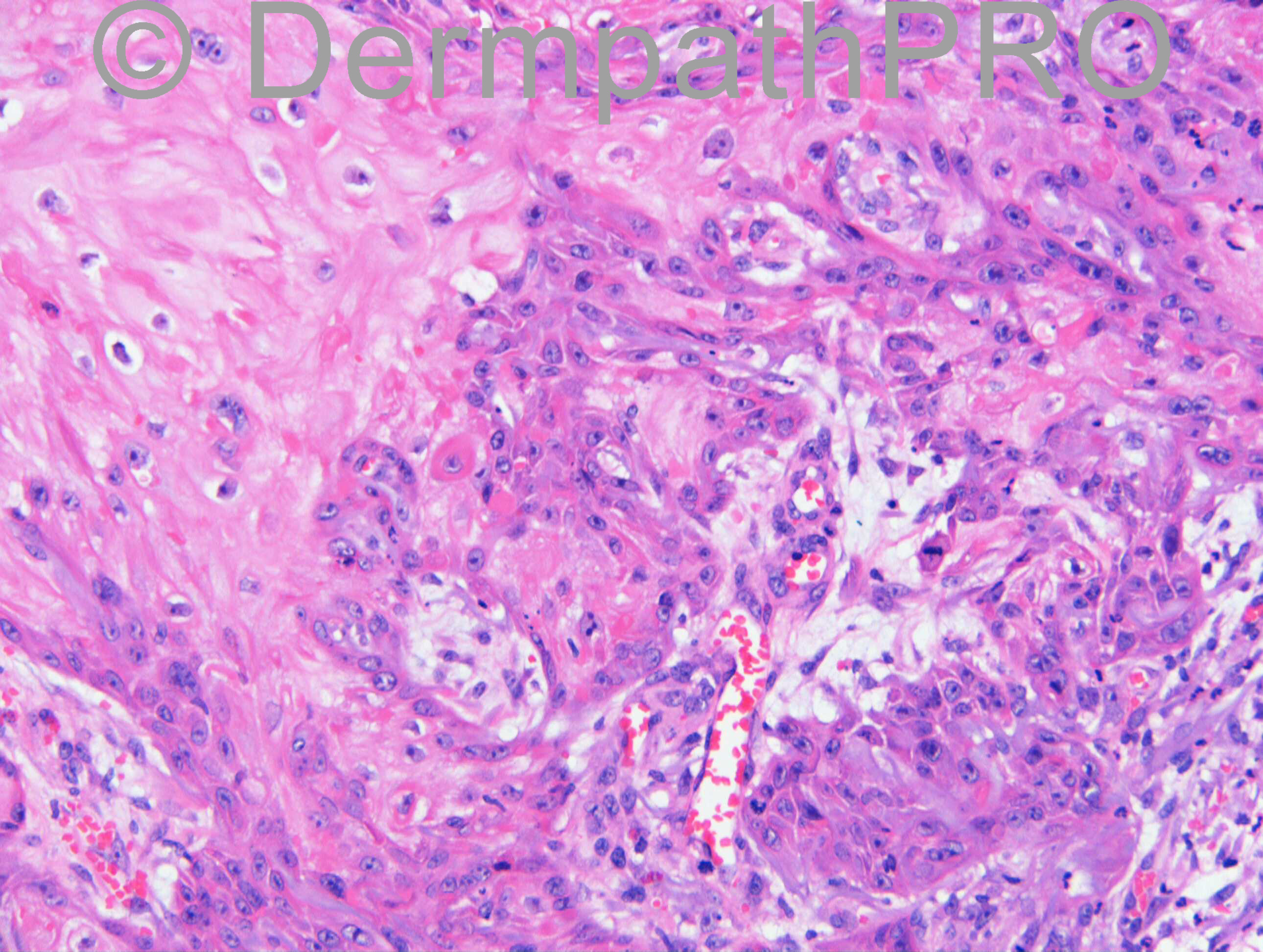
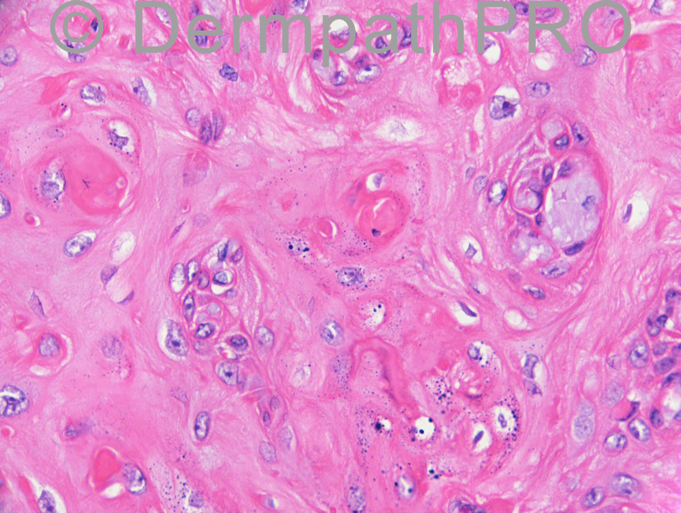
F75. Left upper arm.

Case Posted by Dr.Richard Carr

  Report Record

User Feedback


SCC. Despite the belief that perineural invasion does not equate malignancy, most, if not all, of the pathologists I have worked under will call this an SCC.

Share this comment


Link to comment
share_externally

Guest Giovanni Falconieri

Posted

KA vs hyperkeratotic SCC, favor the latter due to the deep front of invasion (i.e. beyond the eccrine gland coils) and the growth pattern undermining the adjacent epidermis (? cuniculate carcinoma)

Share this comment


Link to comment
share_externally

Sasi Attili

Posted

Favour KA, though I never make a diagnosis of KA without CPC, particularly given the age of the patient!

Share this comment


Link to comment
share_externally

Robledo F. Rocha

Posted

I'd prefer to call it squamous cell carcinoma. Neoplastic tongues have infiltrative pattern of growth and they extend from the base below the level of the eccrine gland coils.

Share this comment


Link to comment
share_externally

Guest Tiberiu Tebeica

Posted

I would call this well differentiated squamous cell carcinoma.

Share this comment


Link to comment
share_externally

Dr. Richard Carr

Posted

This feels a little bit like deja vu (still happy to provide my review on follicular SCC to anyone who wants it [email="richard.carr@swft.nhs.uk"]richard.carr@swft.nhs.uk[/email]). I did not see any features that really favoured SCC over KA histologically and even without a clinical history the appearances strongly support KA. I would draw your attention to the nice arciform lower border around the level of the sweat gland coils but no deeper (excluding the perineural invasion), strikingly infiltrative / proliferative borders with elastic entrapment but fairly abrupt central full maturation [u][b]in all areas[/b][/u] and a clue that is exceedingly helpful in the last image of elastic entrapped in fully mature squamous epithelium (to me indicating prior remarkable proliferation around the connective tissue elements without destruction and subsequent maturation). Note the absence of the more typical rounded pushing borders with peripheral palisading of well differentiated follicular SCC, and asbence of spontaneous acantholysis or intra-epithelial pools of follicular mucin. Poorly differentiated fSCC, although frequently crateriform with superficial well differentiated areas that can be treacherous the deeper portion tends to be more vertical going straight down through subcutis with asymmetry and solid areas that run through the tissue with elastic entrapment occasionally [u][b]but lacks full maturation in all areas[/b][/u]. In my experience neither well or poorly differentiated subtypes of fSCC has elastic entrapment in the fully matured central areas although you might occasionally see a simmulant of that in some types of follicular pseudoepitheliomatous hyperplasia with perforation of elastic and collagen. Because of the absence of history I have used the term "in keeping with" but could easily in this case exchanged "with typical histological features of". The case should be discussed in multi-disciplinary team meeting but I think a wait and watch could be adopted in this instance according to literature available perineural invasion in otherwise typical KA does not carry any untoward clinical implication.
My report as follows:
Well differentiated squamoproliferative lesion in keeping with [u][b]keratoacanthoma[/b][/u].
Depth: 4 mm.
Radial margin: 2 mm.
Deep margin: 1 mm.
Clark level: 4.
Widespread perineural invasion present.
No vascular invasion.
MDM discussion advised.

[b]Reference[/b]
Godbolt AM, Sullivan JJ, Weedon D. Keratoacanthoma with perineural invasion: a report of 40 cases. Australas J Dermatol. 2001 Aug;42(3):168-71.

Share this comment


Link to comment
share_externally


Join the conversation

You can post now and register later. If you have an account, sign in now to post with your account.

Guest
Add a comment...

×   Pasted as rich text.   Paste as plain text instead

  Only 75 emoji are allowed.

×   Your link has been automatically embedded.   Display as a link instead

×   Your previous content has been restored.   Clear editor

×   You cannot paste images directly. Upload or insert images from URL.

Loading...

×
×
  • Create New...