Jump to content
In this section we have spot diagnoses posted on a daily basis since June 2010, now over 4000! You can review the archived cases and read the suggested diagnoses by users and the final comment by the contributors.
Case are uploaded each week day by 10 am UK time with the correct diagnosis will generally be posted at 8 pm UK time. Why not view the most recent spot diagnosis and proffer a diagnosis?

Case Number : Case 1177 - 26th December Posted By: Guest

Please read the clinical history and view the images by clicking on them before you proffer your diagnosis.
Submitted Date :
   (0 reviews)

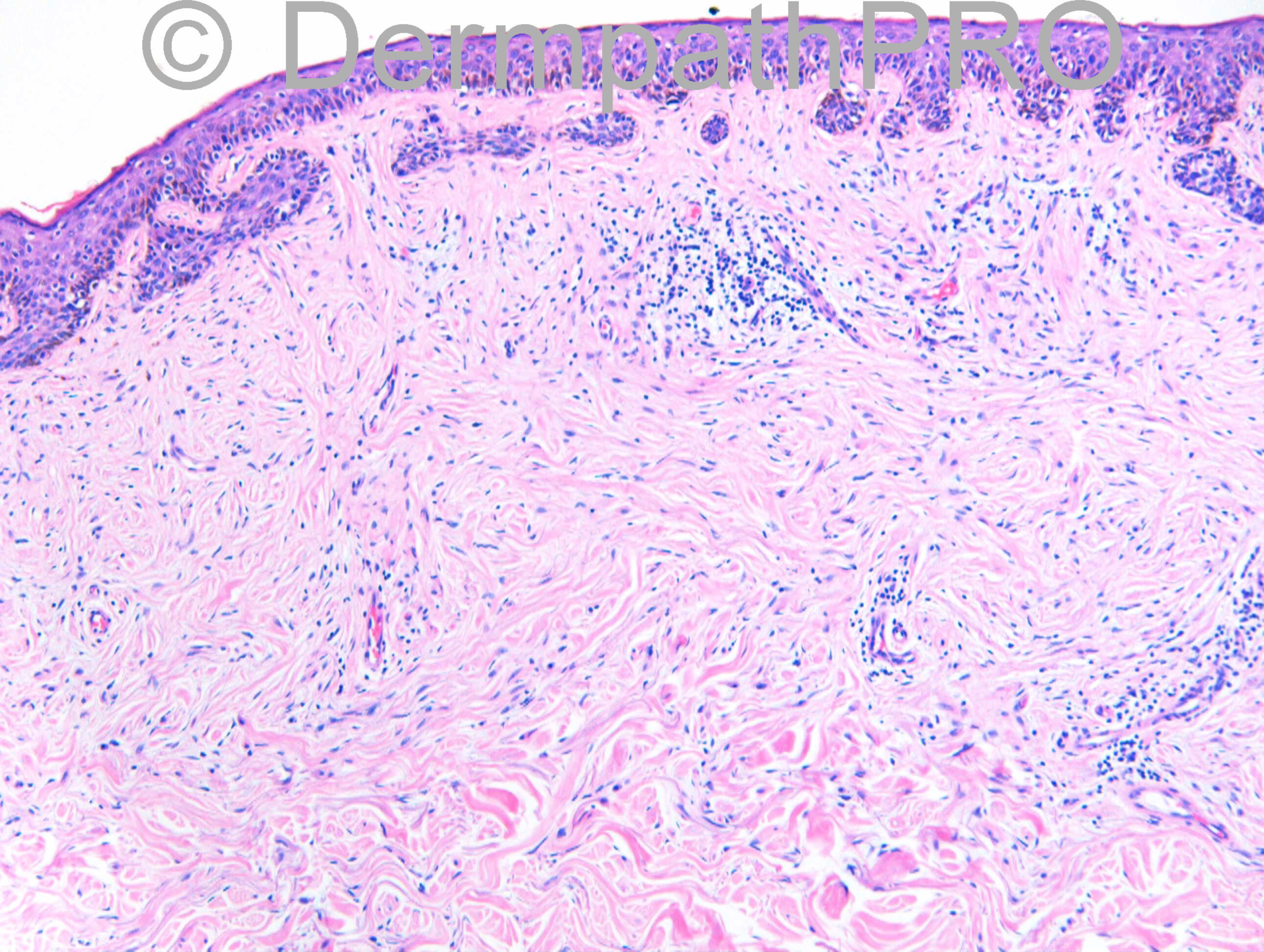
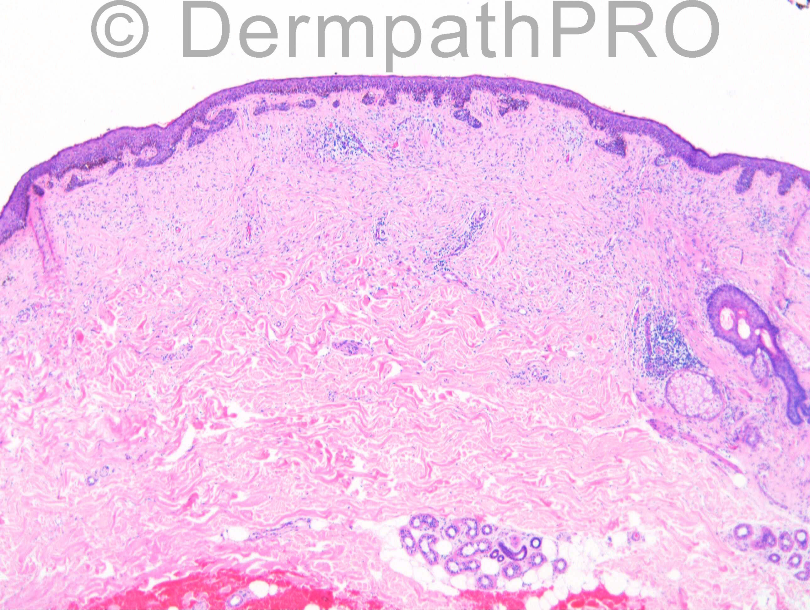
M70 Lesion upper chest.

Case posted by Dr Richard Carr


  Report Record

User Feedback


Dr. Mona Abdel-Halim

Posted

Thinking of desmoplastic melanoma...

Share this comment


Link to comment
share_externally

Dr. Richard Carr

Posted

[img]https://dermpathpro.com/uploads/spot_diagnosis_comment_img/Case%201177%20-%20RAC6936x10_S100.jpg[/img]

[img]https://dermpathpro.com/uploads/spot_diagnosis_comment_img/Case%201177%20-%20RAC6936x20_MelanA.jpg[/img]

Share this comment


Link to comment
share_externally

Eman El-Nabarawy

Posted

So it's desmoplastic melanoma (as Mona suggested) with involvement of the overlying epidermis by superficial spreading melanoma like changes.

Share this comment


Link to comment
share_externally

Guest Romualdo

Posted

Although desmoplastic melanomas may be deceptively bland usually at least some cells with slight to moderate atypia are present, as well as occasional mitotic figures. These findings are not seen here. By the other side, desmoplastic melanomas do not usually express positivity to melanocytic markers such as Melan-A and HMB-45. Only S-100 protein should be positive. I think this is a desmoplastic nevus. A very low Ki-67 index (2 % or less), if present, would be an important clue to this diagnosis.

Share this comment


Link to comment
share_externally

Eman El-Nabarawy

Posted

What about the considerable junctional activity and lymphoid aggregates?age?site?

Share this comment


Link to comment
share_externally

Guest Romualdo

Posted

[font=arial,helvetica,sans-serif][size=4]Dr. Eman, in relation to the presence of lymphoid aggregates in desmoplastic nevus and location on the chest I selected the following extractions from the book "Pathology of Challenging Melanocytic Neoplasms Diagnosis and Management", edited by Shea, Reed and Prieto:
"Similar to desmoplastic melanoma, desmoplastic
nevi show small aggregates of lymphocytes
in the dermis, next to the tumor cells, as
well as perineural involvement. However, in
contrast, dermal mitotic figures are exceptional
in desmoplastic nevi."


"In general, a diagnosis of desmoplastic nevus is favored in
younger individuals, outside the head and neck
area, without mitotic figures and very low Ki-67
expression."[/size][/font][font=GjhlnjXglrfdCrjrknTimes-Roman][size=2][font=GjhlnjXglrfdCrjrknTimes-Roman][size=2] [/size][/font][/size][/font]

Share this comment


Link to comment
share_externally

Guest Giovanni Falconieri

Posted

By a knee-jerk reflex, melanoma goes obviously to the top of the differential due to patient age and the patchy lymphoid infiltrate, yet Melan A seems to stain melanin in keratinocytes rather than melanocytic cells. I would therefore favor desmoplastic nevus based on the available information. As an integration to Romualdo's note above, focal lymphoid infiltrates in desmoplastic melanocytic nevi have been addressed by kiuru et al in their paper published in JCP 2012; 39(10):940-4
[b]http://tinyurl.com/pyrp9pm[/b]

Share this comment


Link to comment
share_externally

Guest Jim Davie MD

Posted

[size=4]
[size=4]
Agree with Desmoplastic melanoma. [/size][size=4]
Age is advanced (70). [size=4] [/size][/size][size=4]
[size=4] The epidermal junction looks 'busy' with focally confluent lentiginous and nested haloed hyperchromatic melanocytes with poor circumscription and prominent pagetoid scatter. There may be adnexal/follicular involvement by lentiginous melanocytes in the last, low power H&E image. This would be consistent with an in-situ melanoma component. [/size][/size][size=4]
The neurofibroma-like dermis has slightly thickened collagen fibers, apparent mucin accumulation, and predominantly single spindled cells with moderate nuclear hyperchromatism and pleomorphism. [size=4] The broad sweeping, branching fascicles of S100+ spindled cells -- prominent in the upper dermis-- shows less prominent staining on the MelanA stain. There is a patchy chronic inflammatory reaction.[/size][/size]
[size=4]
MelanA is insensitive but not infrequently positive in desmoplastic melanoma; one review cites [font=arial,helvetica,sans-serif]21% [size=4](8/37)[/size][size=4] of desmoplastic melanomas as Melan-A positive. [/size][/font][/size][size=4]
[font=arial,helvetica,sans-serif][size=4] [[/size][size=4] Immunohistochemical characteristics of melanoma. [/size][size=4]Ohsie, Steven J.; Sarantopoulos, G. Peter; Cochran, Alistair J.; Binder, Scott W. [/size][size=4] Journal of Cutaneous Pathology , Volume 35 (5)[/size][size=4] [/size][size=4]–[/size][size=4] [/size][size=4]May 1, 2008].[/size][/font][/size][/size]

Share this comment


Link to comment
share_externally

Guest Tiberiu Tebeica

Posted

The silhouette of this lesion points me rather towards a desmoplastic nevus.

Share this comment


Link to comment
share_externally

Dr. Richard Carr

Posted

This was a referred case to my colleague Dr Scott Sanders and it turns out I only photographerd the slides and did not keep a copy to refer back to. We went for the default diagnosis of desmoplastic melanoma for reasons indicted by Jim although based on images submitted I fully understand the dichotomy of opinions here and the presence of lymphoid aggregates and older age are certainly no barrier to the diagnosis of an intradermal Spitz naevus (Plaza et al 2014 abstract below). My colleague Dr Scott Sanders' report was as follows:

[u][color="#800080"] Many thanks for asking me to look at this case. I agree with your
interpretation of the features. In two cross sections there are
scattered subtle S-100 and Melan-A +ve elongated cells in a fibrous
stroma in the dermis overlying which there is a lentigenous and focally
confluent junctional melanocytic proliferation (essentially lentigo
maligna). The appearances are essentially typical of early desmoplastic
melanoma, radial growth phase. (interestingly desmoplastic melanoma is
more often Melan-A -ve). The desmoplastic dermal component is excised
by 2mm but the junctional proliferation extends to within 0.1mm of an
inked radial margin. (seen also by Dr Carr) [/color] [/u]

[u]Reference[/u]
[u][color="#800080"]Plaza JA, De Stefano D, Suster S, Prieto VG, Kacerovska D, Michal M, Sangueza M, Kazakov DV. Intradermal spitz nevi: a rare subtype of spitz nevi analyzed in a clinicopathologic study of 74 cases. Am J Dermatopathol. 2014 Apr;36(4):283-94; quiz 295-7.[/color][/u]

[u][color="#800080"][u][color="#800080"]Spitz nevi are acquired melanocytic lesions with a wide histomorphological spectrum; reliable distinction from spitzoid melanoma is often difficult. Misdiagnoses of benign spitzoid tumors as spitzoid melanomas and vice versa are attributable to a frequently disturbing morphology and inconsistent or poorly defined histological criteria for diagnosis. Many recognized histological variants of Spitz nevi have been described, including the intradermal Spitz. Histopathologic descriptions of intradermal Spitz nevi have been done in the past; however, large studies addressing their histological spectrum have been lacking. We have retrospectively assessed the morphological features in 74 cases of intradermal Spitz nevi, excluding tumors clearly defined as atypical Spitz nevi and Spitzoid melanomas, to further delineate their histological spectrum. The patients' ages ranged from 5 to 81 years (median: 27). Anatomic location included: the upper extremities (27 cases), followed by head and neck (22 cases), lower extremities (9 cases), back (8 cases), buttock (5 cases), chest (1 case), and vulva (1 case). In 1 case, the anatomic location of the lesion was not available. Different histological variants were observed including hyalinized, polypoid, desmoplastic, angiomatoid, and halo Spitz. Morphological features evaluated included symmetry (100%), cell type (epithelioid 42%, spindle 16%, mixed 42%), maturation (85%), pigmentation (26%), chronic inflammation (24%), and mitotic activity (38%). Mild atypia was seen in 36 cases (49%), moderate atypia was seen in 28 cases (38%), and severe atypia was seen in 10 cases (14%). Intradermal Spitz nevus is a distinctive type of Spitz nevus that sometimes can be difficult to define given the unusual features that these lesions can show; thus, strict application of well-defined histological criteria and awareness of their morphological spectrum will facilitate definitive diagnosis.[/color][/u][/color][/u]

Share this comment


Link to comment
share_externally


Join the conversation

You can post now and register later. If you have an account, sign in now to post with your account.

Guest
Add a comment...

×   Pasted as rich text.   Paste as plain text instead

  Only 75 emoji are allowed.

×   Your link has been automatically embedded.   Display as a link instead

×   Your previous content has been restored.   Clear editor

×   You cannot paste images directly. Upload or insert images from URL.

Loading...

×
×
  • Create New...