Jump to content
In this section we have spot diagnoses posted on a daily basis since June 2010, now over 4000! You can review the archived cases and read the suggested diagnoses by users and the final comment by the contributors.
Case are uploaded each week day by 10 am UK time with the correct diagnosis will generally be posted at 8 pm UK time. Why not view the most recent spot diagnosis and proffer a diagnosis?

Case Number : Case 952 - 14th February Posted By: Guest

Please read the clinical history and view the images by clicking on them before you proffer your diagnosis.
Submitted Date :
   (0 reviews)

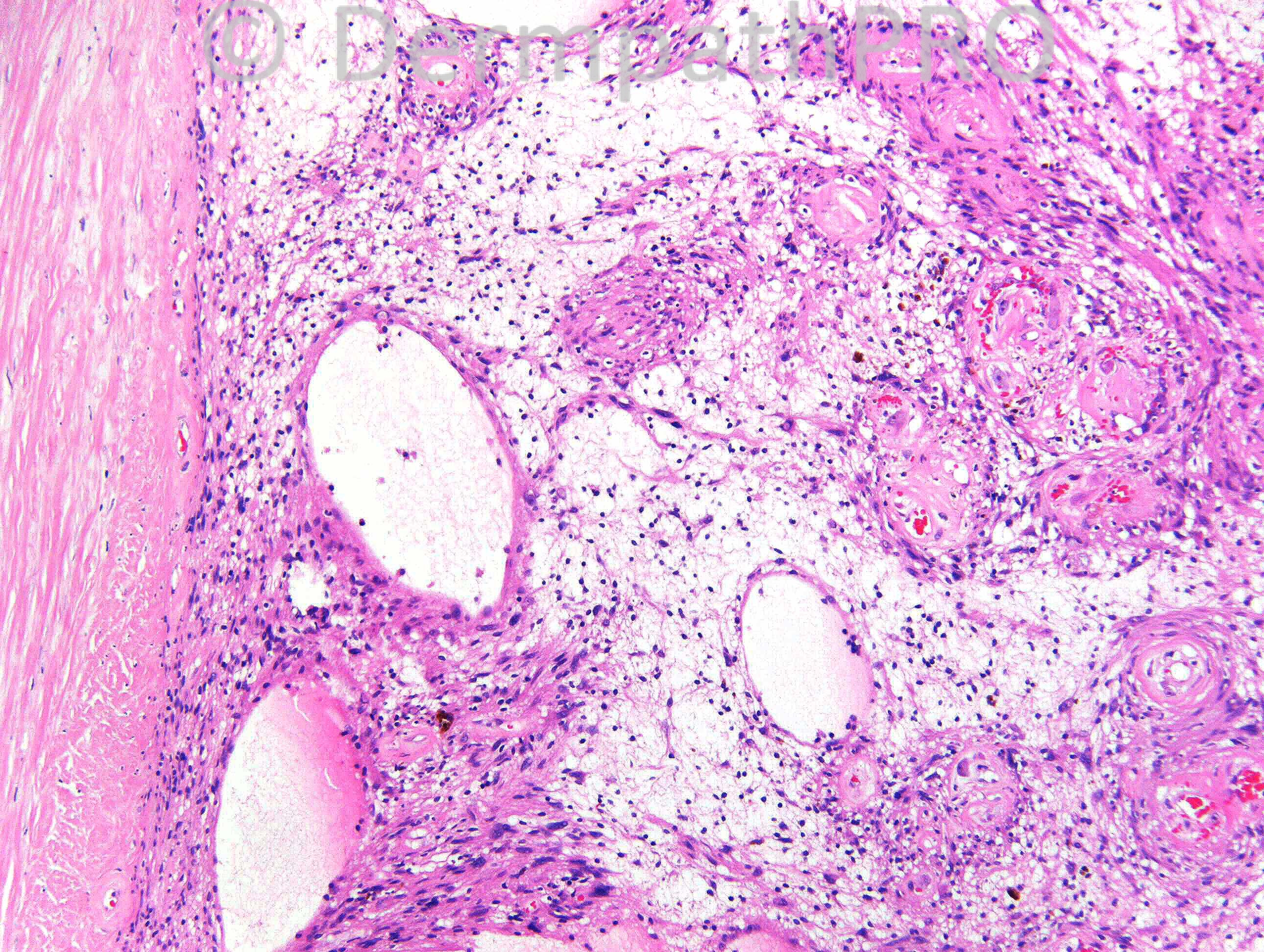
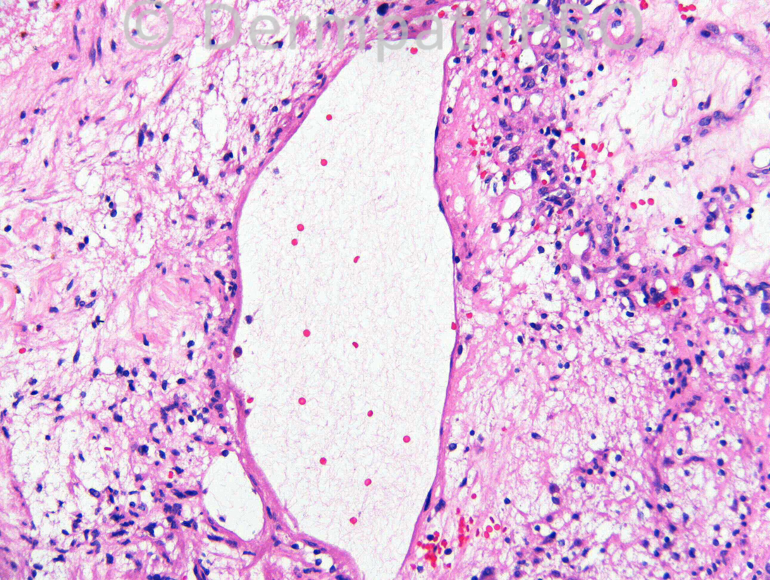
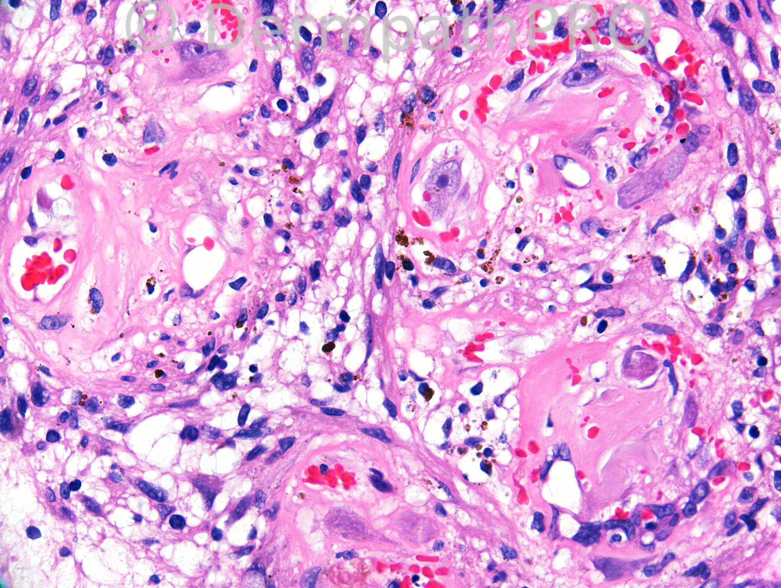
F67. Swelling left forearm increased in size ?lipoma ?haematomatous organised


Case posted by Dr. Richard Carr.

  Report Record

User Feedback


Dr. Mona Abdel-Halim

Posted

Symplastic hemangioma (developing in a pre-existing intravascular hemangioma).

Share this comment


Link to comment
share_externally

Eman El-Nabarawy

Posted

Phosphaturic mesenchymal tumor(mixed connective tissue variant).

Share this comment


Link to comment
share_externally

Guest Maria George

Posted

this is Encapsulated like traumaic fat cyst with SCC inside

Share this comment


Link to comment
share_externally

Guest Maria George

Posted

Another possibility is liposarc with squamous differentiation.Need IHC.

Share this comment


Link to comment
share_externally

Guest Romualdo

Posted

Ancient schwannoma. If S100 protein is negative (I don't believe it will be!) then pleomorphic hyalinizing angiectatic tumor as a second possibility.

Share this comment


Link to comment
share_externally

Dr. Mona Abdel-Halim

Posted

Ancient schawnnoma is an excellent possibility,,, I agree with it as a differential diagnosis...

Share this comment


Link to comment
share_externally

Robledo F. Rocha

Posted

Ancient schwannoma in an unusual superficial site. Antoni B areas flaunting extensive cystic change hold sway over Antoni A areas displaying degenerated Schwann cells with large and dark nuclei.

Share this comment


Link to comment
share_externally

Dr. Richard Carr

Posted

I was hoping for the IHC to have been posted but all I can say is well done Romauldo!! This was diffusely S100 positive with a nice EMA stained capsule and your diagnosis of ancient schwannoma is correct. The slightly active looking cells are probabaly just reactive histiocytes - they were S100 negative.

Enjoy your weekends.

Share this comment


Link to comment
share_externally

Dr. Richard Carr

Posted

[img]https://dermpathpro.com/uploads/spot_diagnosis_comment_img/CASE952-RAC6665x5_EMA_MR%20(1).jpg[/img]

Share this comment


Link to comment
share_externally

Dr. Richard Carr

Posted

[img]https://dermpathpro.com/uploads/spot_diagnosis_comment_img/Case%20952-RAC6665x40_S100_MR%20copy.jpg[/img]

Share this comment


Link to comment
share_externally

Dr. Mona Abdel-Halim

Posted

Very nice case... A great lesson,,, I must write a note on my desk: DO NOT FORGET ANCIENT SCHWANNOMA .... I think because most of these tumors present in deep soft tissue, we as dermatopathologists in Egypt do not see them, this is why I am not aquinted with them and I always miss them... Thanks Dr Carr,,, Really nice...

Share this comment


Link to comment
share_externally


Join the conversation

You can post now and register later. If you have an account, sign in now to post with your account.

Guest
Add a comment...

×   Pasted as rich text.   Paste as plain text instead

  Only 75 emoji are allowed.

×   Your link has been automatically embedded.   Display as a link instead

×   Your previous content has been restored.   Clear editor

×   You cannot paste images directly. Upload or insert images from URL.

Loading...

×
×
  • Create New...