Jump to content
In this section we have spot diagnoses posted on a daily basis since June 2010, now over 4000! You can review the archived cases and read the suggested diagnoses by users and the final comment by the contributors.
Case are uploaded each week day by 10 am UK time with the correct diagnosis will generally be posted at 8 pm UK time. Why not view the most recent spot diagnosis and proffer a diagnosis?

Case Number : Case 961 - 27th February Posted By: Guest

Please read the clinical history and view the images by clicking on them before you proffer your diagnosis.
Submitted Date :
   (0 reviews)

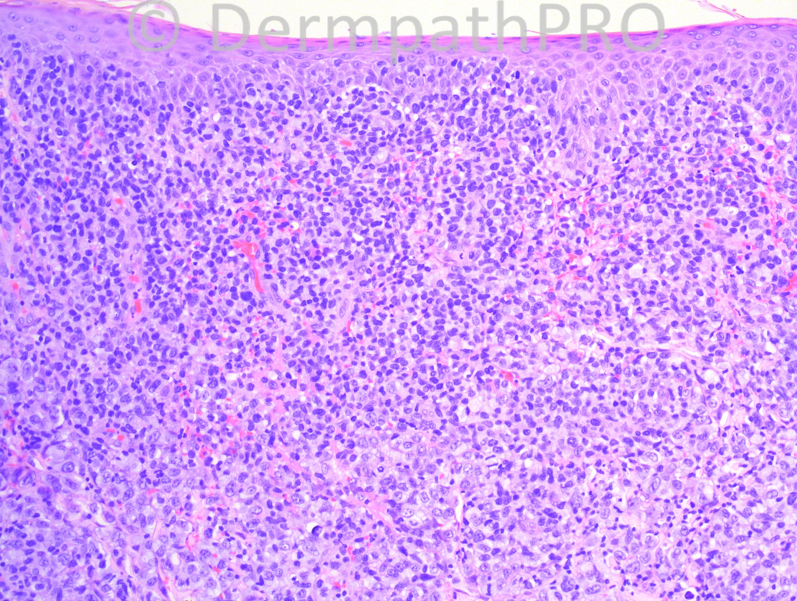
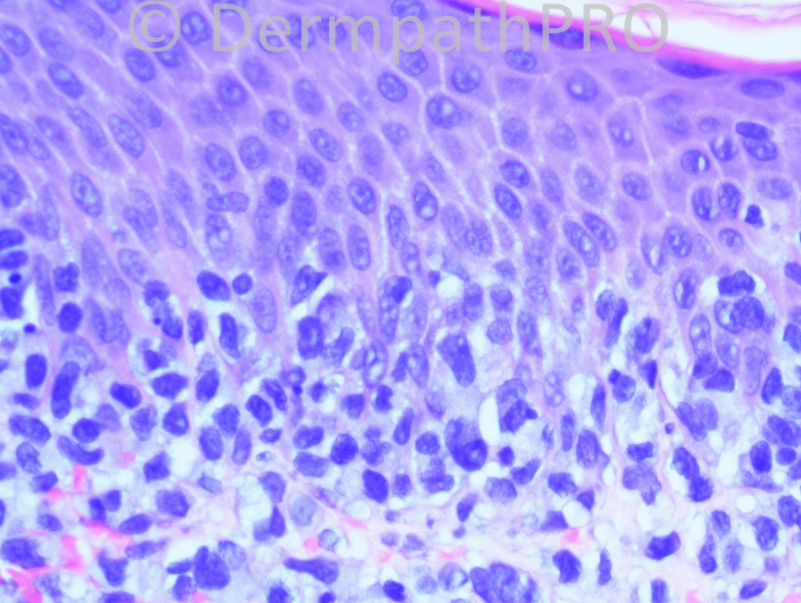
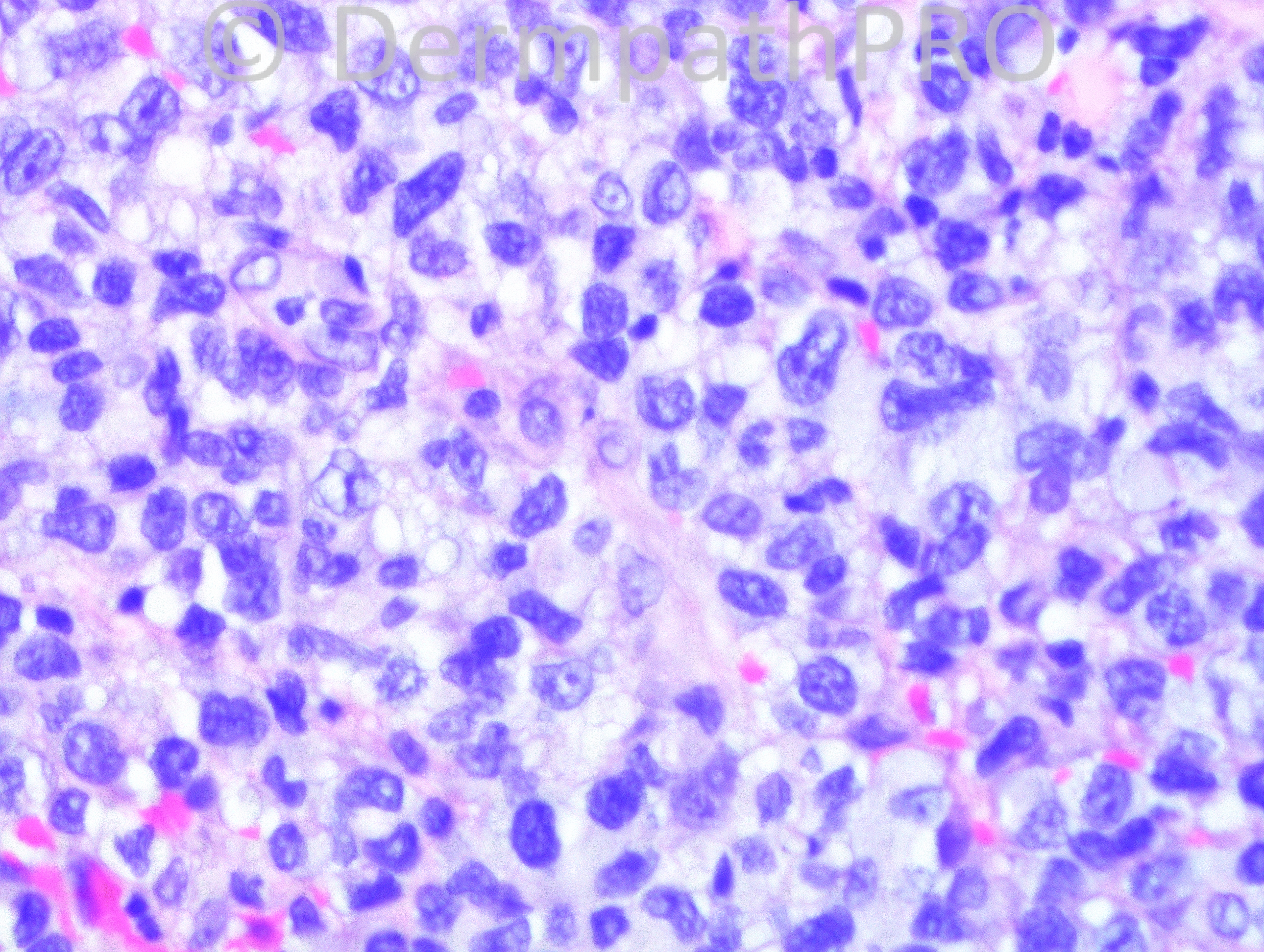
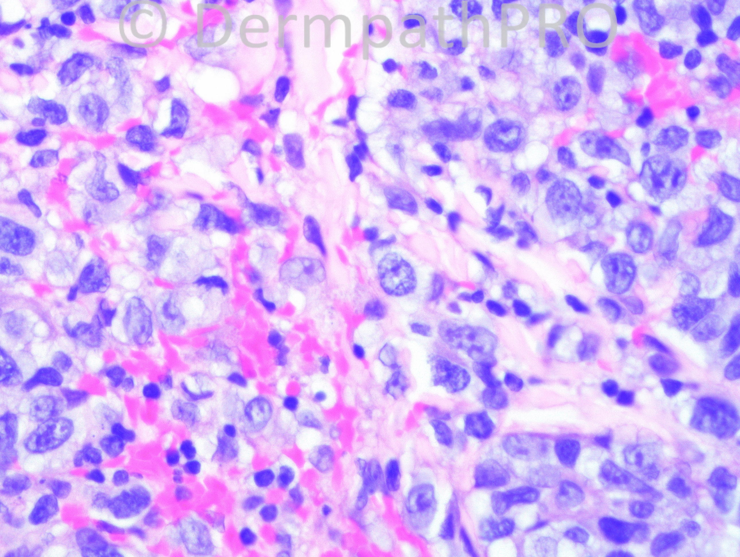
73 year-old male with flank lesions.

Case posted by Dr. Hafeez Diwan.


  Report Record

User Feedback


Epidermotropic lymphoma. Possibly mycosis fungoides, tumour stage + transformation.

Share this comment


Link to comment
share_externally

Guest Hazem Hamed

Posted

I agree with Dr Abdul-Kadir. However, would like to exclude adult T cell leukaemia/lymphoma before suggesting MF. Immuno including CD25 would be essential. Has the patient been investigated for HTLV1?

Share this comment


Link to comment
share_externally

Epidermotropic lymphoma, probably some kind of T-cell Lymphoma. The lesions are located in a typical location for MF, the age is also typical and there´s more than 25% of large cells in the infiltrate, so I think this is probably MF with blastic transformation.
Clinical history is essential. If the patient had this lesion for some years, maybe associated with plaques or patches, it must be MF. If this lesion is of rapid onset, could be CD8+ epidermotropic agressive T-cell cutaneous lymphoma. Adult T cell leukemia/lymphoma is also a good hypothesis.
Do not forget that if these lesions were recurrent papules with crust formation this case could be Type B Lymphomatoid Papulosis.
Rare cases of B-cell lymphoma are epidermotropic. Always necessary histology, clinical information and immunoprofile for a correct diagnosis.

Share this comment


Link to comment
share_externally

Eman El-Nabarawy

Posted

Tumor stage MF with large cell transformation.

Share this comment


Link to comment
share_externally

Robledo F. Rocha

Posted

[color=#1C2837][font=arial, verdana, tahoma, sans-serif][size=4]Epidermotropic lymphoma, probably tumor stage of mycosis fungoides.[/size][/font][/color]

Share this comment


Link to comment
share_externally

Guest Jim Davie MD

Posted

Agree with the above excellent differentials and need for stains. Very high mitotic activity with some immunoblastic features.
I would favor ALCL.

Share this comment


Link to comment
share_externally

Dr. Mona Abdel-Halim

Posted

Agree with the differentials, Immunos are mandatory..

Share this comment


Link to comment
share_externally

Dr. Hafeez Diwan

Posted

This was mycosis fungoides with large cell transformation. CD4 positivity was much greater than CD8. The large cells were CD30 positive. Clinically the patient had the full spectrum of lesions (patches, plaques and tumors).

Share this comment


Link to comment
share_externally

Robledo F. Rocha

Posted

Dra. Halim have recommeded an excellent article about this issue in the [url="https://dermpathpro.com/index.html/_/latest-news/large-cell-transformation-of-mycosis-fungoides-r53"]News section[/url].

Share this comment


Link to comment
share_externally


Join the conversation

You can post now and register later. If you have an account, sign in now to post with your account.

Guest
Add a comment...

×   Pasted as rich text.   Paste as plain text instead

  Only 75 emoji are allowed.

×   Your link has been automatically embedded.   Display as a link instead

×   Your previous content has been restored.   Clear editor

×   You cannot paste images directly. Upload or insert images from URL.

Loading...

×
×
  • Create New...