Jump to content
In this section we have spot diagnoses posted on a daily basis since June 2010, now over 4000! You can review the archived cases and read the suggested diagnoses by users and the final comment by the contributors.
Case are uploaded each week day by 10 am UK time with the correct diagnosis will generally be posted at 8 pm UK time. Why not view the most recent spot diagnosis and proffer a diagnosis?

Case Number : Case 923 - 6th January Posted By: Guest

Please read the clinical history and view the images by clicking on them before you proffer your diagnosis.
Submitted Date :
   (0 reviews)

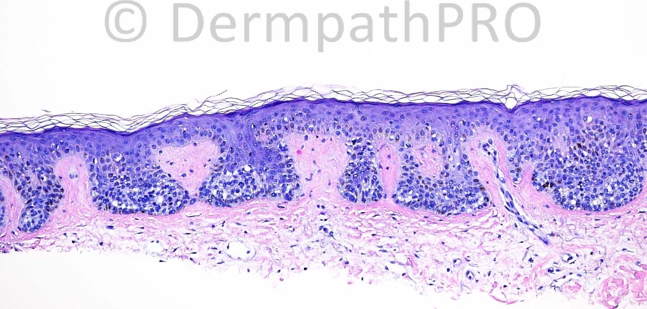
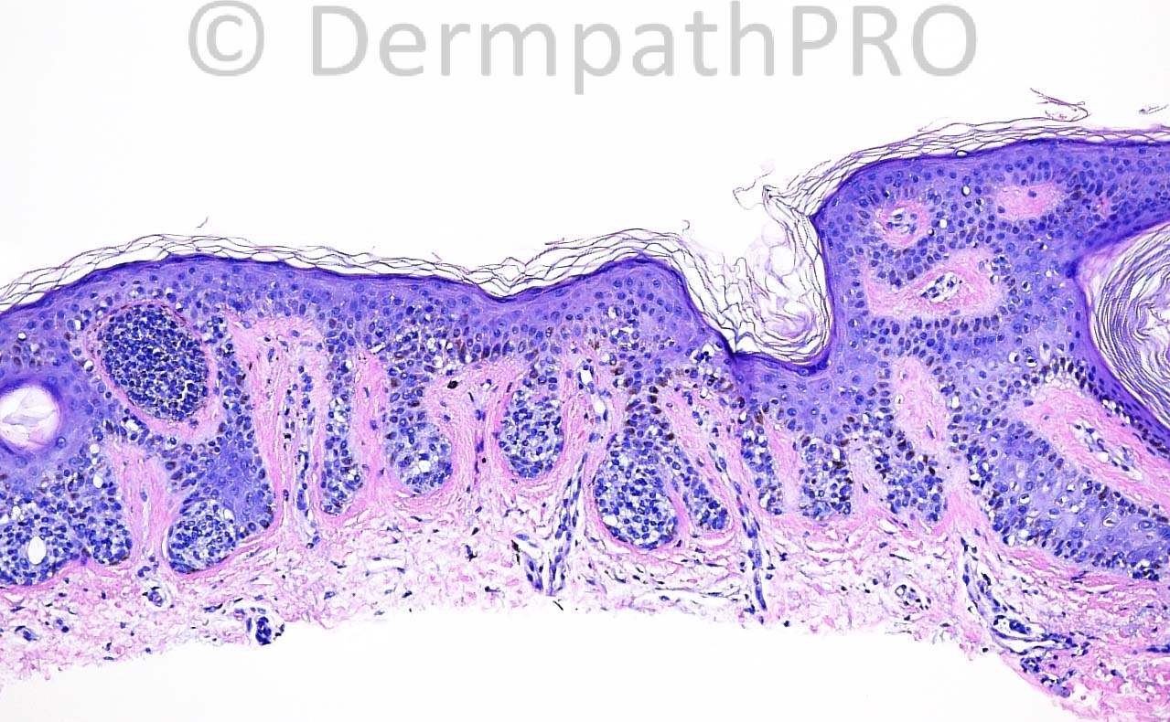
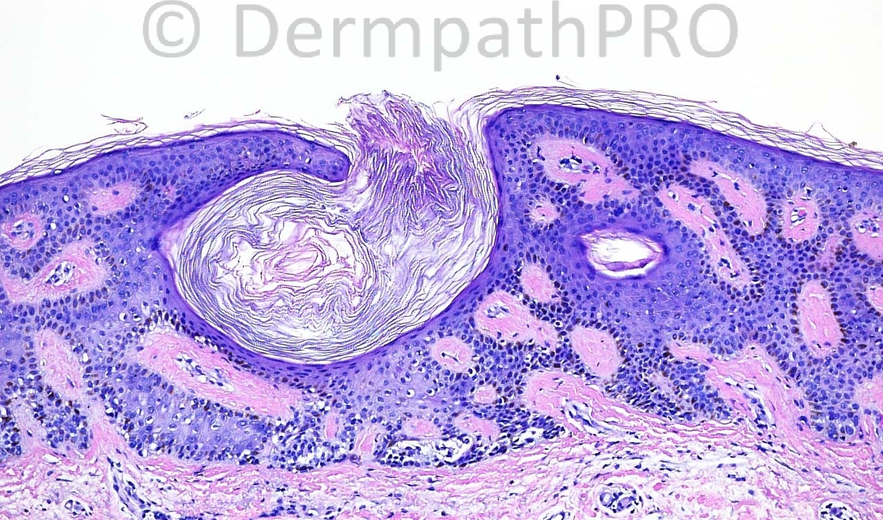
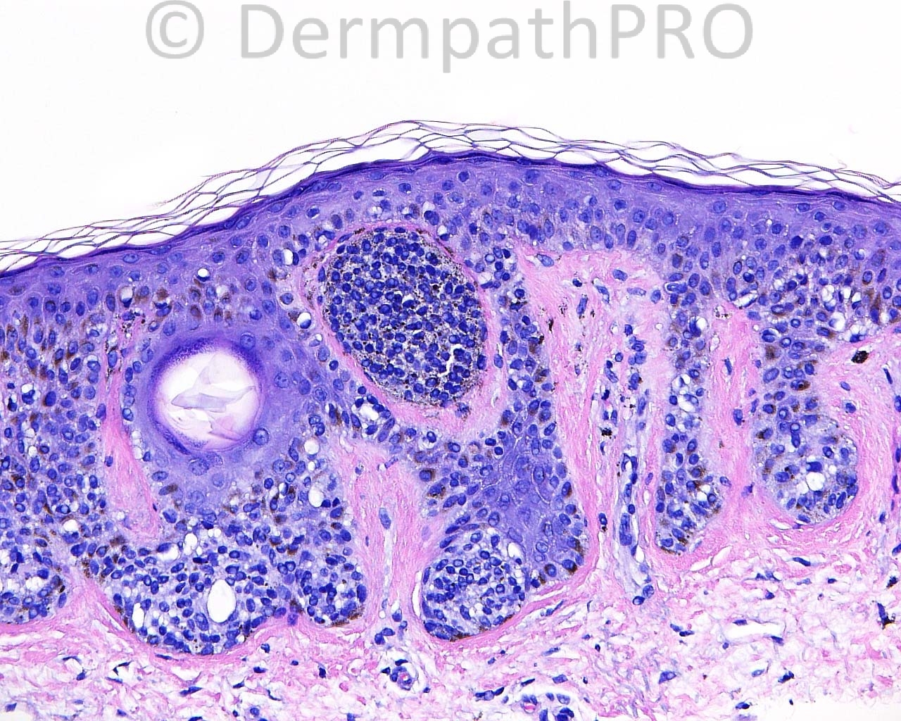
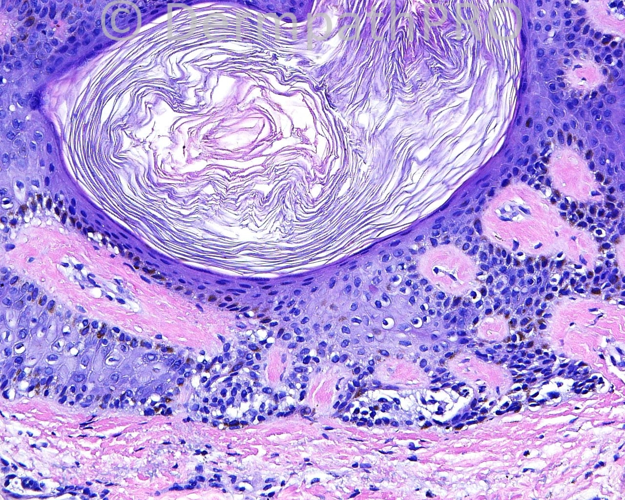
The patient is a 67 year old woman with a shave biopsy of an irregular macule taken from the superior aspect of the right scapula

Case posted by Dr. Mark Hurt


  Report Record

User Feedback


Guest Engin Sezer

Posted

Pigmented clonal seborrheic keratoses

Share this comment


Link to comment
share_externally

Guest Maria George

Posted

Take it easy Mark.Junctional nevus+milia+Seb.Keratosis.

Share this comment


Link to comment
share_externally

Junctional naevus (left) + reticulated seborrhoeic keratosis (right).

Share this comment


Link to comment
share_externally

Eman El-Nabarawy

Posted

Junctional nevus with seborrheic keratosis-like appearance of the overlying epidermis.

Share this comment


Link to comment
share_externally

Guest Ali Y. Hashim

Posted

[b]Clonal seborrheic keratosis[/b]

Share this comment


Link to comment
share_externally

Robledo F. Rocha

Posted

[font=arial,helvetica,sans-serif][size=4][color=#000000]Collision of junctional melanocytic nevus and reticulated seborrheic keratosis, a common association that seems [url="http://www.derm101.com/wp-content/uploads/dpc0202a09.pdf"]one of the two lesions have induced the other[/url].[/color][/size][/font]

Share this comment


Link to comment
share_externally

Guest Jim Davie MD

Posted

Pigmented seborrheic keratosis, with coexistent compound melanocytic nevus. (The pigment increase in the papillary dermal nest of bottom left photo hints that it is intradermal, in my opinion).

Share this comment


Link to comment
share_externally

Guest Romualdo

Posted

Lentiginous junctional melanocytic nevus.

Share this comment


Link to comment
share_externally

Guest Tiberiu Tebeica

Posted

Collision between compound melanocytic nevus with dysplastic features / Clark's nevus and seborrheic keratosis

Share this comment


Link to comment
share_externally

Eman El-Nabarawy

Posted

Lentiginous junctional melanocytic nevus with overlying epidermal seb k like features.

Share this comment


Link to comment
share_externally

Dr. Mona Abdel-Halim

Posted

Just arrived home... Lentiginous junctional nevus in conjunction with a reticulated seb K.

Share this comment


Link to comment
share_externally


Join the conversation

You can post now and register later. If you have an account, sign in now to post with your account.

Guest
Add a comment...

×   Pasted as rich text.   Paste as plain text instead

  Only 75 emoji are allowed.

×   Your link has been automatically embedded.   Display as a link instead

×   Your previous content has been restored.   Clear editor

×   You cannot paste images directly. Upload or insert images from URL.

Loading...

×
×
  • Create New...