Jump to content
In this section we have spot diagnoses posted on a daily basis since June 2010, now over 4000! You can review the archived cases and read the suggested diagnoses by users and the final comment by the contributors.
Case are uploaded each week day by 10 am UK time with the correct diagnosis will generally be posted at 8 pm UK time. Why not view the most recent spot diagnosis and proffer a diagnosis?

Case Number : Case 926 - 9th January Posted By: Guest

Please read the clinical history and view the images by clicking on them before you proffer your diagnosis.
Submitted Date :
   (0 reviews)

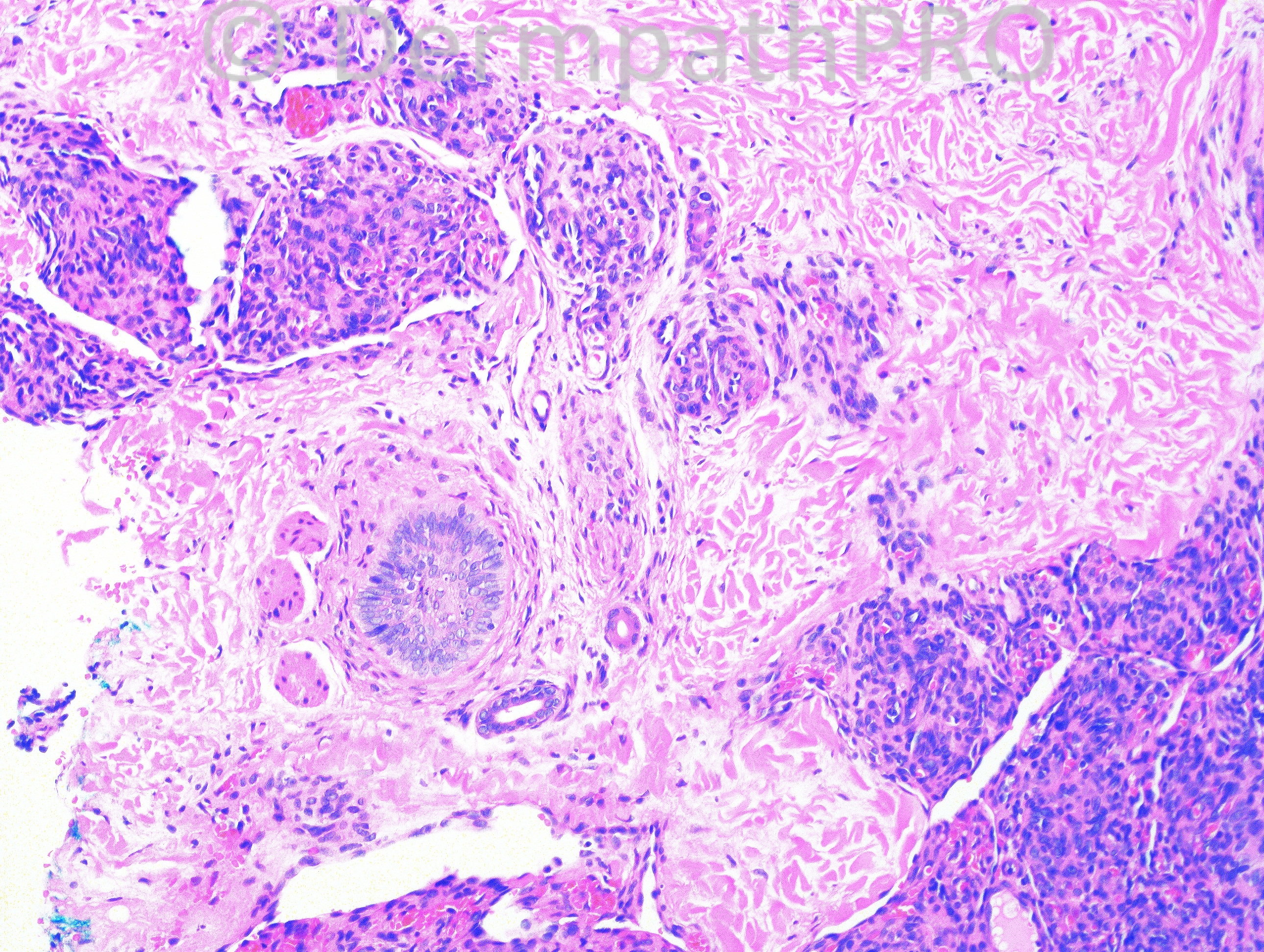
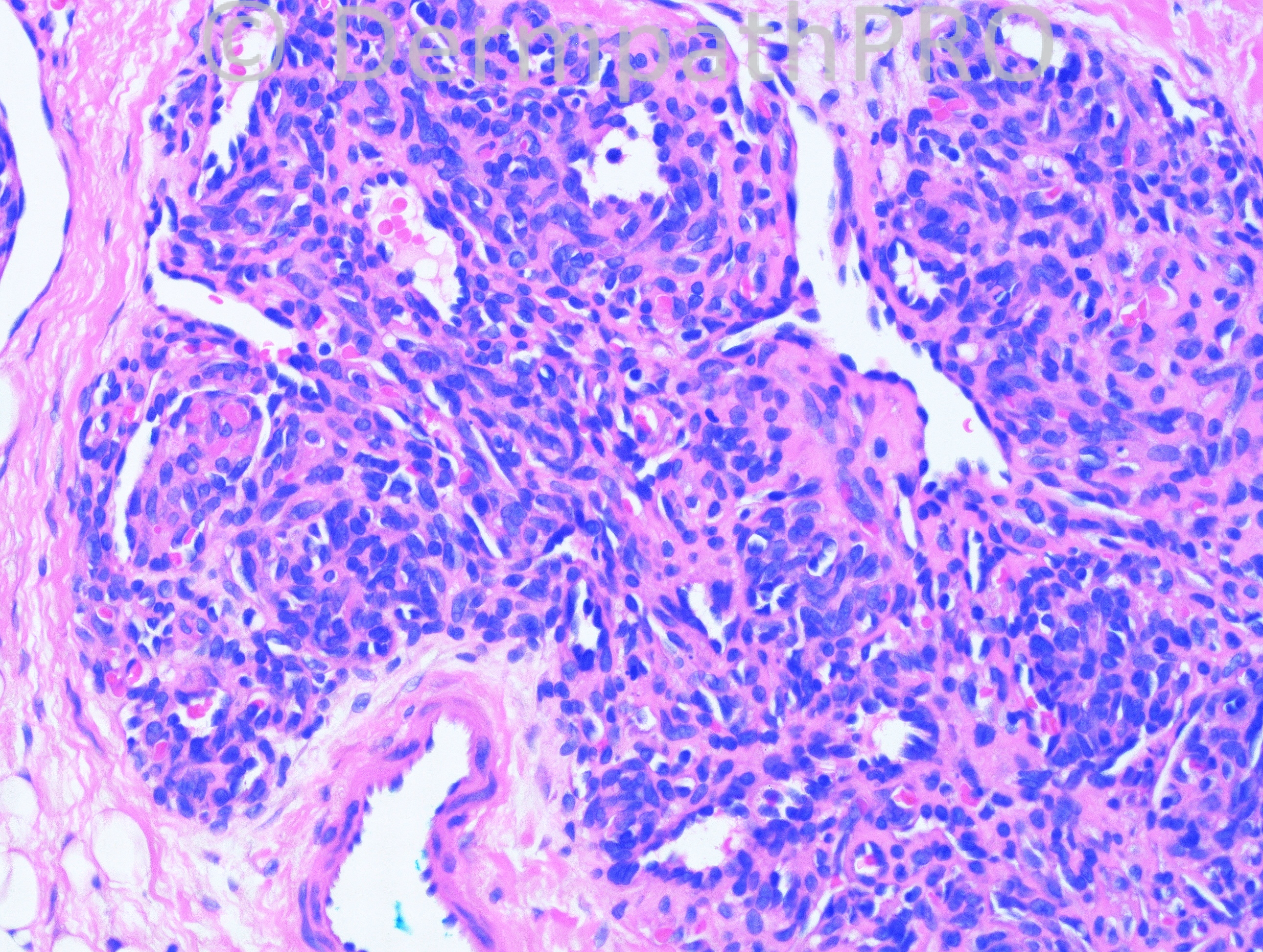
3 month-old boy with lesion on right medial knee.

Case posted by Dr. Hafeez Diwan.

  Report Record

User Feedback


Guest Tiberiu Tebeica

Posted

Kaposiform hemangioendothelioma

Share this comment


Link to comment
share_externally

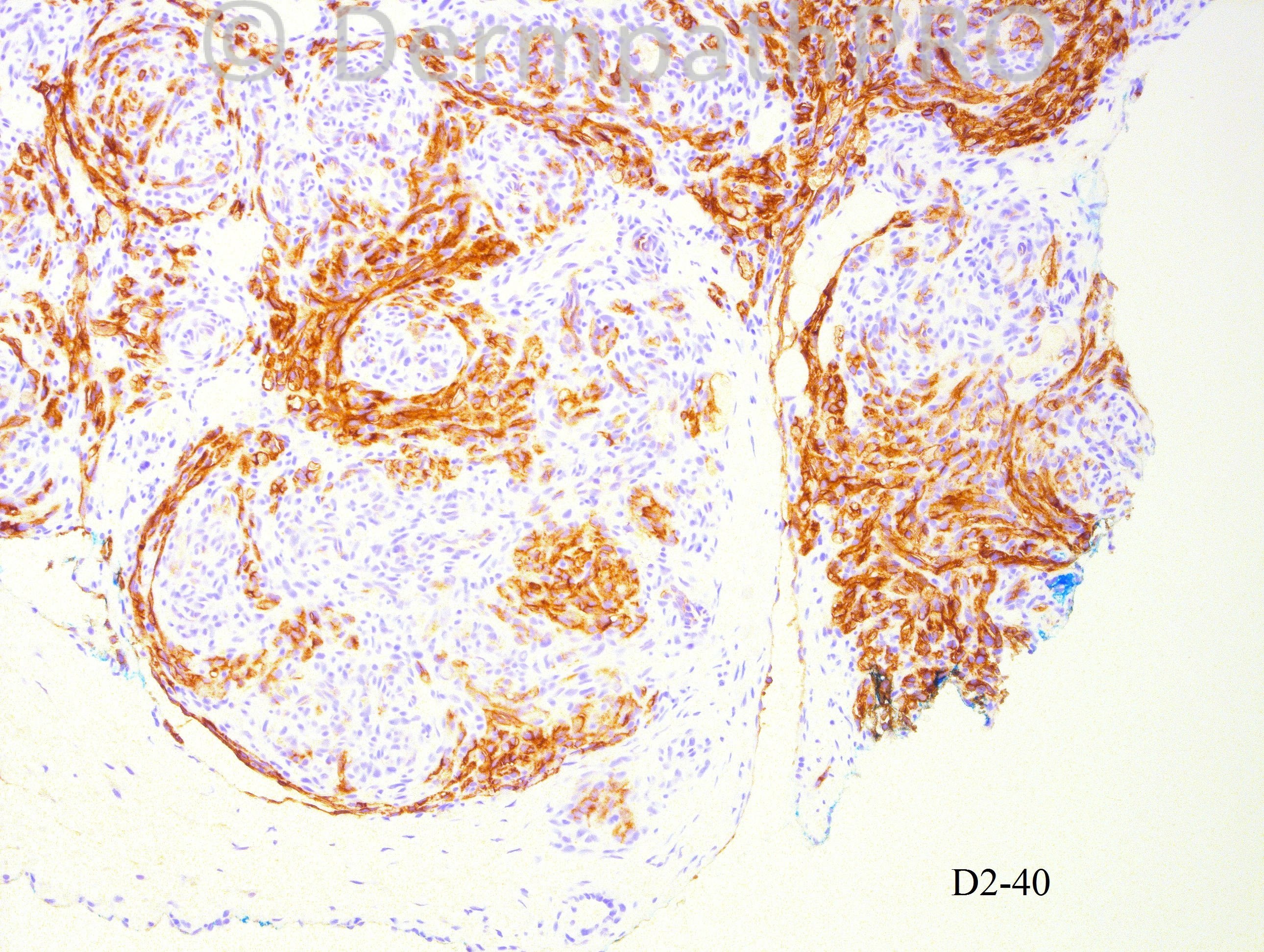
Tufted angioma. It is nice to see the proliferative nodules inside the vascular spaces negative for D2-40.

Share this comment


Link to comment
share_externally

Guest Giovanni Falconieri

Posted

With some delay, to say the least, happy new hear from Italy! I would go for tufted hemangioma. There are some nice crescentic spaces.

Share this comment


Link to comment
share_externally

Guest Dr.Yüksel Okumuş

Posted

Agree with Eman El-Nabarawy. The presence of lymphatic channels (positive with D2-40) around the lobules is the distinctive feature of Tufted Angioma.

Share this comment


Link to comment
share_externally

Guest Romualdo

Posted

[font=Arial, sans-serif][size=2]Tufted angioma and kaposiform hemangioendothelioma are vascular tumors of the same neoplastic spectrum. Kaposiform hemangioendothelioma may have tufted angioma like areas. I think the D2-40 pattern of staining, disclosing a predominance of spindle cells at the periphery of the cannonballs, is more in keeping with kaposiform hemangioendotelioma. In tufted angioma the positivity should be limited to dilated vesssels present in the stroma and in the cannonballs and, at most, in some of the slit-like capillaries in the cannonballs.[/size][/font]

Share this comment


Link to comment
share_externally

Robledo F. Rocha

Posted

The cannonball-like vascular lobules are small and well circumscribed, and the expression of D2-40 around, not inside, the vascular lobules are characteristics that make me favor tufted angioma. Patient should be screened for Kasabach-Merritt syndrome.

Share this comment


Link to comment
share_externally

Dr. Mona Abdel-Halim

Posted

Tufted angioma, typical D2 -40 pattern,,,

Share this comment


Link to comment
share_externally

Sasi Attili

Posted

[color=#1C2837][font=arial, verdana, tahoma, sans-serif][size=4]Tufted angioma[/size][/font][/color]

Share this comment


Link to comment
share_externally

Dr. Hafeez Diwan

Posted

Yes. I called this tufted angioma.

Share this comment


Link to comment
share_externally


Join the conversation

You can post now and register later. If you have an account, sign in now to post with your account.

Guest
Add a comment...

×   Pasted as rich text.   Paste as plain text instead

  Only 75 emoji are allowed.

×   Your link has been automatically embedded.   Display as a link instead

×   Your previous content has been restored.   Clear editor

×   You cannot paste images directly. Upload or insert images from URL.

Loading...

×
×
  • Create New...