Jump to content
In this section we have spot diagnoses posted on a daily basis since June 2010, now over 4000! You can review the archived cases and read the suggested diagnoses by users and the final comment by the contributors.
Case are uploaded each week day by 10 am UK time with the correct diagnosis will generally be posted at 8 pm UK time. Why not view the most recent spot diagnosis and proffer a diagnosis?

Case Number : Case 928 - 13th January Posted By: Guest

Please read the clinical history and view the images by clicking on them before you proffer your diagnosis.
Submitted Date :
   (0 reviews)

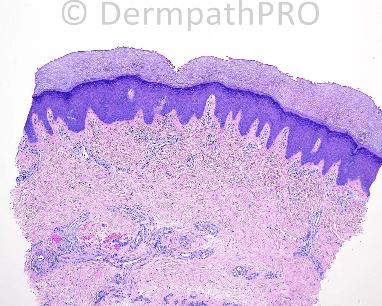
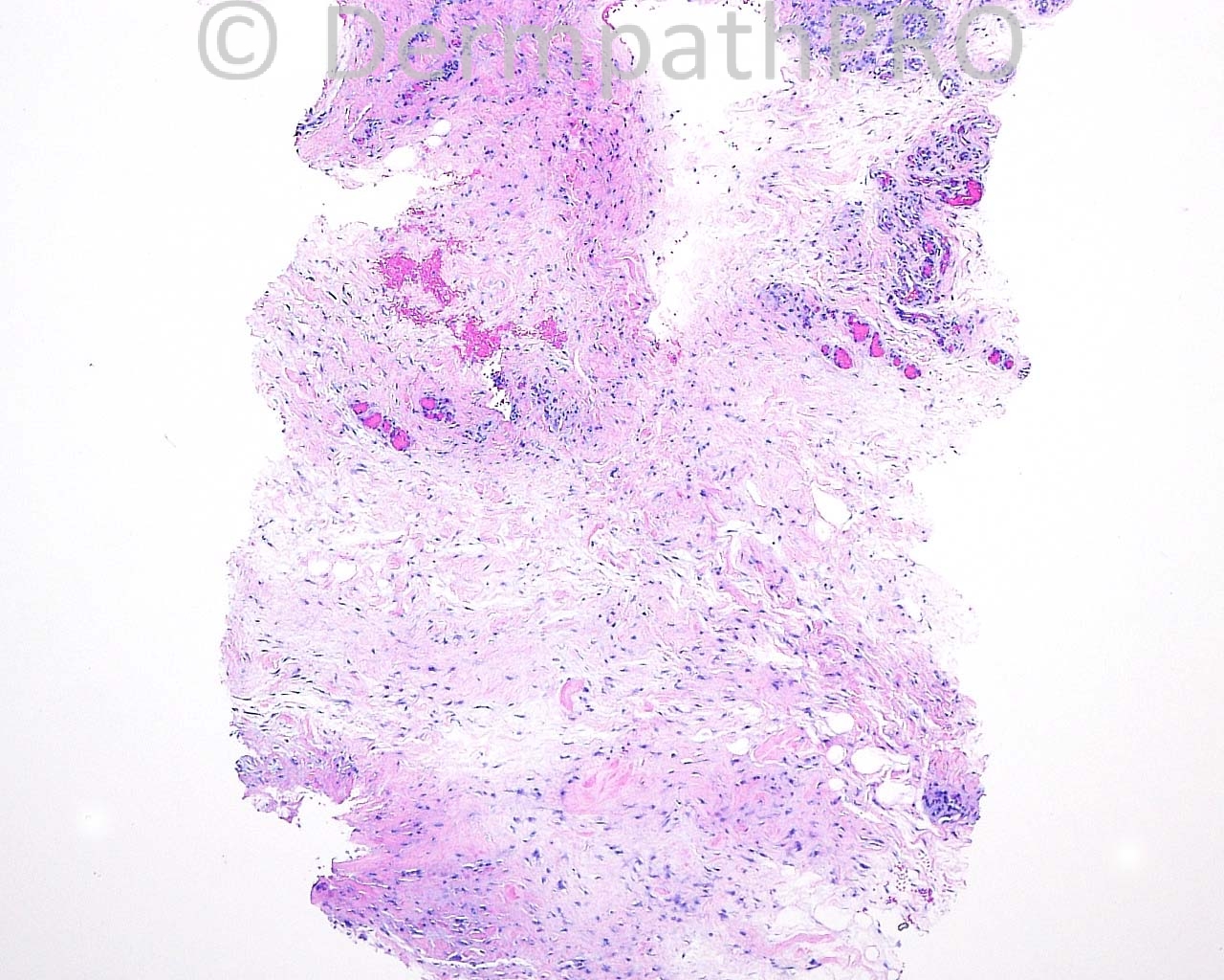
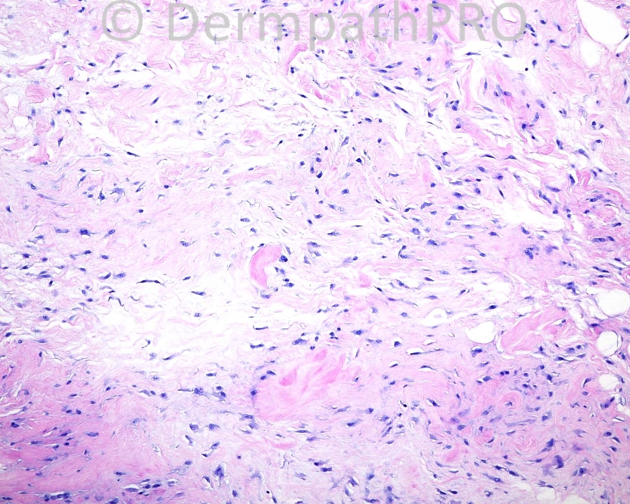
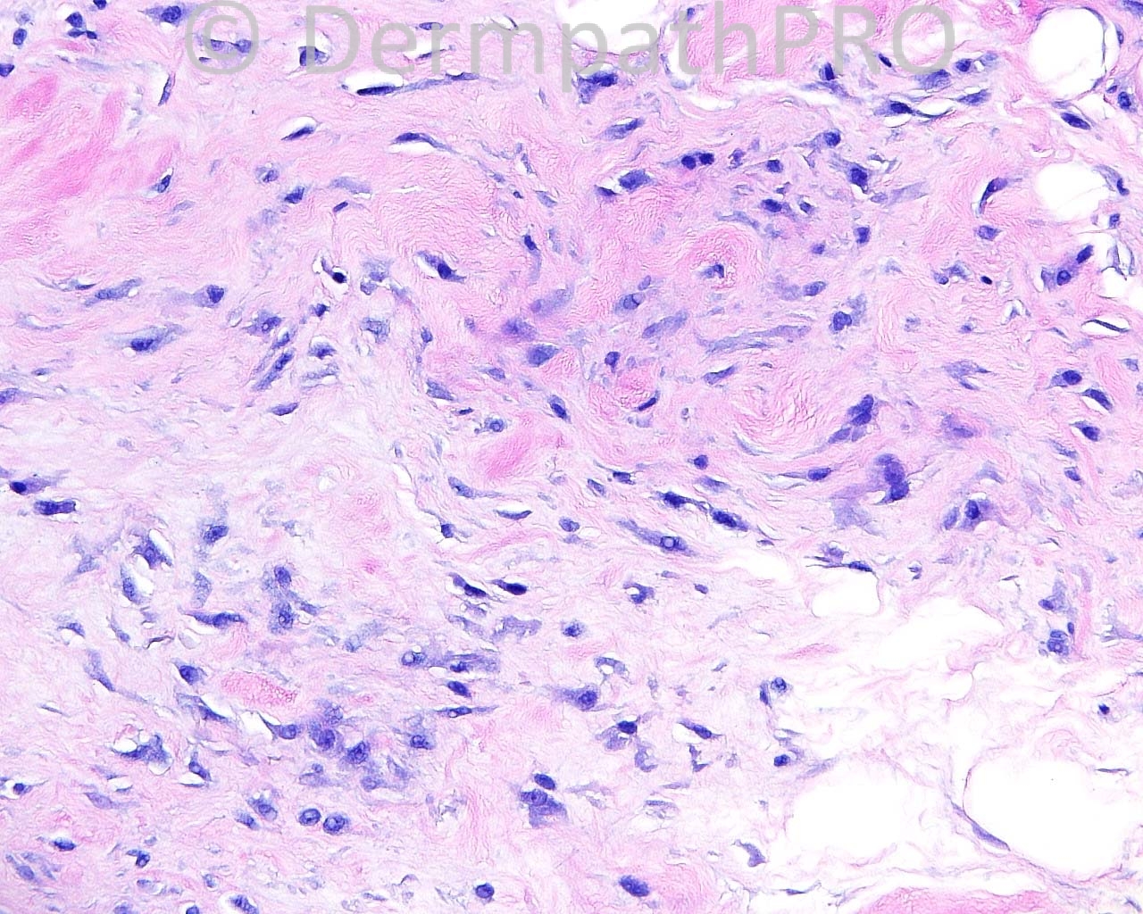
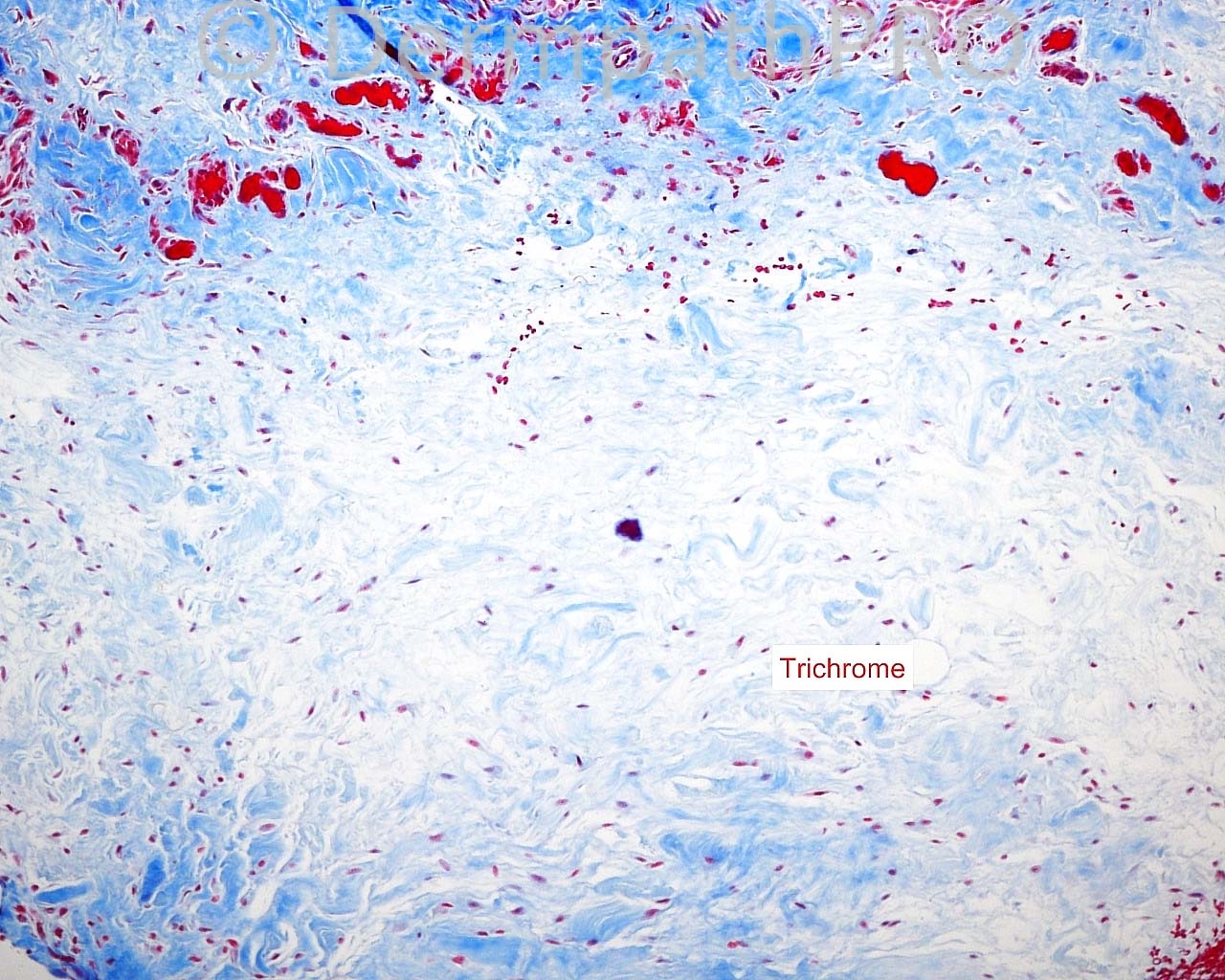
The patient is a 45 year old woman with a punch biopsy taken from the left middle finger over the PIP knuckle.

Case posted by Dr. Mark Hurt


  Report Record

User Feedback


I see diminished elastic fibers in a background of sparse cellular inflammation. My first hypothesis was Acrokeratoelastoidosis, but this is typically seen over the margins of the hands and feet. I also think this could be a late stage if Granuloma annulare.

Share this comment


Link to comment
share_externally

Eman El-Nabarawy

Posted

Degenerative collagenous plaques of the hands vs acrokeratoelastoidosis presenting as knuckle padlike lesions (as a part of marginal papular acrokeratoderma disorders).

Share this comment


Link to comment
share_externally

Guest Tiberiu Tebeica

Posted

The first impression I had looking at the low power picture was nephrogenis systemic fibrosis, but taking into account the clinical information and the special stains, I think acrokeratoelastoidosis fits best. There is a case of acrokeratoelastoidosis described in literature, which presented clinically as [url="http://www.ncbi.nlm.nih.gov/pubmed/?term=3891801"]small papules located mainly over the interphalangeal joints[/url].

Share this comment


Link to comment
share_externally

Guest Maria George

Posted

ITo e there is mcinous degeneration.Could be surface of synviial cyst , i.e mucus cyst .I would order deeeeep secttion.

Share this comment


Link to comment
share_externally

Robledo F. Rocha

Posted

Acrokeratoelastoidosis is a genodermatotic keratoderma and, as such, I would expect to find hyperkeratosis overlying a slightly pushed-down epidermis. To me, stratum corneum appears normal for an acral site. Furthermore, the basophilic fragmented elastic fibers are too deep located and escorted by interstitial histiocytes immersed in increased connective tissue mucin, findings that suggest subcutaneous granuloma annulare.

Share this comment


Link to comment
share_externally

Guest Jim Davie MD

Posted

I agree with Maria and Robledo. There is some raggedness at the lower edge that may suggest a synovial cyst edge, and there is increased dermal mucin. Synovial cyst vs. GA.

Share this comment


Link to comment
share_externally

Guest Mazen charaf iddin

Posted

[right][font="arial, verdana, tahoma, sans-serif"][color="#1c2837"][size=4]I agree with Dr. Robledo F. Rocha for the epidermal changes, and the lesion is deeper than [/size][/color][/font][/right][color=#1C2837][font=arial, verdana, tahoma, sans-serif][size=4]acrokeratoelastoidosis.[/size][/font][/color][right][font="arial, verdana, tahoma, sans-serif"][color="#1c2837"][size=4] If it's only one lesion, I'll considered it a resolving (fibrotic, [/size][/color][/font][/right]scarred ) [color=#1C2837][font=arial, verdana, tahoma, sans-serif][size=4][right]fibrous histiocytoma ( giant cell tumor of tendon sheath) with degenrative changes in nucli of fibroblast. Nuclear changes like this [/right][/size][/font][/color]aren't[color=#1C2837][font=arial, verdana, tahoma, sans-serif][size=4][right] seen in [/right][/size][/font][/color][color=#1C2837][font=arial, verdana, tahoma, sans-serif][size=4]acrokeratoelastoidosis[/size][/font][/color]

Share this comment


Link to comment
share_externally


Join the conversation

You can post now and register later. If you have an account, sign in now to post with your account.

Guest
Add a comment...

×   Pasted as rich text.   Paste as plain text instead

  Only 75 emoji are allowed.

×   Your link has been automatically embedded.   Display as a link instead

×   Your previous content has been restored.   Clear editor

×   You cannot paste images directly. Upload or insert images from URL.

Loading...

×
×
  • Create New...