Jump to content
In this section we have spot diagnoses posted on a daily basis since June 2010, now over 4000! You can review the archived cases and read the suggested diagnoses by users and the final comment by the contributors.
Case are uploaded each week day by 10 am UK time with the correct diagnosis will generally be posted at 8 pm UK time. Why not view the most recent spot diagnosis and proffer a diagnosis?

Case Number : Case 932 - 17th January Posted By: Guest

Please read the clinical history and view the images by clicking on them before you proffer your diagnosis.
Submitted Date :
   (0 reviews)

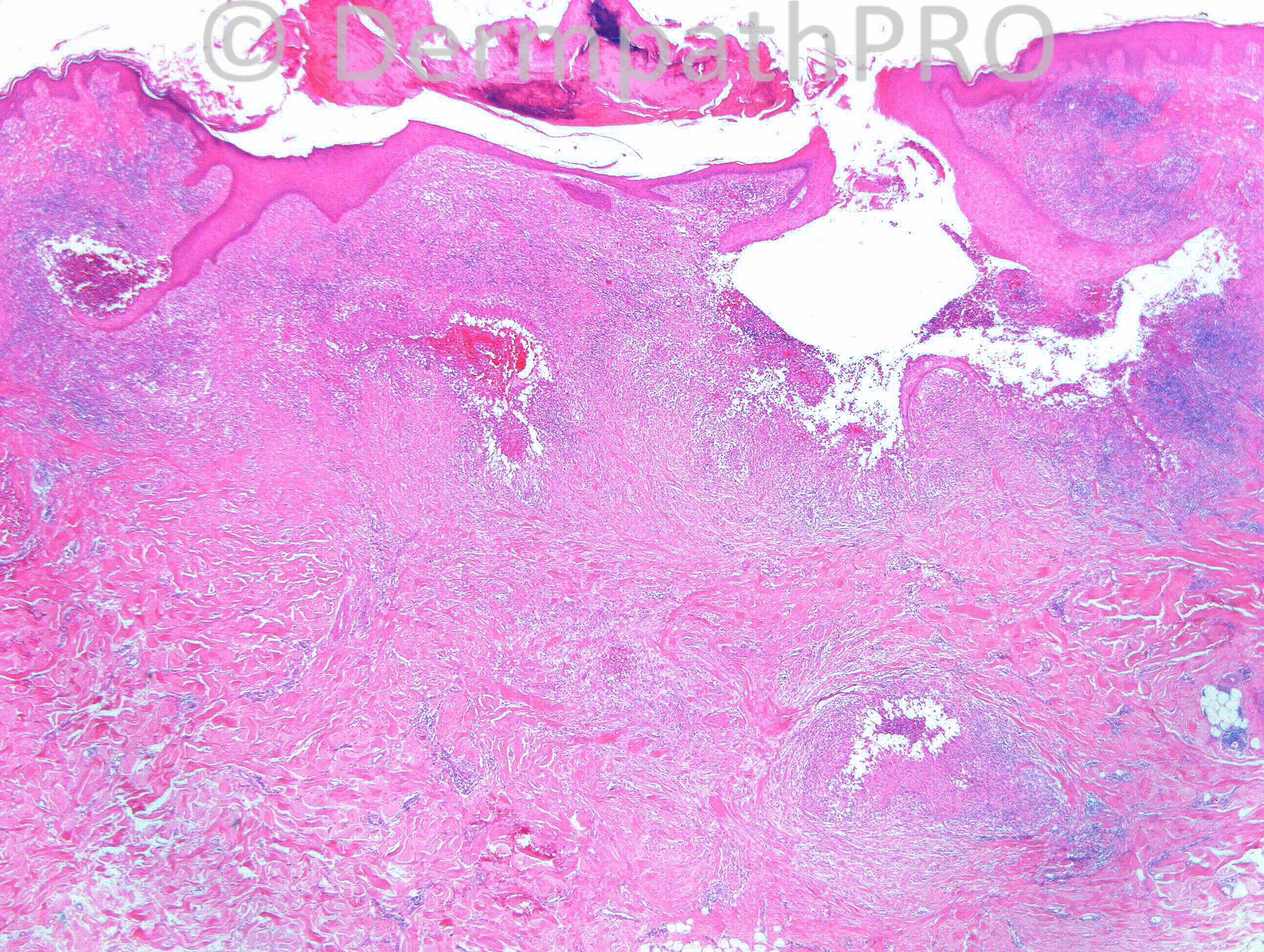
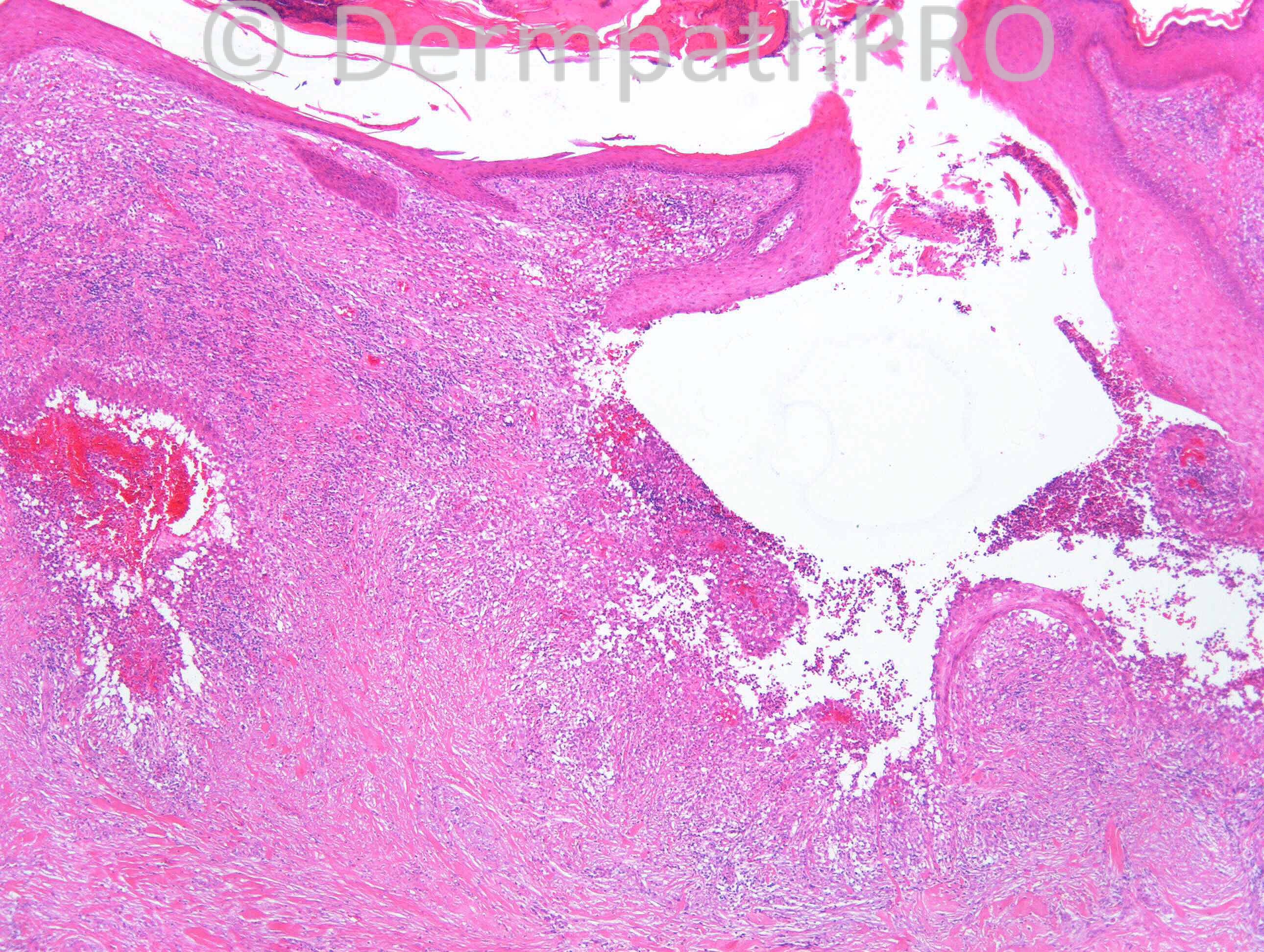
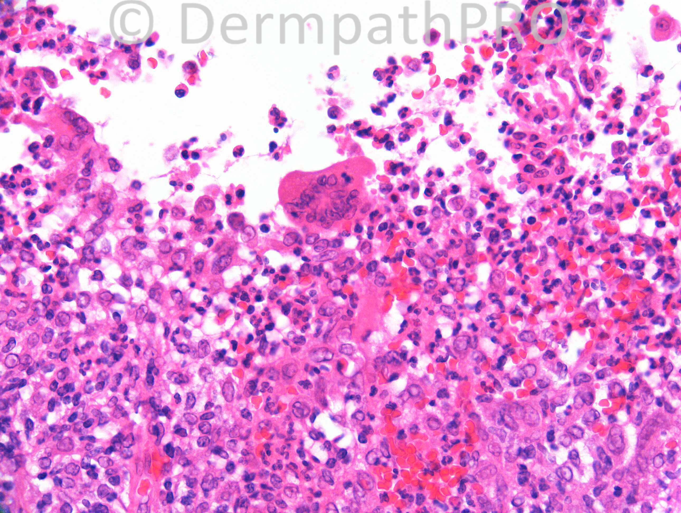
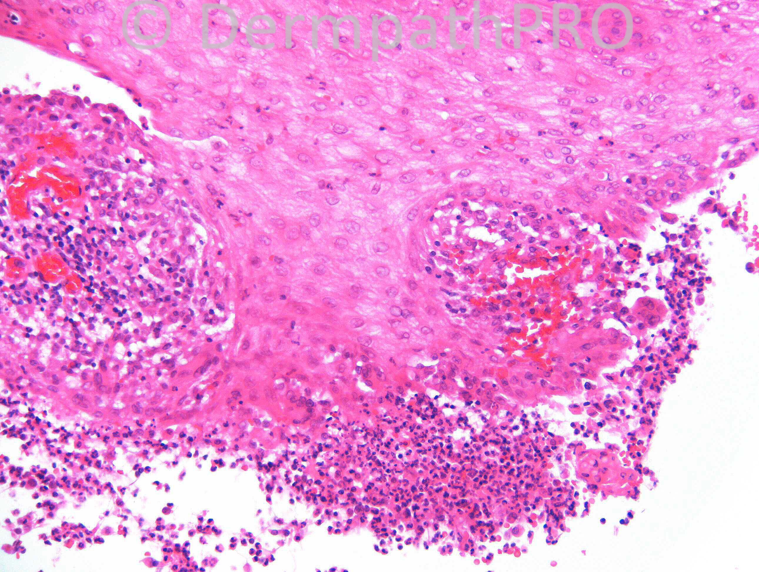
49 years old male. Shoulder.

Case posted by Dr. Richard Carr.

  Report Record

User Feedback


Guest Maria George

Posted

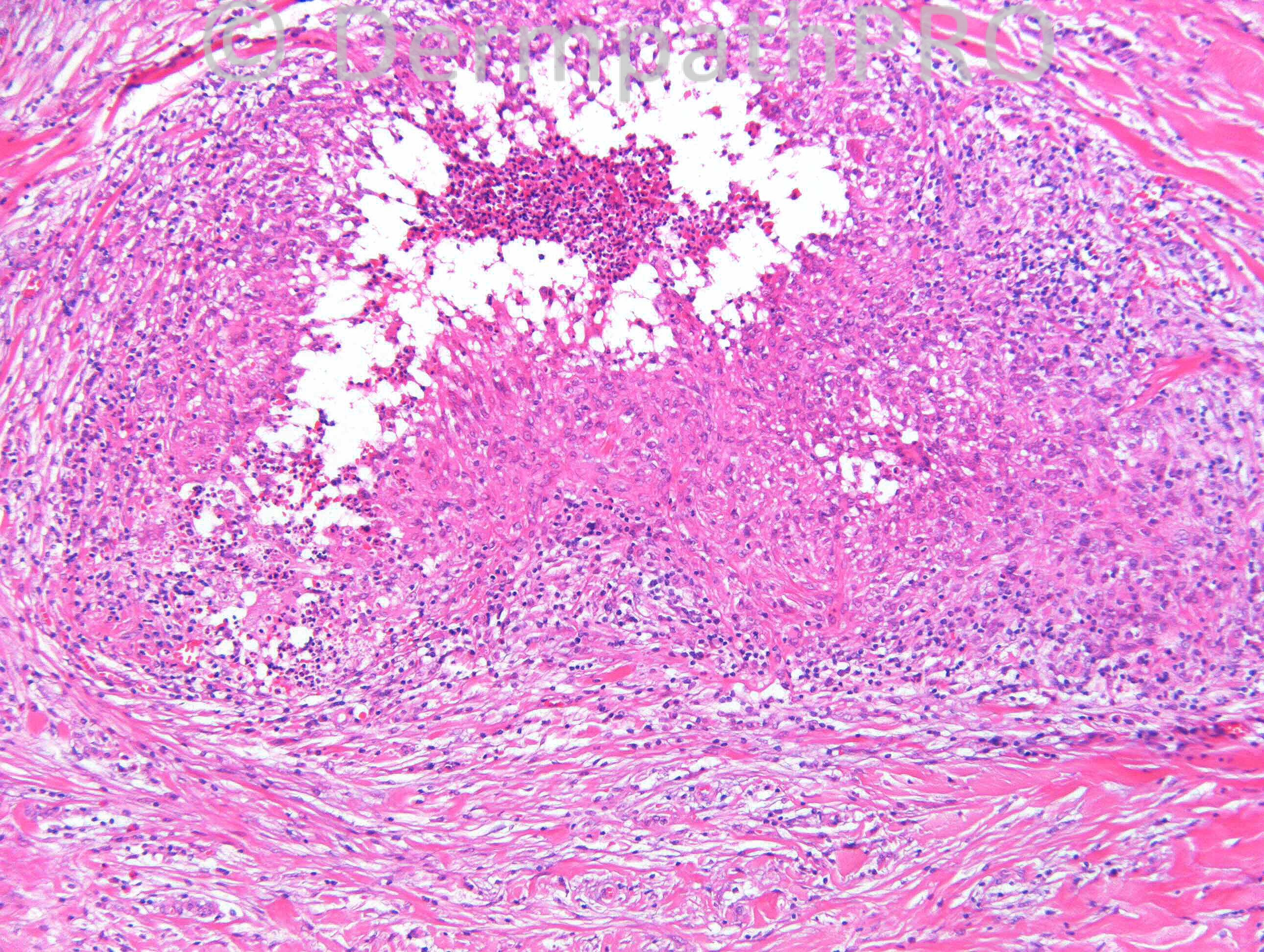
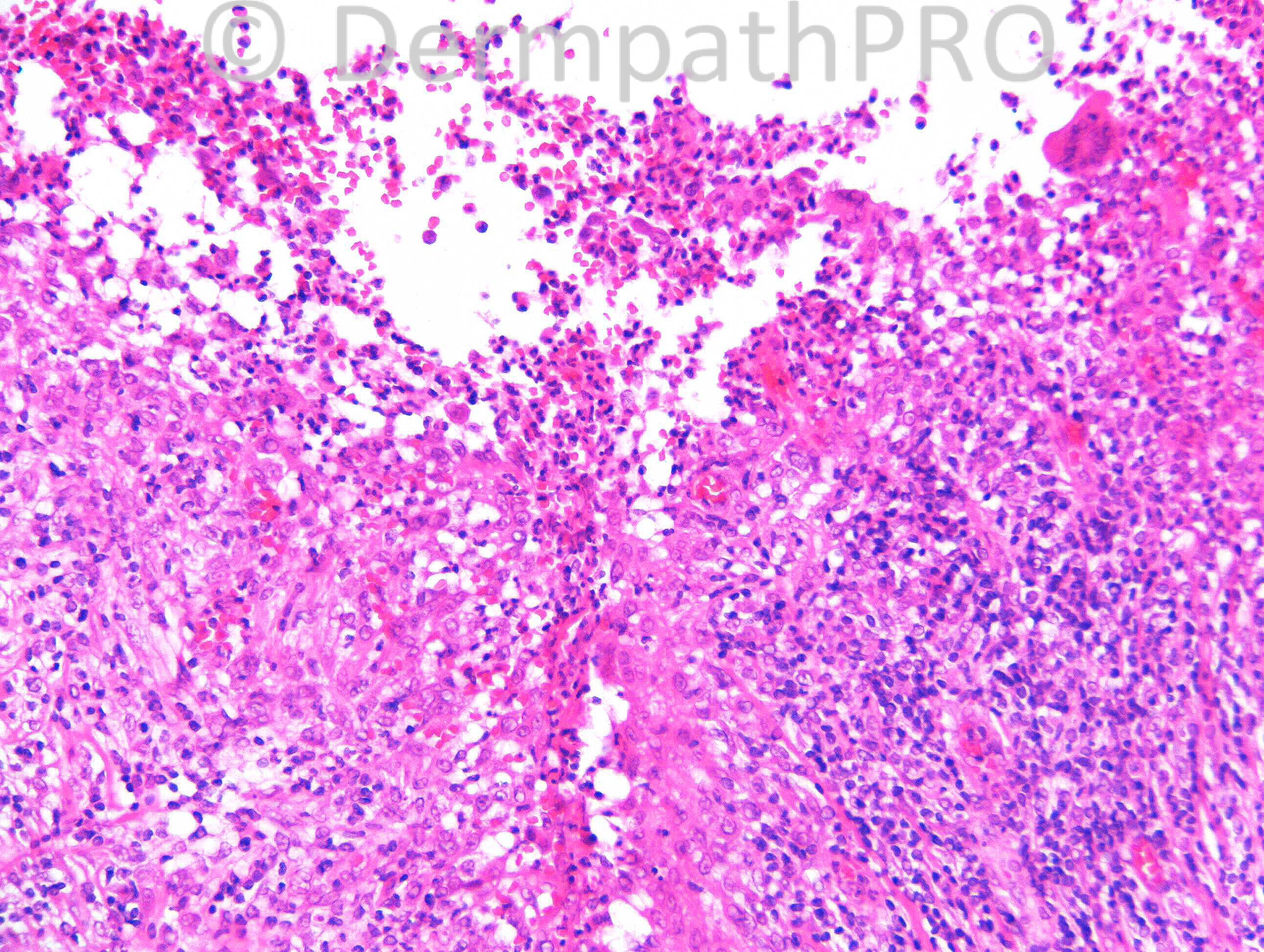
In this landscape of ulcer with multiple granulomas with microabscess and Warthin Finkedley type of giant cell woud go for iinfetious process first.Since no organism seen.I may favor sporo or myobact.
Secial stains please.

Share this comment


Link to comment
share_externally

Dr. Mona Abdel-Halim

Posted

Supprative granulomatous reaction mostly related to ruptured folliculitis. Stains should be carried out to exclude infectious causes such as deep mycoses and atypical mycobacteria. Pyoderma gangrenosum can also give this picture.

Share this comment


Link to comment
share_externally

Guest Dr.Yüksel Okumuş

Posted

Atypical mycobacterial enfection or Blastomycosis

Share this comment


Link to comment
share_externally

Guest Romualdo

Posted

If special stains exclude infectious causes such as mycobacteriosis and sporotrichosis I would suggest acne conglobata. CPC is needed.

Share this comment


Link to comment
share_externally

Sasi Attili

Posted

I agree with above differentials (wide). CPC needed.

Share this comment


Link to comment
share_externally

Eman El-Nabarawy

Posted

Infection: sporotrichosis, blastomycosis, mycobacteriosis top the list of DD.

Share this comment


Link to comment
share_externally

Robledo F. Rocha

Posted

Multiple suppurative ruptured folliculitis. If special stains rule out infectious causes, than those excellent differential diagnoses suggested here, to which I would add halogenoderma, can be confirmed by clinicopathological correlation.

Share this comment


Link to comment
share_externally

Guest Tiberiu Tebeica

Posted

Suppurative granulomatous dermatitis with pseudoepitheliomatous hyperplasia and ruptured folliculitis should favor an infectious process. I like pyoderma gangrenosum if stains are negative, but CPC needed.

Share this comment


Link to comment
share_externally

Guest Jim Davie MD

Posted

Infectious etiology. Agree with the excellent differentials! Stains and CPC required.
HSV/VZV in background of immunosuppression was an initial thought, based on hint of nuclear clearing and dense multinucleation in some sections (difficult to evaluate in these images), but the architecture of necrotic/suppurative granulomas would not be typical and make it a long shot.

Share this comment


Link to comment
share_externally

Dr. Richard Carr

Posted

All bug stains negative. Note the pattern here is of serpiginous superficial and deep ulcerative process with neutrophils coating a band-like layer of macrophages. There is psuedoepitheliomatous hyperplasia. Note the history - odd location. This is quite a characteristic histology for the diagnosis. Jim I interpreted those cells as multinucleate macrophage that are also quite typical (in the diagnosis) but nice try.

Share this comment


Link to comment
share_externally

Dr. Mona Abdel-Halim

Posted

Did the negative bug stains included also silver stains for bartonella to R/O cat scratch disease??? Or may be monoclonal antibodies will be better here... I think this explains the odd site.

Share this comment


Link to comment
share_externally

Dr. Richard Carr

Posted

I did not consider of Cat Scratch to be honest.

Share this comment


Link to comment
share_externally

Nice hypotheses. I still think this is an infectious disease. Without more clinical information and with negative special stains, maybe a PCR for microorganisms like Mycobacterium could be helpfull.
I have never seen Cat Scratch disease outside lymph node, but the "geografic" necrotic granulomas seems quite the same.
If cultures are positive we may be dealing with a Blastomycosis-like pyoderma.

Share this comment


Link to comment
share_externally

Dr. Mona Abdel-Halim

Posted

If infections were excluded by all possible means, then we r left with superficial vegetative pyoderma (a form of pyoderma gangrenosum)... My third suggested DD...

Share this comment


Link to comment
share_externally

Guest Maria George

Posted

Acute Milliarisa Diss Facie.A form of granulomatous rosacea is also a possibility.

Share this comment


Link to comment
share_externally

Guest Romualdo

Posted

Superficial granulomatous pyoderma: a localized vegetative form of pyoderma gangrenosum. Well done Mona and the other colleagues who suggested pyoderma gangrenosum!

Share this comment


Link to comment
share_externally

Eman El-Nabarawy

Posted

Yes I think it fits in superficial granulomatous pyoderma gangrenousum.

Share this comment


Link to comment
share_externally

Guest Maria George

Posted

Necrobiotic xanthogran. may be added to differential

Share this comment


Link to comment
share_externally

Guest Dr. Yüksel Okumuş

Posted

Injection (vaccine) side reaction ? Swimming pool granuloma ?

Share this comment


Link to comment
share_externally

Dr. Richard Carr

Posted

Thanks all and yes well done Mona. This is in my experience a typical example of superficial granulomatous pyoderma. Try to remember this pattern. There was a prior history of pyoderma gangrenosum. You must always exclude infections but please do remember this case and think of this diagnosis that usually responds to steroids. I think the name is not ideal as clealy the ulceration can extend deeply into the dermis as in this case. In my experience the diagnosis is often missed. One patient I will never forget. We received a specimen (taken by a surgeon) as excision of a chronic wound / sinus on the back of the arm of a middle aged womman. I received the biopsy and after negative bug stains suggested the possibility of superficial granulomatous pyoderma and advised referral to a dermatologist. The surgeon rang me to ask about the report of the patient with granuloma pyogenicum because the wound had broken down, extending, gaping and coated in pus. I asked the surgeon to read back the words in the report carefully and explained that the patient probably had a variant of pyoderma gangrenosum (not pyogenic granuloma). The lesson for me would be to ring the clinician next time and explain in the report this is considered to be a variant of pyoderma gangrenosum. Turns out that patient had had the wound for a year and had been repeatedly treated for infection. The excellent dermatologist on reading the same histopathology report immediately put the patient on moderately high dose oral steroids. The now extensive wound responded very well and the patient was greatly relieved to finally get a diagnosis.
Enjoy the rest of your weekends.

Share this comment


Link to comment
share_externally


Join the conversation

You can post now and register later. If you have an account, sign in now to post with your account.

Guest
Add a comment...

×   Pasted as rich text.   Paste as plain text instead

  Only 75 emoji are allowed.

×   Your link has been automatically embedded.   Display as a link instead

×   Your previous content has been restored.   Clear editor

×   You cannot paste images directly. Upload or insert images from URL.

Loading...

×
×
  • Create New...