Jump to content
In this section we have spot diagnoses posted on a daily basis since June 2010, now over 4000! You can review the archived cases and read the suggested diagnoses by users and the final comment by the contributors.
Case are uploaded each week day by 10 am UK time with the correct diagnosis will generally be posted at 8 pm UK time. Why not view the most recent spot diagnosis and proffer a diagnosis?

Case Number : Case 934 - 21st January Posted By: Guest

Please read the clinical history and view the images by clicking on them before you proffer your diagnosis.
Submitted Date :
   (0 reviews)

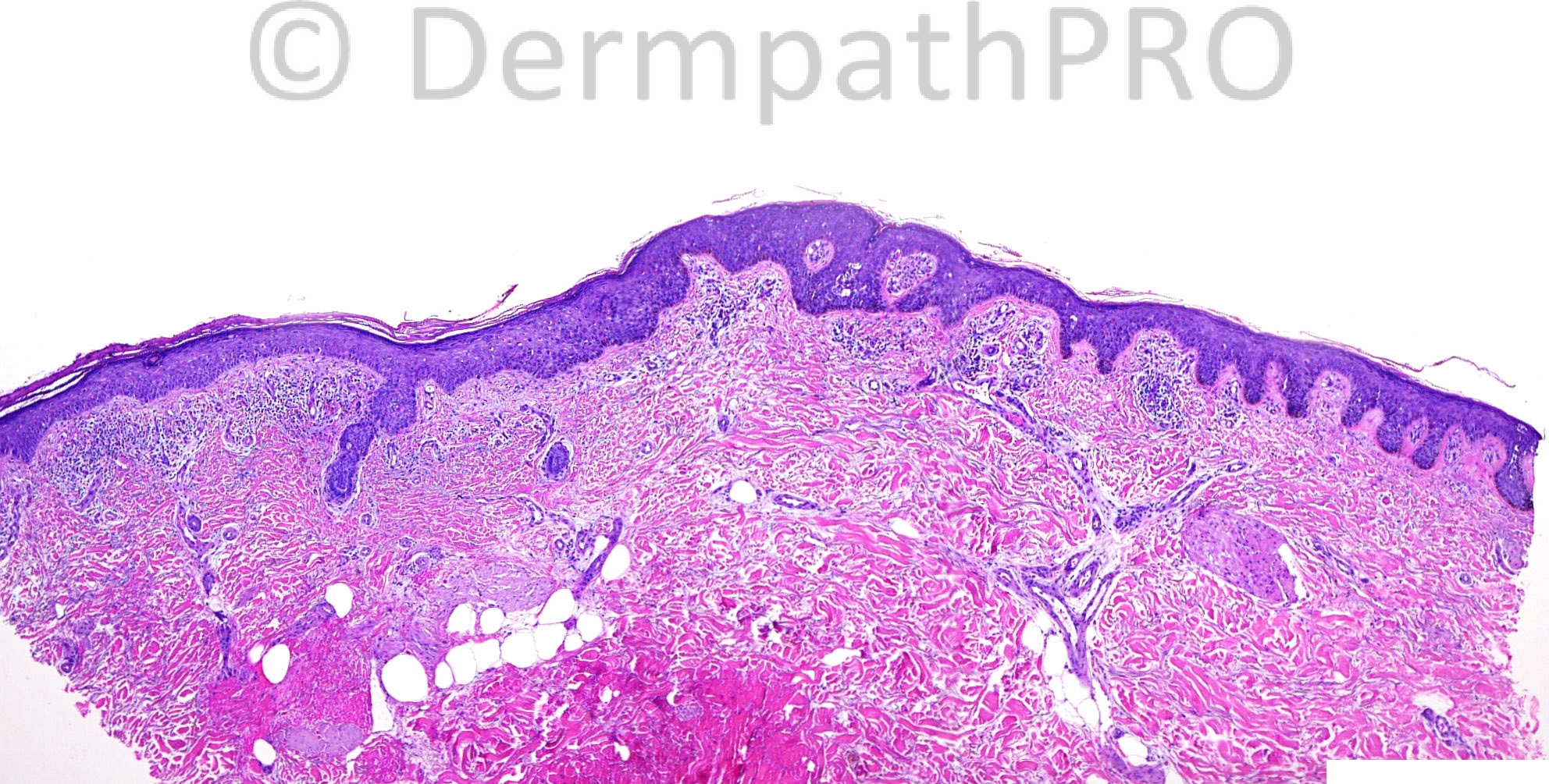
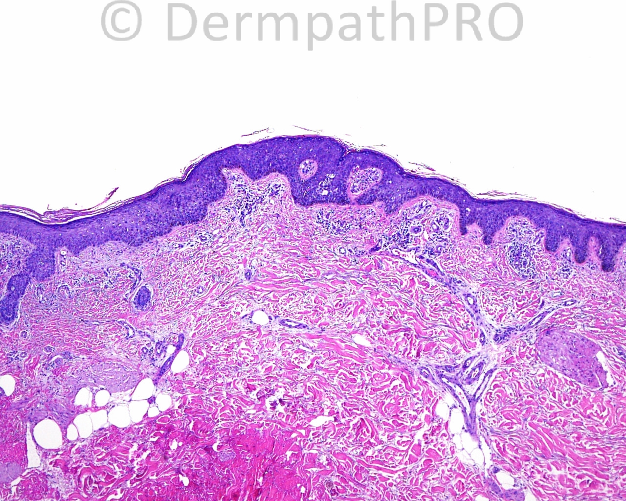
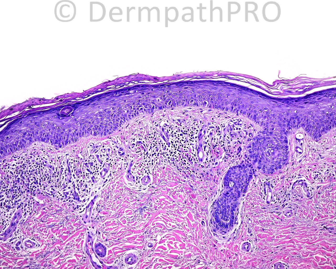
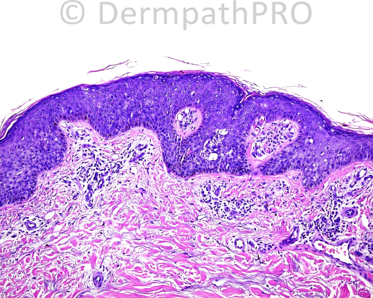
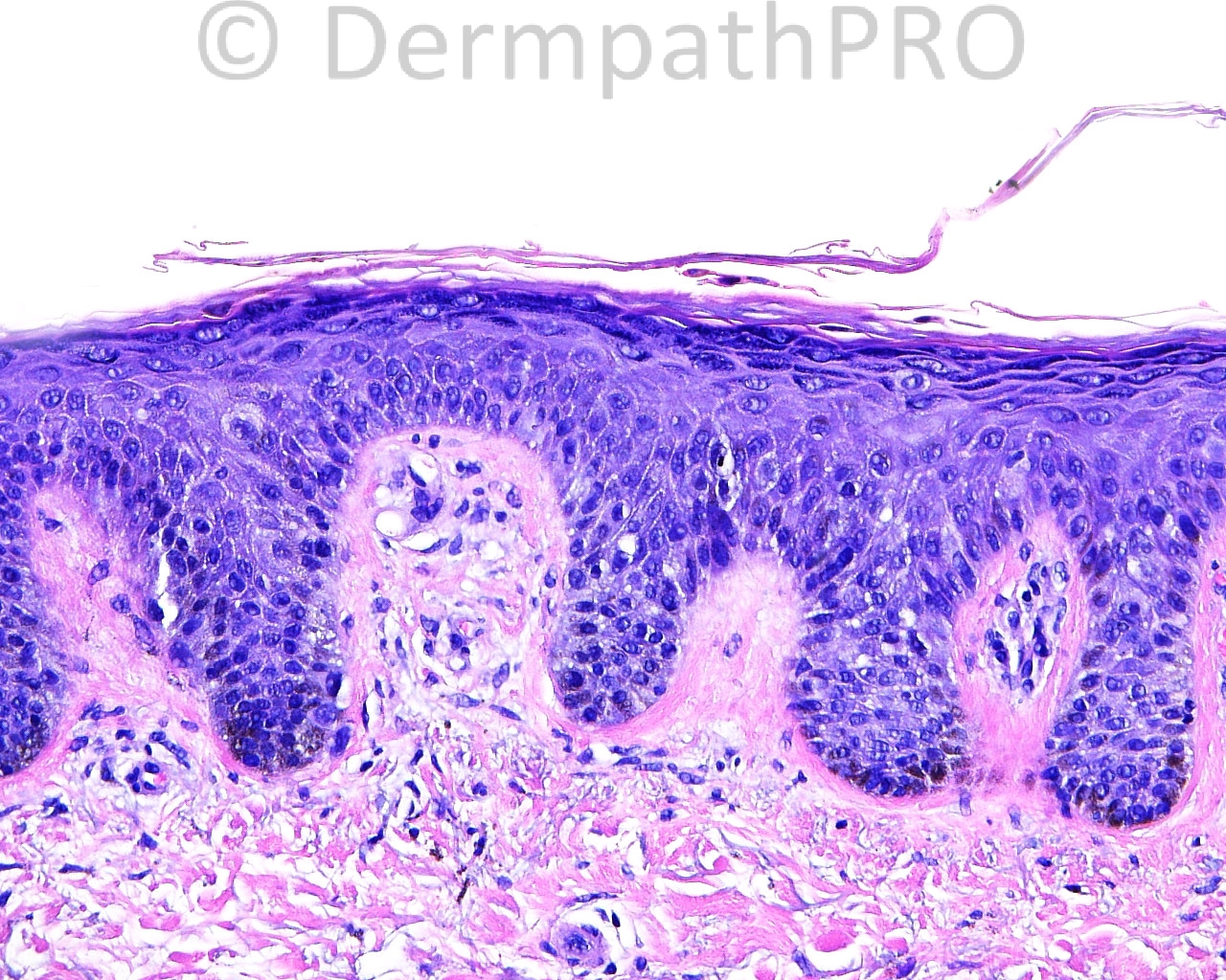
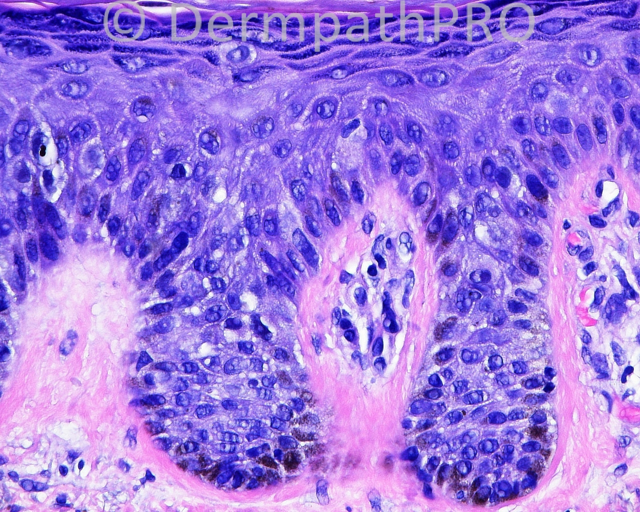
The patient is a 53-year-old woman with a punch biopsy of a 1.0 x 0.6 cm irregularly hyperpigmented and pink plaque taken from the left calf.

Case posted by Dr. Mark Hurt


  • Like 1
  Report Record

User Feedback


Guest Saleem Taibjee

Posted

I wondered about a subtle bowenoid lesion, I don't think there is a melanocytic proliferation. Might also want a PAS to rule out dermatophyte.

Share this comment


Link to comment
share_externally

Eman El-Nabarawy

Posted

Bowen's disease vs large cell acanthoma.

Share this comment


Link to comment
share_externally

Dr. Mona Abdel-Halim

Posted

Difficult case. The scanning image shows that the right side is different from the left side. Maybe the biopsy was taken from both the hyperpigmented (right side) and the pink (left side)???? I am thinking of a subtle Bowen's developing in collision or within a lentigo?? Need Melan stain to confirm the nature of the increased pigmentation on the right side. Need proliferation markers to assess the subtle bowenoid changes. Interesting case..

Share this comment


Link to comment
share_externally

Sasi Attili

Posted

agree with Mona.....Subtle Bowens. But would like to see a Melan-A, to be on the safe side.

Share this comment


Link to comment
share_externally

Guest Maria George

Posted

Pigmented Actinic keratosis is also a possibility here.

Share this comment


Link to comment
share_externally

Eman El-Nabarawy

Posted

Actinic keratosis (atrophic and bowenoid types) vs actinic keratosis with Bowen's disease.

Share this comment


Link to comment
share_externally

Guest Jim Davie MD

Posted

I favor atypical junctional melanocytic proliferation with pagetoid scatter, rule out MMIS.

I see variably atypical and/or enlarged epithelioid cells at the junction, and also in the epidermis with high-level pagetoid scatter. There is parakeratosis (with flattened hyperpigmented cells embedded therein) and reactive keratinocyte changes that may suggest a non-neoplastic etiology (such as previous trauma) for melanocyte scatter.

Share this comment


Link to comment
share_externally

Guest Dr.Yüksel Okumuş

Posted

In addition to epidermal Bowenoid features, is there dermal hamartomatous (smooth muscle proliferation) and nevus lipomatosus like changes ? Especially at the right side, increased basal layer pigmentation, hyperkeratosis and elongation of rete ridges maybe the signs of Becker’s melanosis, although the localization and the patient age is contrary.

Share this comment


Link to comment
share_externally

Eman El-Nabarawy

Posted

Melanoma in situ in collision with Bowen's disease.

Share this comment


Link to comment
share_externally

Dr. Mona Abdel-Halim

Posted

So after the Immunos, Bowen's in collision with a melanom in situ..

Share this comment


Link to comment
share_externally

Guest Jim Davie MD

Posted

My opinion:
1. [b]MMIS[/b]. The large epithelioid cells at the junction and pagetoid within the dermis stain with Melan-A. The flattened hyperpigmented cells embedded in the parakeratosis are also strongly Melan-A positive, as expected. There is downwards pagetoid extension along adnexa.
2. [b]Incidental clonal-pattern SK[/b]. I think it's too discrete and well-circumscribed to be Bowen's. Ki67 (and mitotic activity) can be deceitfully high in SKs.

Share this comment


Link to comment
share_externally


Join the conversation

You can post now and register later. If you have an account, sign in now to post with your account.

Guest
Add a comment...

×   Pasted as rich text.   Paste as plain text instead

  Only 75 emoji are allowed.

×   Your link has been automatically embedded.   Display as a link instead

×   Your previous content has been restored.   Clear editor

×   You cannot paste images directly. Upload or insert images from URL.

Loading...

×
×
  • Create New...