Jump to content
In this section we have spot diagnoses posted on a daily basis since June 2010, now over 4000! You can review the archived cases and read the suggested diagnoses by users and the final comment by the contributors.
Case are uploaded each week day by 10 am UK time with the correct diagnosis will generally be posted at 8 pm UK time. Why not view the most recent spot diagnosis and proffer a diagnosis?

Case Number : Case 937 - 24th January Posted By: Guest

Please read the clinical history and view the images by clicking on them before you proffer your diagnosis.
Submitted Date :
   (0 reviews)

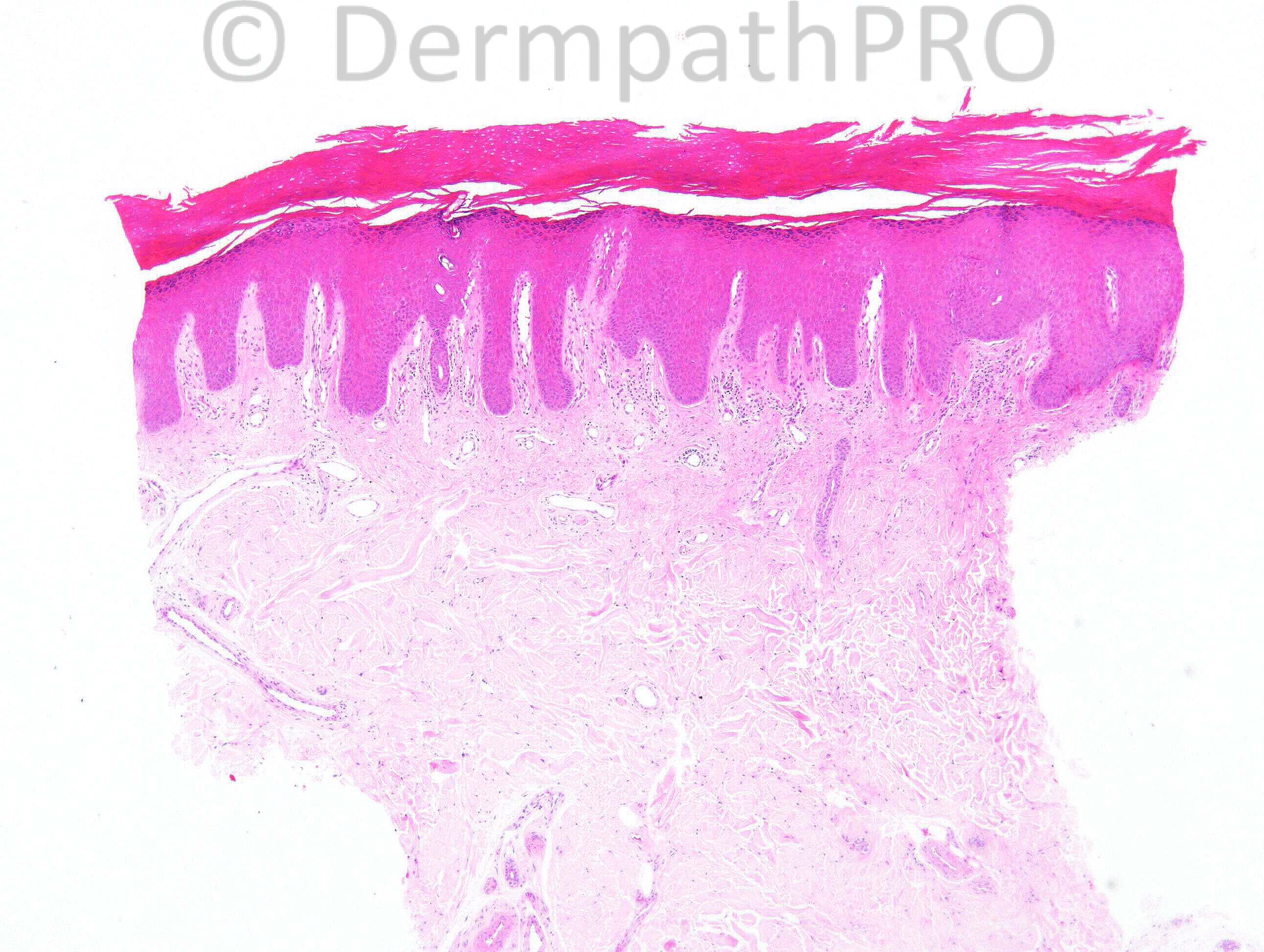
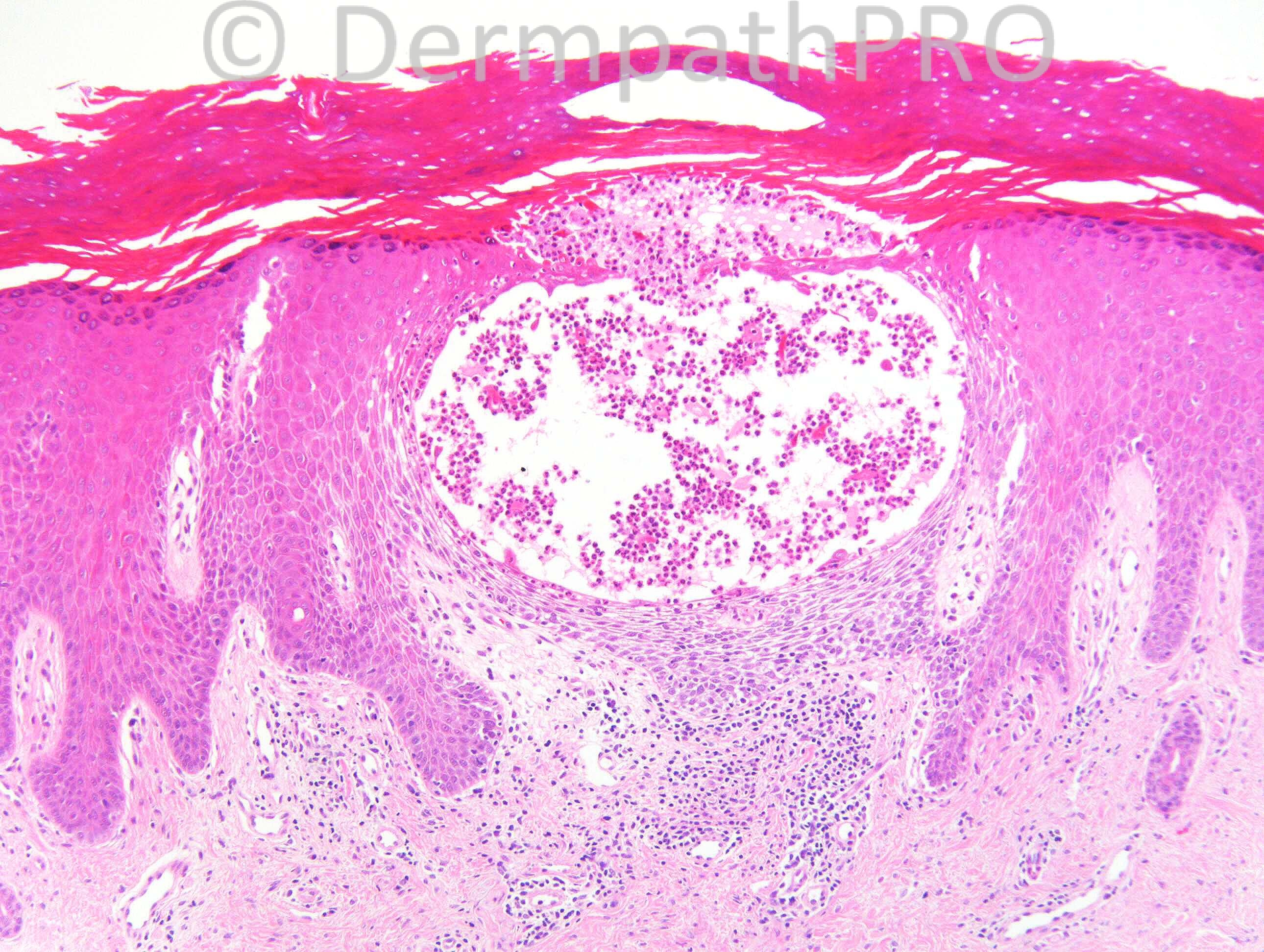
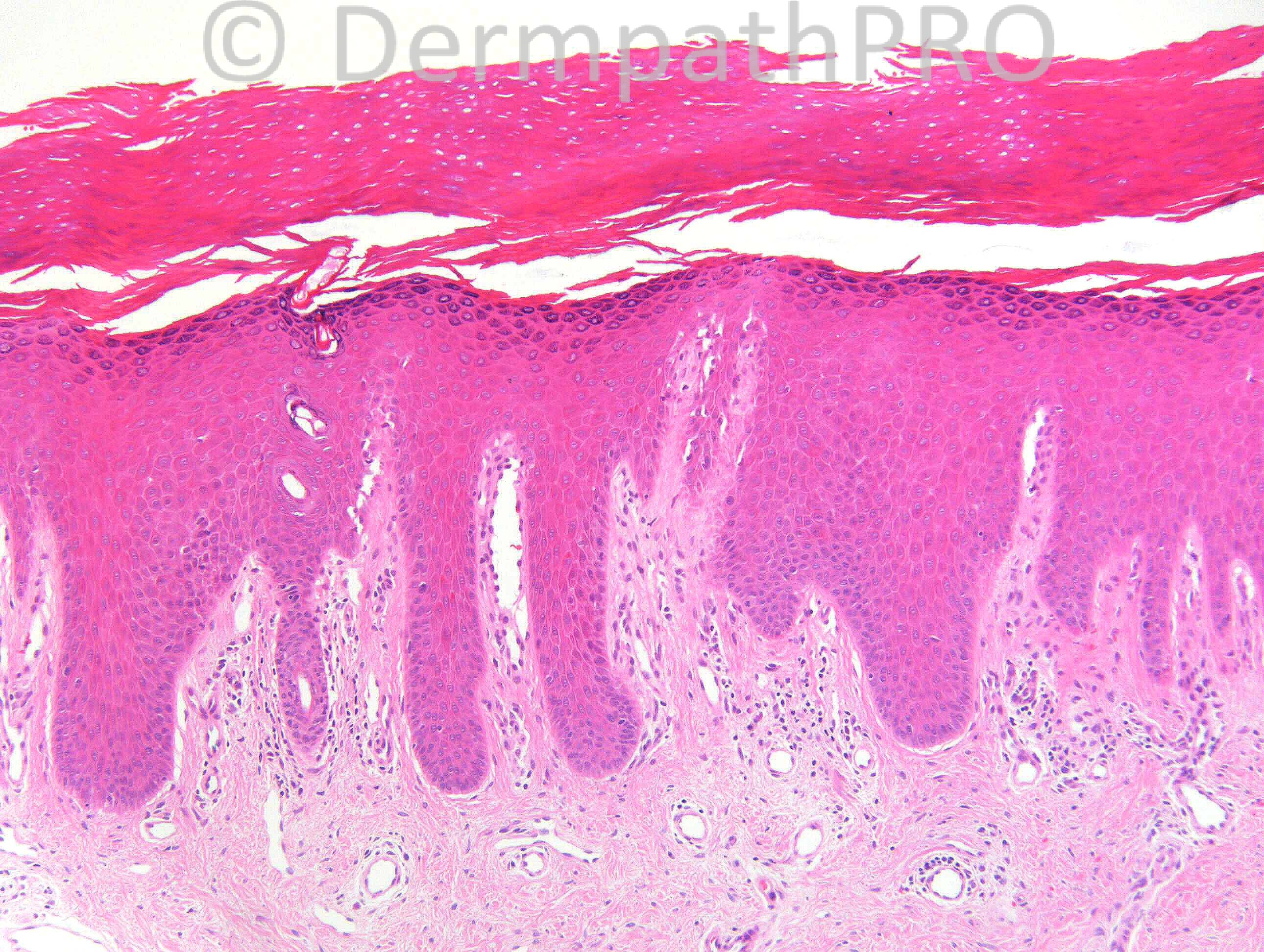
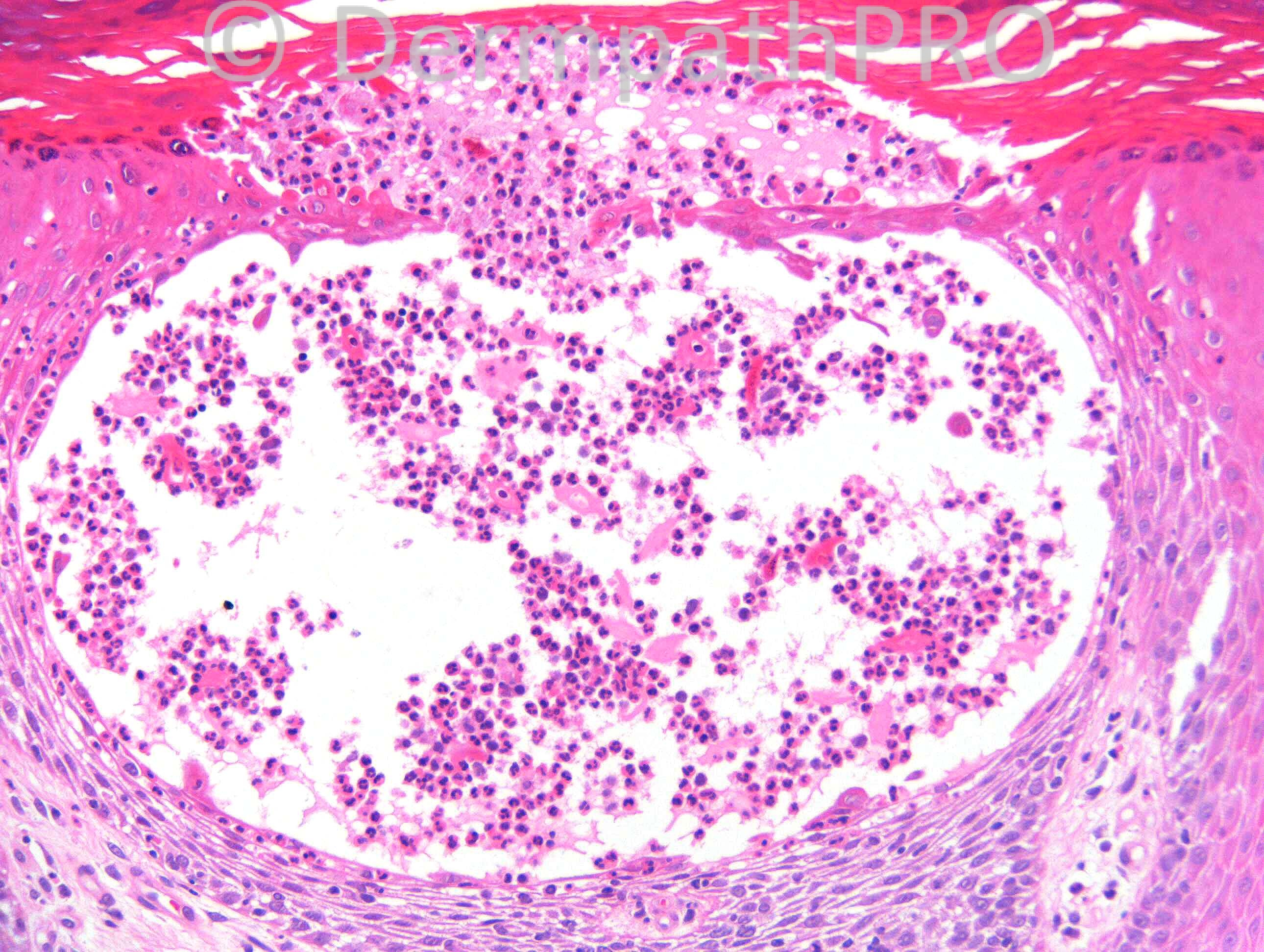
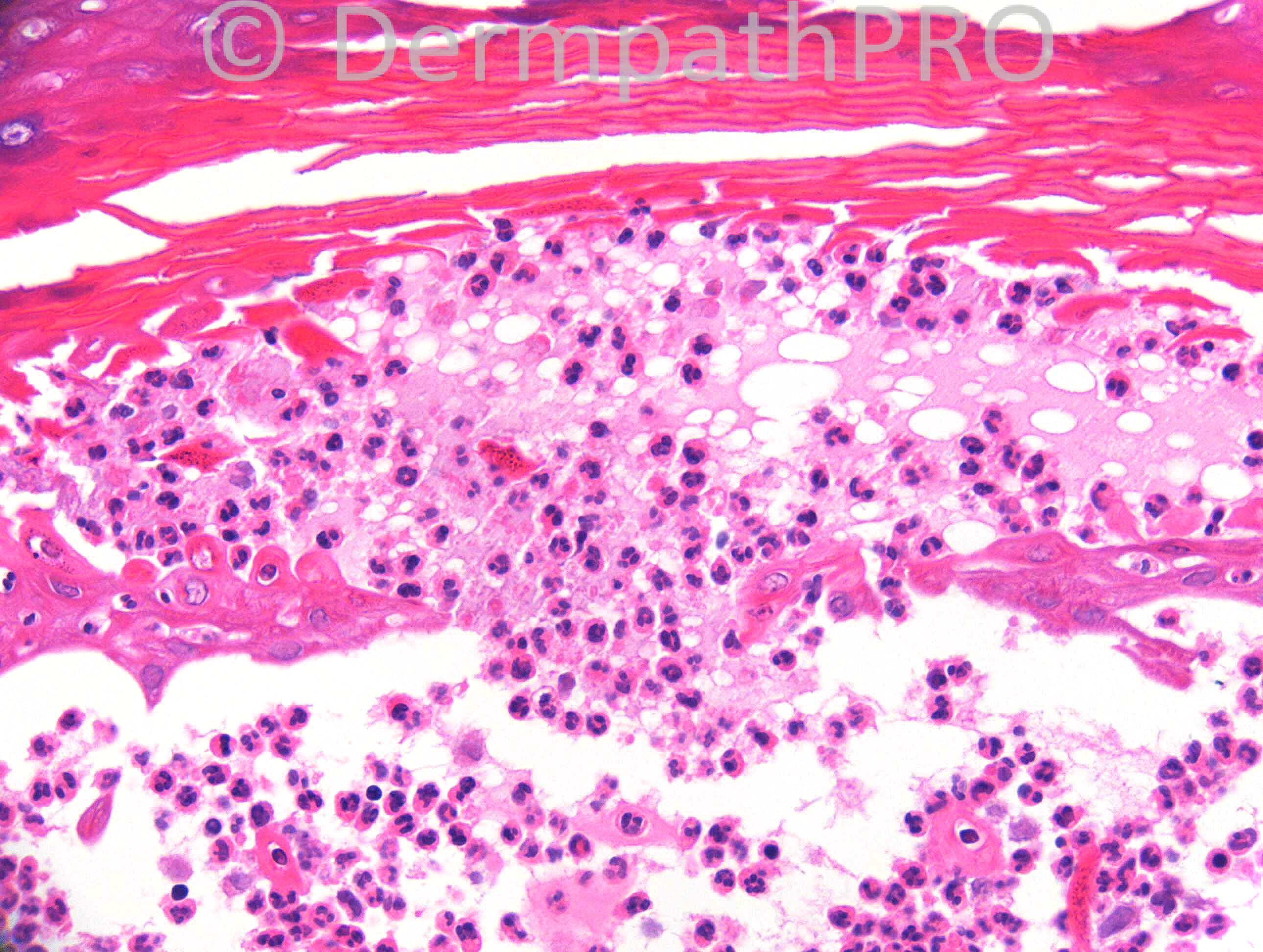
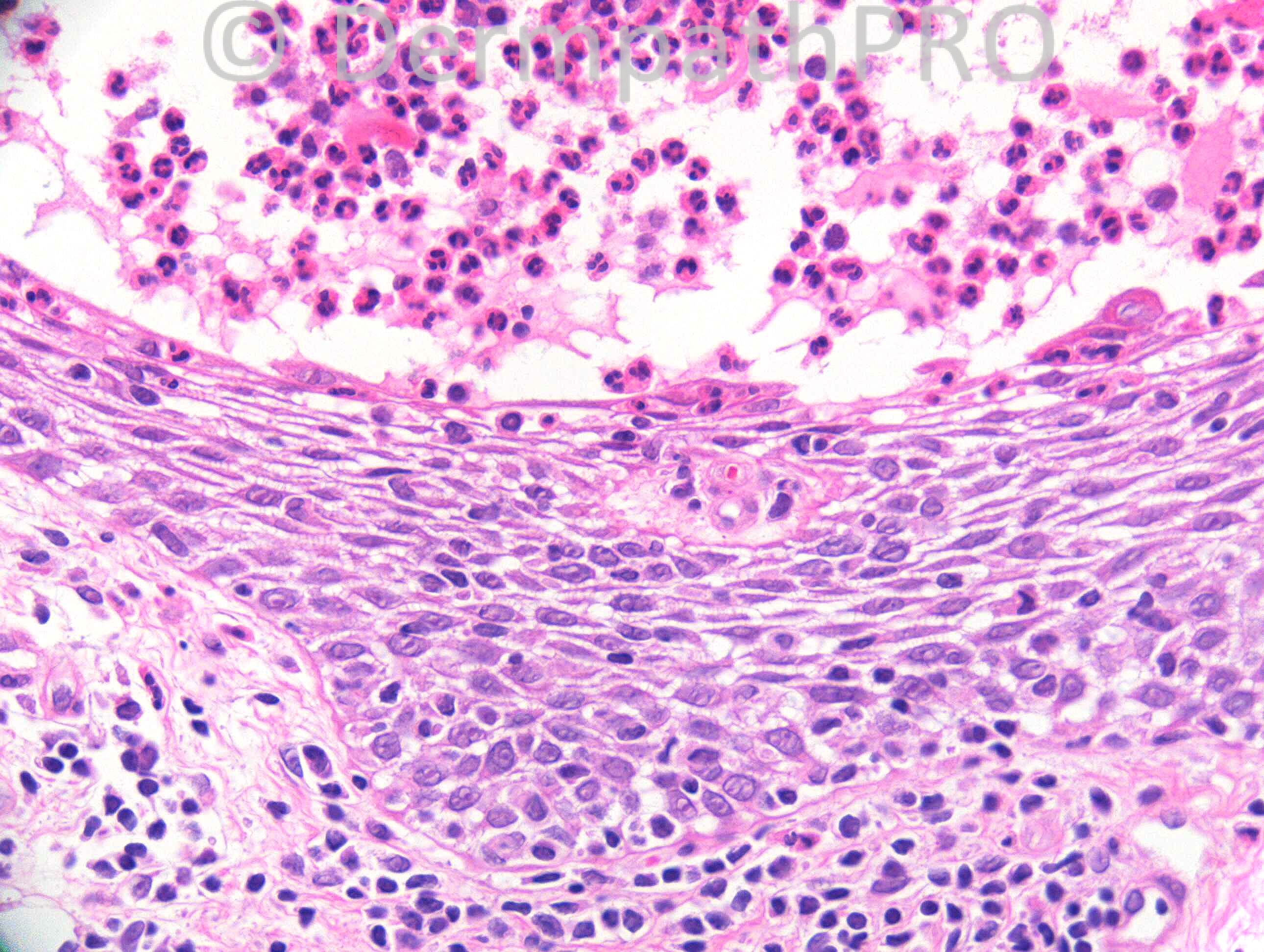
52 years old female. 2 yrs, scaly, thickened plaque & pustules isolated to right hand.

Case posted by Dr. Richard Carr.


  Report Record

User Feedback


Sasi Attili

Posted

There is a typical flask shaped (pompholyx like) vesicle. However, the predominance of neutrophils and the localised nature of the 'plaque', made me initially favour psoriasis as pompholyx is not usually localised to one area and does not present as a plaque. Secondly, LSC like changes are often seen in acral pustular psoriasis which can be itchy.There is a slight loss of granular layer and parakeratosis but these are not specific enough. Focal spongiosis in one of the pictures, could be an evolving psoriatic pustule.
But, I think it is a difficult case without CPC. Pompholyx can have pustules in some cases i.e. has been described to be neutrophil rich. Don't see any evidence of fungi, though PAS is advisable.

Share this comment


Link to comment
share_externally

Eman El-Nabarawy

Posted

Pomphylex. Acute on top of chronic dermatitis (lichen simplex chronicus).

Share this comment


Link to comment
share_externally

I agree with Dr El-Nabarawy, that the background is that of lichen simplex chronicus, rather than psoriasis, & the intraepidermal pustule is that of spongiotic dermatitis (unless PAS for fungi is positive).

Share this comment


Link to comment
share_externally

Guest Saleem Taibjee

Posted

Definitely need to exclude fungal given unilateral.
Otherwise, presence of pustules for me would favour psoriasis rather than eczema clinically.
One of my previous teachers used to insist that blisters (but not pustules) was a discriminating point for eczema rather than psoriasis. However over the years, I am beginning to think there is a spectrum/overlap between spongiotic psoriasis and hyperkeratotic eczema, and evolution over time sometimes makes things clearer.

Share this comment


Link to comment
share_externally

Sasi Attili

Posted

Agree Saleem- I think clinically and histologically PPP can be difficult to distinguish from Pompholyx on occasions, and CPC/ history important.

Share this comment


Link to comment
share_externally

Guest Engin Sezer

Posted

Pustular psoriasis/palmoplantar pustulosis

Share this comment


Link to comment
share_externally

Dr. Mona Abdel-Halim

Posted

This pathological picture in the context of a unilateral lesion presenting as a thickened erythematous plaque with pustules necessitates doing PAS before signing out to exclude Tinea incognito. If no fungus is detected, then this could be localized pustular psoriasis.

Share this comment


Link to comment
share_externally

Robledo F. Rocha

Posted

I go with localized pustular psoriasis. On acral sites, psoriasis usually manifests as a spongiotic dermatitis so much that even simulates eczema.

Share this comment


Link to comment
share_externally

Dr. King-Chung Lee

Posted

Agree with localized pustular psoriasis subject to exclusion of infection. Apart from fungal infection, would also like to exclude bacterial infection by Gram stain. I previously had a case with clinical differential between pompholyx and pustular psoriasis which turns out to be Gram stain positive. The neutrophilic infiltrate is much more dense, though. Hope that you can see the photomicrographs.
[url="http://candc.familyds.net/S12-17206_0314.jpg"]H&E showing pustule[/url]
[url="http://candc.familyds.net/S12-17206_0318.jpg"]Gram stain[/url]

Share this comment


Link to comment
share_externally

Dr. Richard Carr

Posted

Additional Information: The clinical suggestions were ?palmo-plantar pustulosis, ?tinea manuum. Gram and PAS stains were negative.

Share this comment


Link to comment
share_externally

Guest Maria George

Posted

Tinea is number 1 for me best seen by GMS.
ID reaction is a possblity.[list=1]
[*]I got this DDX from the net for [b]pustules...pustulosis[/b][b]:[/b] some more [url="http://www.palpath.com/MedicalTestPages/skiner.htm#pustulosis1"]DDX HERE[/url].
[*]subcorneal pustular dermatosis (SPD).
[/list][list]
[*]bites...especially fire ant bites.
[*]halogenoderma: (especially bromides) lesions usually of lower extremities and have pseudocarcinomatous downward epidermal growth containing pustules[b][sup][size="2"]5[/size][/sup][/b].
[*]infected eczematous dishydrosis.
[*]pustular psoriasis[b][sup][size="2"]5[/size][/sup][/b]: a key diagnostic finding is polys layered (dried Kogoj's pustule-like perimeter...see [url="http://www.palpath.com/MedicalTestPages/skindermpalm.htm"]DDX[/url]) between keratocytes at perimeter of pustule [L07-2586][b][sup][size="2"]23[/size][/sup][/b]; the following 3 are the same with different presentations...
[list=1]
[*]pustular psoriasis of Zumbusch: when outbreak is in a background of clinical psoriasis.
[*]impetigo herpetiformis: hypocalcemia setting...an uncommon pustular dermatosis that typically occurs during pregnancy (or after loss of parathyroids) with sudden onset of severely pruritic erythema and pustules that, within days to weeks, become erythematosquamous plaques bordered by tiny pustules scattered on trunk and extremities (pustules may be spongiotic).
[*]acrodermatitis continua of Hallopeau: affecting only [url="http://www.palpath.com/MedicalTestPages/skindermpalm.htm"]hands & feet[/url] (dermatitis repens a broader synonym) & like a mix of acral psoriasis and pyoderma.
[*]see pustulosis palmaris et plantaris, [url="http://www.palpath.com/MedicalTestPages/pustulosispalmarisetplantaris"]just below.[/url]
[*]see SAPHO via [url="http://www.palpath.com/MedicalTestPages/skiner.htm#SAPHO"]above[/url].(or [url="http://en.wikipedia.org/wiki/SAPHO_syndrome"]HERE[/url])
[/list][*]acute generalized exanthemous pustulosis (AGEP): often a drug eruption; predilection for distal extremities; may have leukocytoclastic vasculitis.
[*]subcorneal pustulosis like drug eruption: [S-02-10387...subsequently found to have drug-induced hepatitis with skin & liver clearing on stopping the drug; L07-2485].
[*]pustulosis palmaris et plantaris: may not be a variant of psoriasis though some call it a variant of pustular psoriasis; is a deep epidermal unilocular pustule [S07-3373] & underlying dermis with chronic infiltrate & a few polys[b][sup][size="2"]5[/size][/sup][/b].
[*]IgG pemphigus herpetiformis, see below.
[*]IgA vesicopustular dermatosis (IAVPD) (or "intra-epidermal IgA pustulosis", "IgA pemphigus", "intraepidermal neutrophilic IgA dermatosis", "IgA herpetiform pemphigus", "subcorneal pustular dermatosis with IgA deposition") :
[list=1]
[*]intra-epidermal neutrophilic (IEN) dermatosis type: polys accumulate in papillae, then into epidermis, then form intra-epidermal pustules [S-01-10098].
[*]subcorneal pustular dermatosis (SPD) type: superficial epidermal pustule formation and pemphigus-like DIF.
[/list]
[/list][list=1]
[*][b]<a name="spongpust">spongiform pustules ([/b]spongiosis-derived vesiculation[b]):[/b]
[*][url="http://www.palpath.com/MedicalTestPages/skiner.htm#pustulosis"]pustular psoriasis[/url] (acrodermatitis continua; impetigo herpetiformis...see above, "pustules")...spongiform pustule of Kogoj (superficial keratinocytes get severely edematous and polys get into them and yet the cell walls form a mesh that breaks down as too many polys accumulate)[b][sup][size="2"]5[/size][/sup][/b]. Psoriasis [b][url="http://www.pathology.med.umich.edu/gynonc/ASCCP/B9_Inflam/psoriasis.htm"]HERE[/url][/b].
[/list][list]
[*]pustular contact dermatotis.
[*]AGEP = Acute Generalized Exanthematous Pustulosis (AGEP) is a drug-induced dermatosis characterized by an acute episode of sterile pustules over erythematous-edematous skin.
[*]Reiter's disease: pustules of glands penis, palms, soles & histology psoriatic[b][sup][size="2"]5[/size][/sup][/b].
[*]rheumatoid neutrophilic dermatitis (RND): severe RA cases, extensor surface papules, plaques, (rarely) vesicles and dermal polys without vasculitis and poly micro-abscesses of papillae.
[*]vesicopustular eruption of ulcerative colitis: intraepidermal & subcorneal pustular foci and a linear BMZ IgG.
[*]very superficial dermatophytosis.
[*]subcorneal pustular dermatosis.
[*]palmoplantar pustulosis.
[*]eczematous dermatitis with impetiginization.
[*]id reaction: autoeczematization or autosensitization is about complicating lesions at a distance from a primary inciting lesion, [url="http://www.palpath.com/MedicalTestPages/skiner.htm#idautosense"]HERE[/url].
[*]dermatitis herpetiformis occasionally has spongiform pustules.
[*]IgA pemphigus
[*]herpetiform pemphigus
[*]infantile acral putulosis
[*]secondarily infected pemphigus foliaceous.
[/list]

Share this comment


Link to comment
share_externally

Guest Jim Davie MD

Posted

Favor pompholyx over psoriasis. There is no parakeratosis in the vicinity of the vescicles or elsewhere. The base of the vescicle shows mononuclear cells in the spongiotic stratum spinosum.

Many of the infiltrates show rare binucleated forms, which may be just section or degenerative changes in neutrophils, but can't make it out for sure.... is there an eosinophil component?

Share this comment


Link to comment
share_externally

Guest Tiberiu Tebeica

Posted

Neutrophils scattered between keratinocytes at the periphery of a tense intraepidermal vesicle / pustule on volar skin represents a clue to pustular psoriasis.

Share this comment


Link to comment
share_externally

Dr. Richard Carr

Posted

This was my report:
Mixed psoriasiform and spongiform pattern with the latter predominating. However, neutrophil rich spongiform pustules are noted in deeper sections. No fungi (PAS) in the multiple levels examined. No bacteria (gram). We favour palmoplantar pustulosis.

I made the following comment in our slide collection database and via e-mail to the clinical colleague: Associations includes smoking (95%), anti-gliadin antibodies, metal sensitizers, response to helicobacter (presume gastritis although I am sure I read a paper recently of helicobacter in the skin), bacterid (to infection elsewhere).

The patient has been prescribed ultra-potent topical steroids and will be followed-up shortly. I will put a reminder in my diary for this case in 3 months time so those of you who subscribe can get an up-date.

Share this comment


Link to comment
share_externally


Join the conversation

You can post now and register later. If you have an account, sign in now to post with your account.

Guest
Add a comment...

×   Pasted as rich text.   Paste as plain text instead

  Only 75 emoji are allowed.

×   Your link has been automatically embedded.   Display as a link instead

×   Your previous content has been restored.   Clear editor

×   You cannot paste images directly. Upload or insert images from URL.

Loading...

×
×
  • Create New...