Jump to content
In this section we have spot diagnoses posted on a daily basis since June 2010, now over 4000! You can review the archived cases and read the suggested diagnoses by users and the final comment by the contributors.
Case are uploaded each week day by 10 am UK time with the correct diagnosis will generally be posted at 8 pm UK time. Why not view the most recent spot diagnosis and proffer a diagnosis?

Case Number : Case 939 - 28th January Posted By: Guest

Please read the clinical history and view the images by clicking on them before you proffer your diagnosis.
Submitted Date :
   (0 reviews)

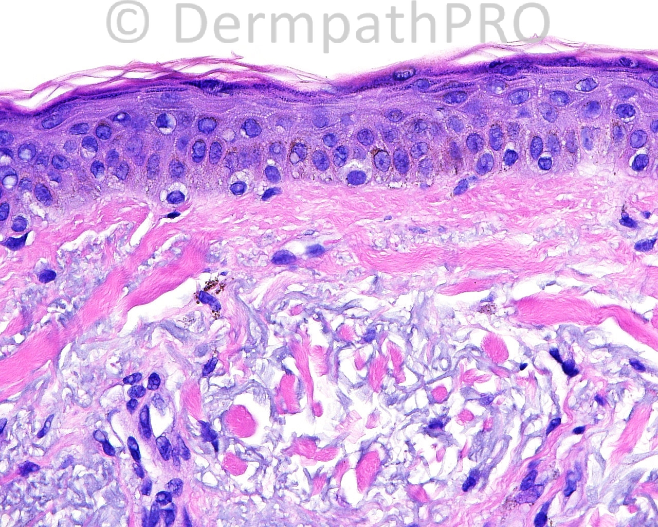
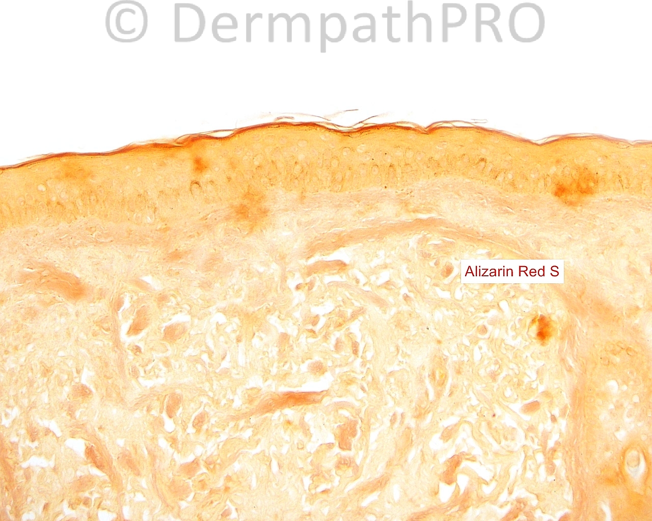
The patient is a 68-year-old woman who takes medication for ocular rosacea. A shave biopsy is taken of asymptomatic, blue-gray, macular pigment on the left cheek.

Case posted by Dr. Mark Hurt


  Report Record

User Feedback


Mark A. Hurt MD

Posted

[img]https://dermpathpro.com/uploads/spot_diagnosis_comment_img/CASE939_Image%2009.jpg[/img]

[img]https://dermpathpro.com/uploads/spot_diagnosis_comment_img/CASE939_Image%2010.jpg[/img]

Share this comment


Link to comment
share_externally

Dr. Mona Abdel-Halim

Posted

Possibly she is taking minocycline, minocycline induced pigmentation.

Share this comment


Link to comment
share_externally

Robledo F. Rocha

Posted

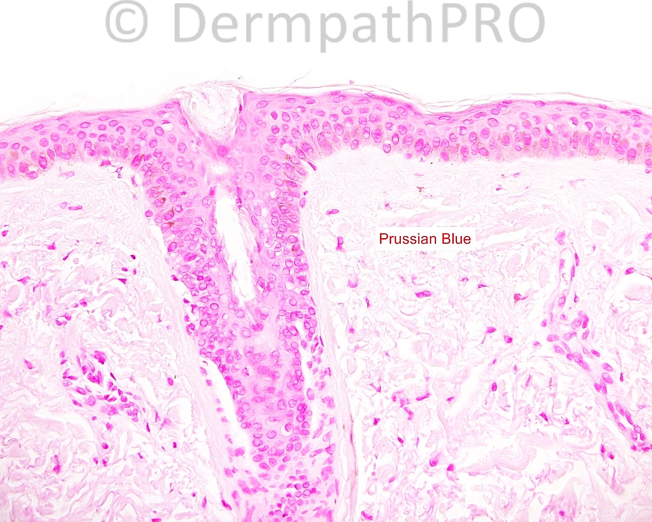
I don’t think this is minocycline-induced pigmentation since it is negative for Prussian blue, but I agree this is postinflammatory pigmentation due to photosensitizing agent.

Share this comment


Link to comment
share_externally

Dr. Mona Abdel-Halim

Posted

There is a report about a type IV minocycline induced pigmentation that was confined to acne scars and was negative for iron stain but positive with calcium stain. Could it be this case.

http://www.ncbi.nlm.nih.gov/pubmed/14723711

Share this comment


Link to comment
share_externally

Mark A. Hurt MD

Posted

I think this is a form of Minocycline hyperpigmentation. By the way, I did not think there was calcium deposition on Alizarin Red S or von Kossa.

from my report:

[b]-- HYPERPIGMENTATION [/b]
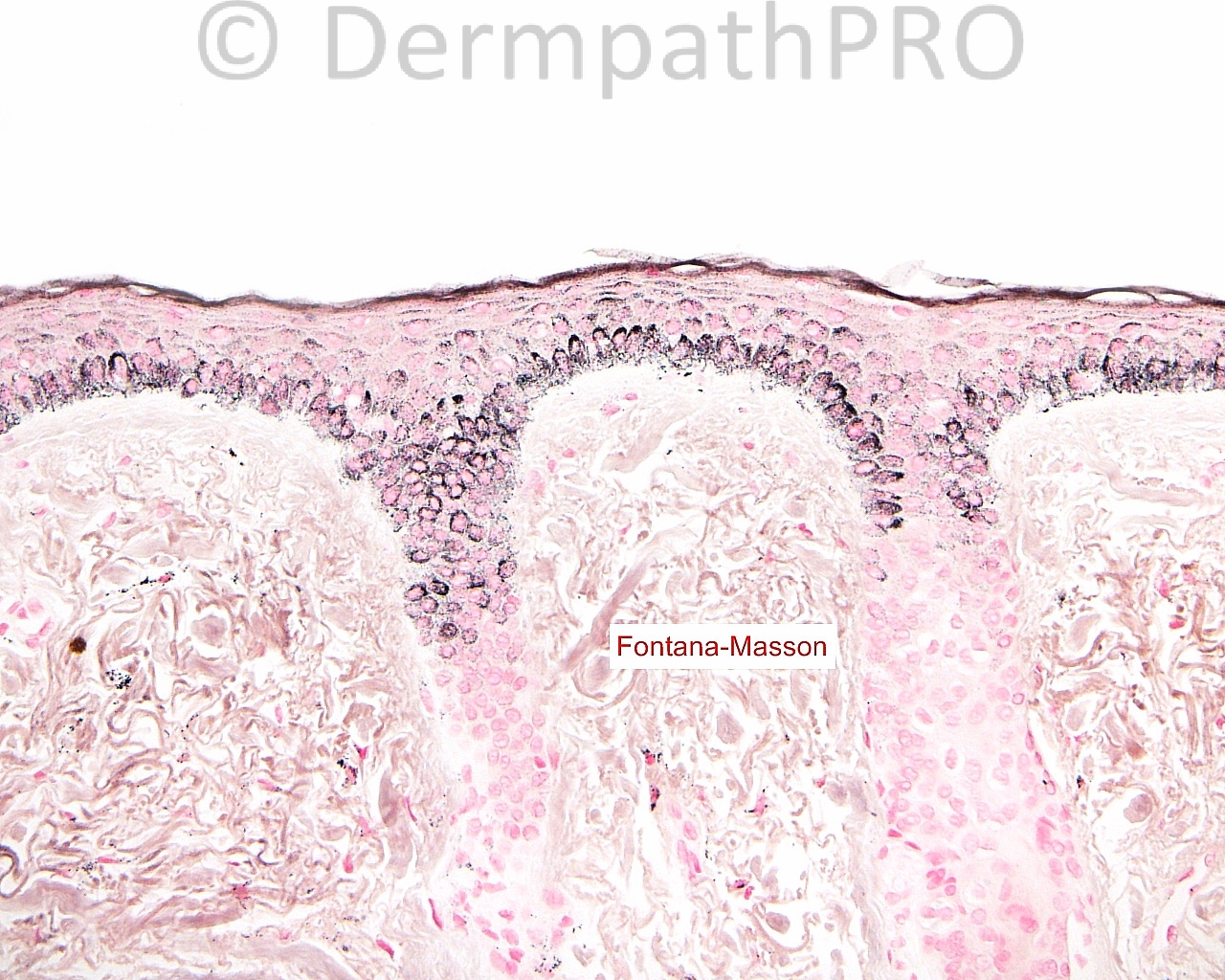
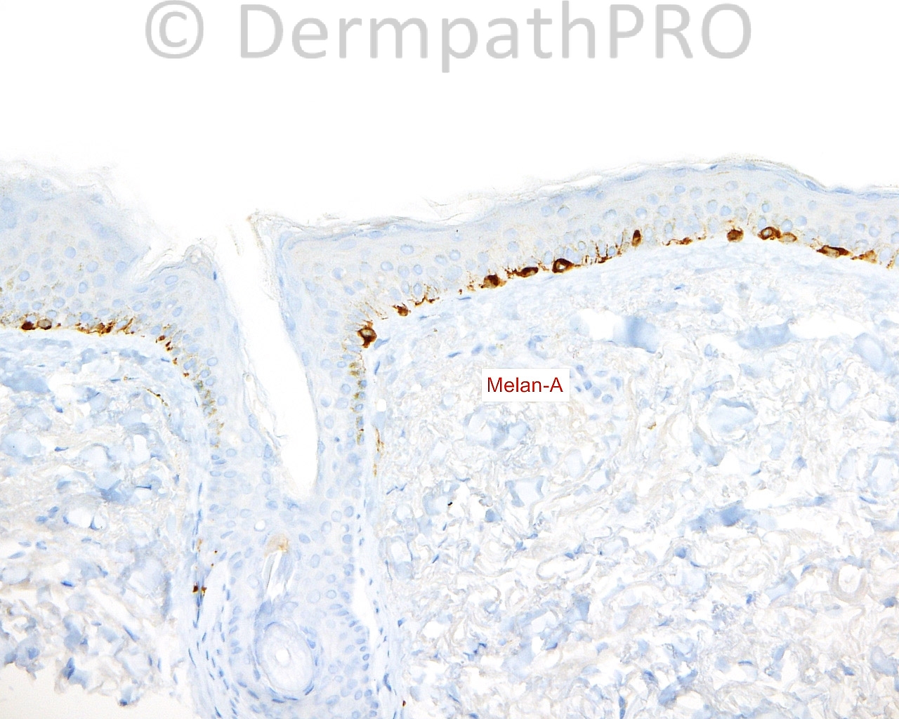
[b]COMMENT:[/b] The presence of hyperpigmentation with Fontana-Masson indicates melanin hyperpigmentation in the basal layer, presumably from minocycline, which has been described as "type III". Other types of pigmentation with minocycline use include a hemosiderin and iron chelate of minocycline (type I) and a metabolite protein complex of chelated iron and calcium (type II). I see neither hemosiderin nor calcium deposition in this skin, and thus presume, by default, that the pigmentation is due to melanin in the basal layer of the epidermis.


[b]And from Weedon (2010):[/b]

[u][b]Type I: [/b][/u] bluish-black pigmentation of scars and old inflammatory foci, including sites of immunobullous diseases, related to hemosiderin or an iron chelate of minocycline, a variant of this [b](proposed type IV)[/b], with blue-gray pigmentation of acne scars on the back was characterized by calcium-containing melanin deposits within dendritic cells and in an extracellular location.

[u][b]Type II:[/b][/u] blue-gray circumscribed pigmentation of the lower legs and arms due to a pigment which is probably a drug metabolite–protein complex chelated with iron and calcium. The recently reported cases with deposits of pigment localized to the subcutaneous fat of the lower extremity appear to be a different type [b](proposed type V)[/b].

[u][b]Type III:[/b][/u] a generalized muddy brown pigmentation due to increased melanin in the basal layer, and accentuated in sun-exposed areas.

Share this comment


Link to comment
share_externally


Join the conversation

You can post now and register later. If you have an account, sign in now to post with your account.

Guest
Add a comment...

×   Pasted as rich text.   Paste as plain text instead

  Only 75 emoji are allowed.

×   Your link has been automatically embedded.   Display as a link instead

×   Your previous content has been restored.   Clear editor

×   You cannot paste images directly. Upload or insert images from URL.

Loading...

×
×
  • Create New...