Jump to content
In this section we have spot diagnoses posted on a daily basis since June 2010, now over 4000! You can review the archived cases and read the suggested diagnoses by users and the final comment by the contributors.
Case are uploaded each week day by 10 am UK time with the correct diagnosis will generally be posted at 8 pm UK time. Why not view the most recent spot diagnosis and proffer a diagnosis?

Case Number : Case 1061 - 17th July Posted By: Guest

Please read the clinical history and view the images by clicking on them before you proffer your diagnosis.
Submitted Date :
   (0 reviews)

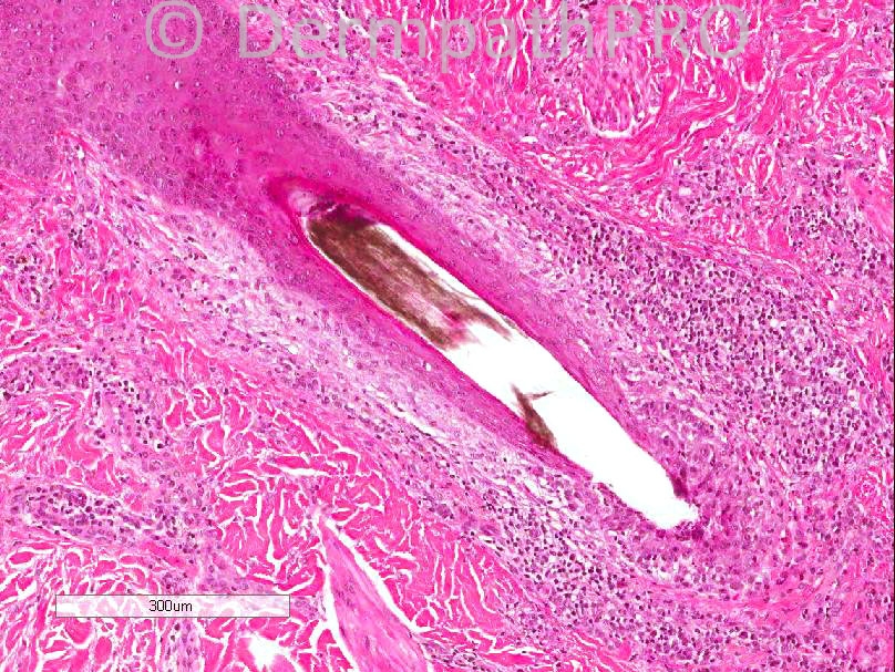
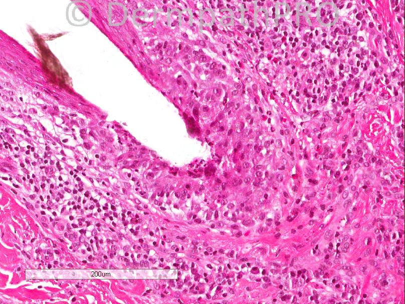
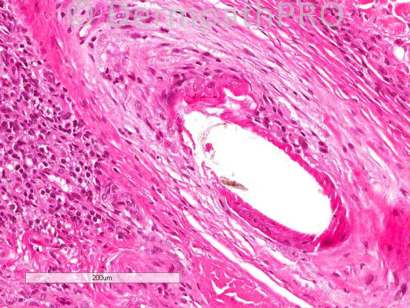
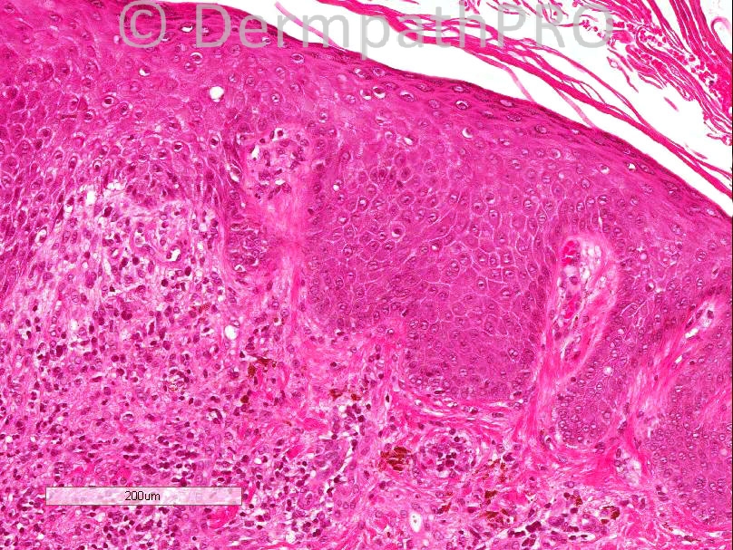
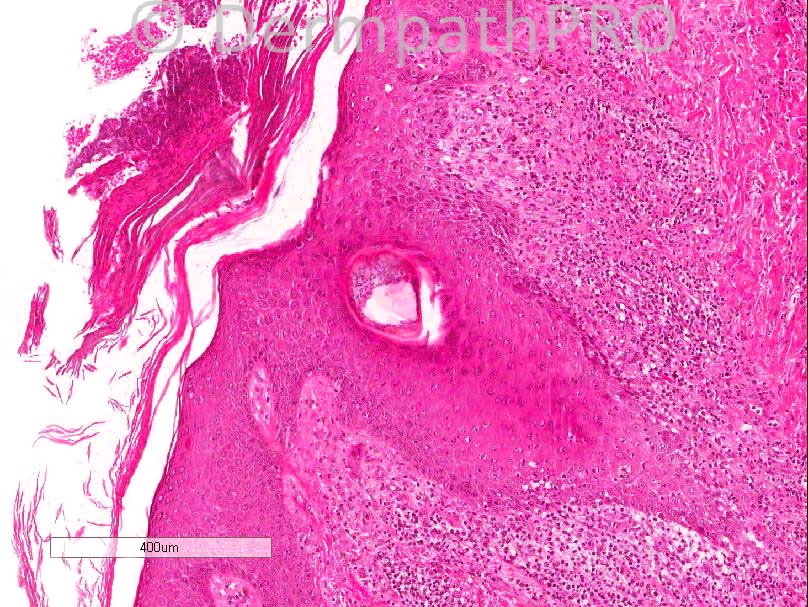
Male, 39 year old, scaly erythema, post inflammatory peri-follicular change. Some scarring ?DLE ?LLP. Punch biopsy. Crown/vertex 6mm punch biopsy


  Report Record

User Feedback


Dr. Mona Abdel-Halim

Posted

The infiltrate is mainly centered around the hair follicles with interface changes in the follicular epithelium. This favours LPP.

Share this comment


Link to comment
share_externally

Degeneration of the ORS and infiltration of eosinophils made me think of follicular mucinosis.

Share this comment


Link to comment
share_externally

I see a mononuclear infiltrate around follicular epithelium with interface changes. The follicular drop out in figures 7 and 8 are consistent with Cicatricial alopecia and there are many plasma cells (figure 5). I think this is a case of DLE. As we are seeing a young male patient with interface and psoriasiform changes and many plasma cells, my DDx. is Syphilitic alopecia.

Share this comment


Link to comment
share_externally

Robledo F. Rocha

Posted

[size=4][font=arial,helvetica,sans-serif]I go with lichen planopilaris. There’s a perifollicular lichenoid infiltrate of lymphocytes and plasma cells with vacuolar interface change. A hair follicle shows concentric lamellar fibrosis around it, which [/font][/size][font=arial, helvetica, sans-serif]moves the infiltrate away from the follicle. C[/font][font=arial, helvetica, sans-serif]lefts reminiscent of Max Joseph spaces between the follicular epithelium and the stroma can be seen.[/font]
[size=4][font=arial,helvetica,sans-serif]Lupus erythematosus is my favorite differential diagnosis, but I didn’t find basement membrane thickening, dermal mucin or infundibular cornified plug.[/font][/size]

Share this comment


Link to comment
share_externally

Iskander H. Chaudhry

Posted

Thank you for your comments. Alopecia cases are always difficult and the final diagnosis in this case was lichen planopilaris. You may have noted the presence of neutrophils for which I raised the possibility of folliculitis decalvans, however, this did not fit the clinical appearances and at our local CPC we concluded the appearance were in keeping with lichen planopilaris.

Share this comment


Link to comment
share_externally


Join the conversation

You can post now and register later. If you have an account, sign in now to post with your account.

Guest
Add a comment...

×   Pasted as rich text.   Paste as plain text instead

  Only 75 emoji are allowed.

×   Your link has been automatically embedded.   Display as a link instead

×   Your previous content has been restored.   Clear editor

×   You cannot paste images directly. Upload or insert images from URL.

Loading...

×
×
  • Create New...