Jump to content
In this section we have spot diagnoses posted on a daily basis since June 2010, now over 4000! You can review the archived cases and read the suggested diagnoses by users and the final comment by the contributors.
Case are uploaded each week day by 10 am UK time with the correct diagnosis will generally be posted at 8 pm UK time. Why not view the most recent spot diagnosis and proffer a diagnosis?

Case Number : Case 1064 - 22nd July Posted By: Guest

Please read the clinical history and view the images by clicking on them before you proffer your diagnosis.
Submitted Date :
   (0 reviews)

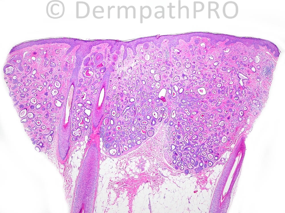
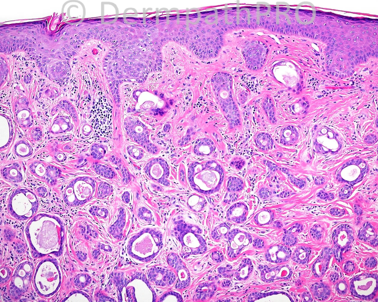
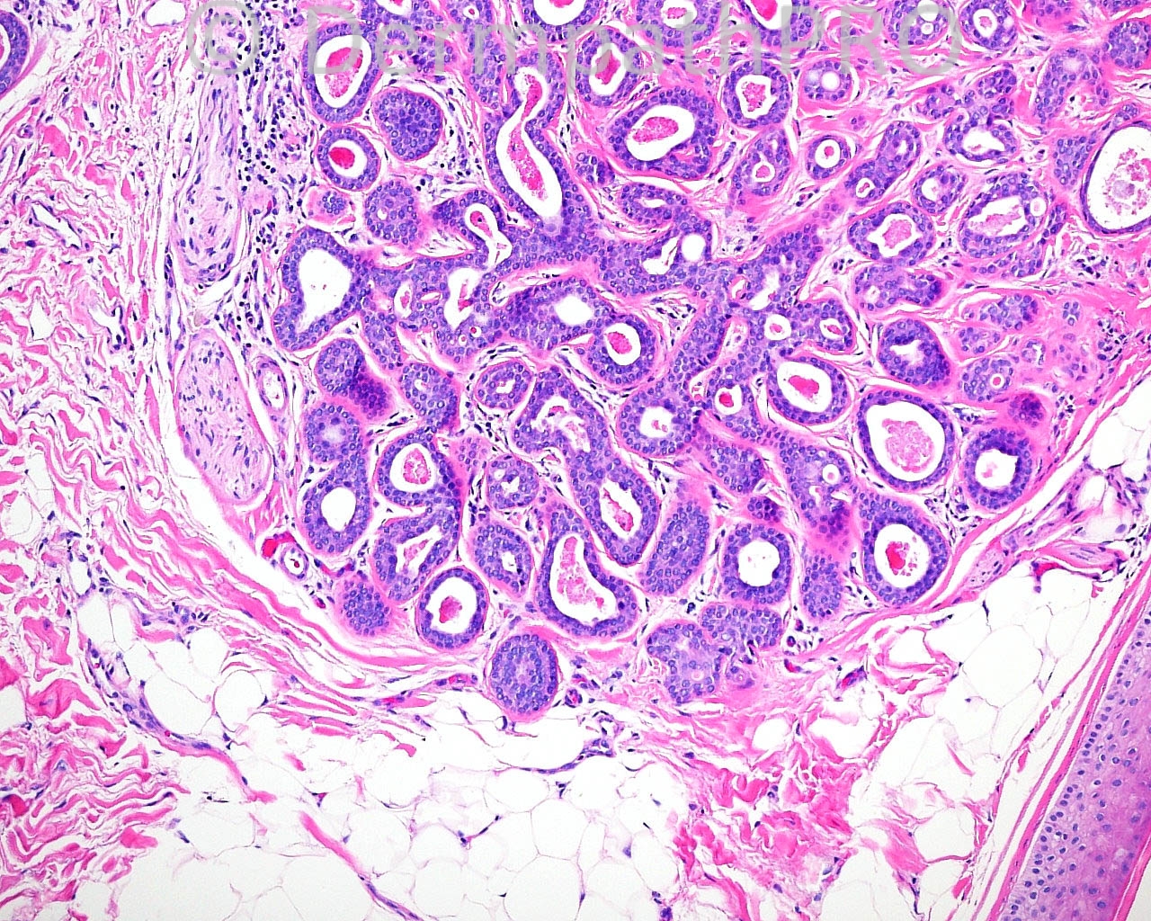
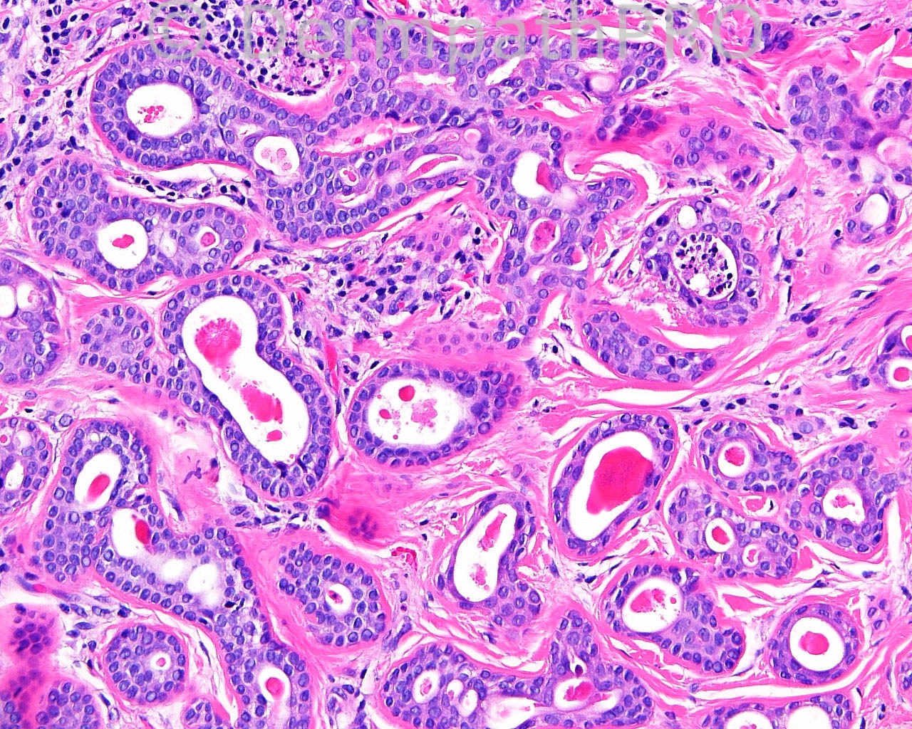
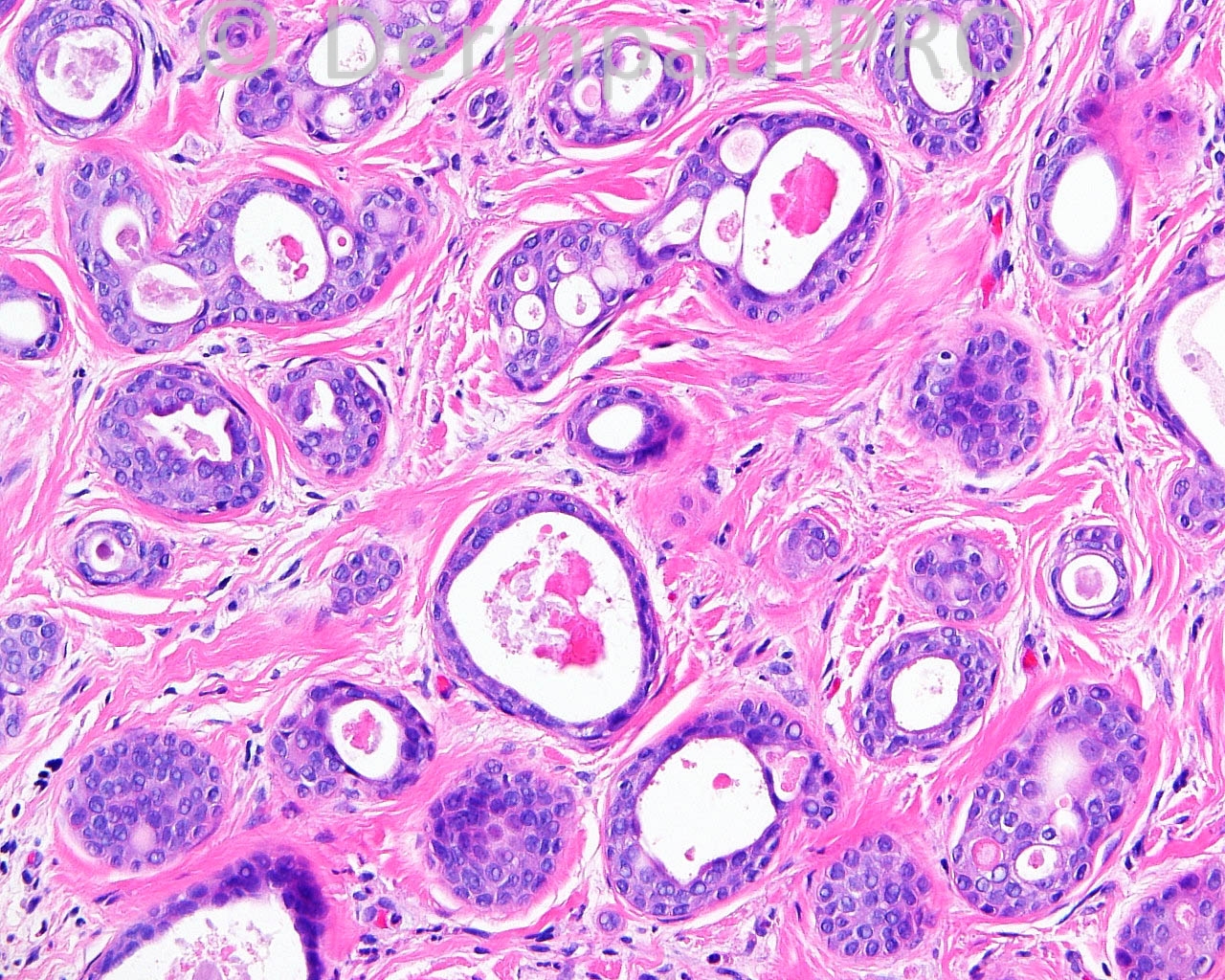
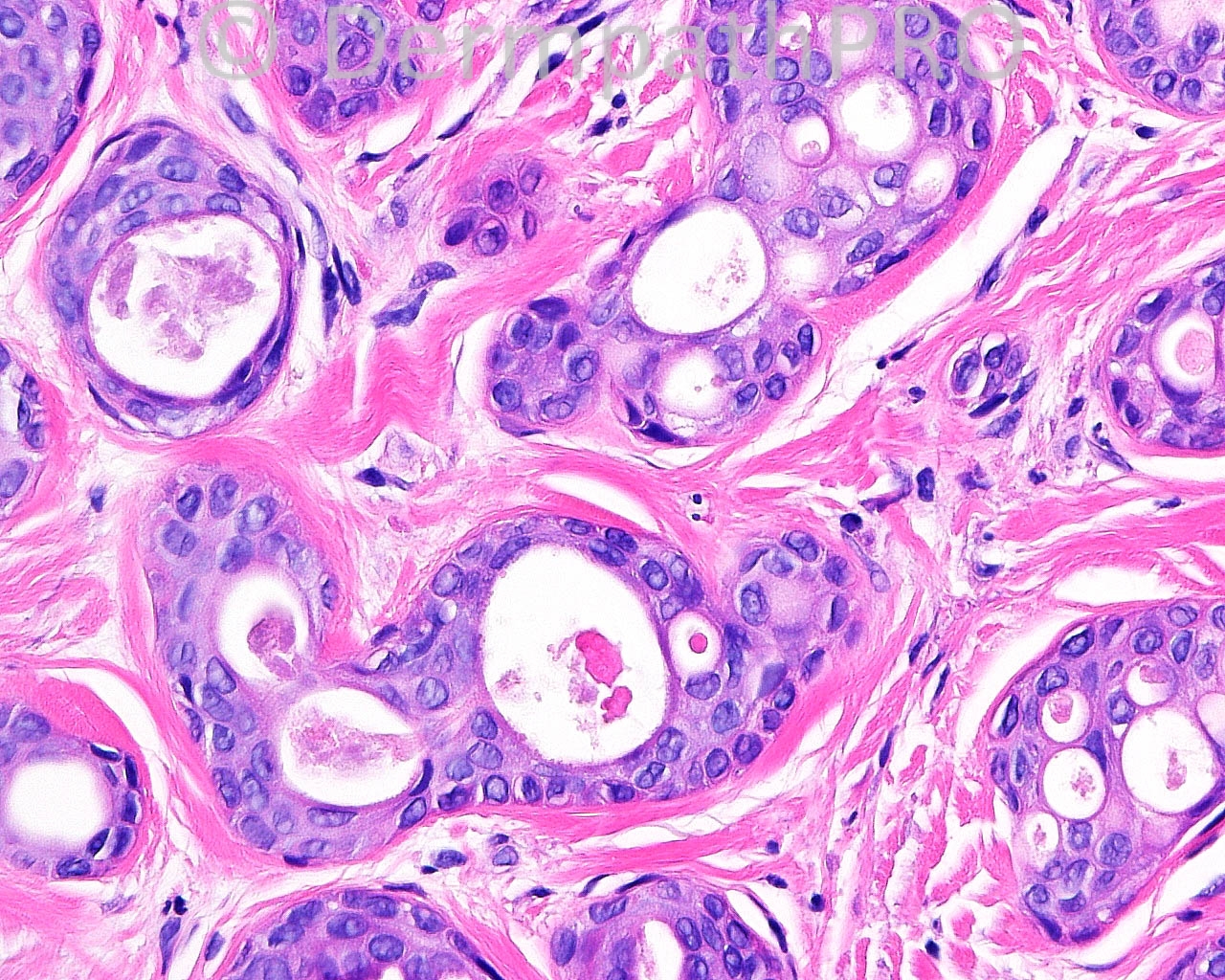
The patient is a 59-year-old woman with a biopsy of a 6 mm tender white, sclerotic-appearing lesion present for one year on the left side of the scalp.

Case posted by Dr. Mark Hurt


  Report Record

User Feedback


Robledo F. Rocha

Posted

[quote name='I. Abdul-kadir' timestamp='1406020719']
Tubular adenoma.
[/quote]
Ditto.

Share this comment


Link to comment
share_externally

Uma Sundram

Posted

Could it be weird breast carcinoma? The cytology is pretty bland.

Share this comment


Link to comment
share_externally

Other differential diagnosis could be Siringoma.

Share this comment


Link to comment
share_externally

Mark A. Hurt MD

Posted

I thought I would be a minority opinion on this case, and I am. Here is my diagnosis and comment:

[b]-- SYRINGOMATOUS CARCINOMA [/b]
[indent=1][b]COMMENT:[/b] Syringomatous carcinoma is the malignant analog of syringoma and presents, in most cases, much as it has presented in this particular case. The main differences between syringomatous carcinoma and microcystic carcinoma are that the microcystic carcinomas tend to have a morula pattern superficially and tend to have a dense collection of infiltrative strands and cords of ductal cells in the deep part – which produces a gradient effect, in contrast with the case for review here. Syringomatous carcinomas usually are indolent biologically, but they do have the capacity to metastasize, as there are some rare examples that have been presented in the literature. “Trichoadenomas” (which are actually infundibular) have much more of an infundibular pattern than a tubular or ductal pattern. I have not seen mucoepidermoid carcinoma much in the skin, but I can say that they are not as organized as is the lesion under review here.[/indent]

[center][u]Reference[/u]:[/center]

Requena L, Kiryu H, Ackerman AB. Syringomatous carcinoma. in [i]Neoplasms With Apocrine Differentiation[/i]. Philadelphia, Lippincott-Raven-Ardor Scriebendi, 1998: 771-783.

-----oOo----

Share this comment


Link to comment
share_externally

Guest Tiberiu Tebeica

Posted

My impression was eccrine ductal carcinoma (syringoid carcinoma). Thank you for sharing a rare one!

Share this comment


Link to comment
share_externally


Join the conversation

You can post now and register later. If you have an account, sign in now to post with your account.

Guest
Add a comment...

×   Pasted as rich text.   Paste as plain text instead

  Only 75 emoji are allowed.

×   Your link has been automatically embedded.   Display as a link instead

×   Your previous content has been restored.   Clear editor

×   You cannot paste images directly. Upload or insert images from URL.

Loading...

×
×
  • Create New...