Jump to content
In this section we have spot diagnoses posted on a daily basis since June 2010, now over 4000! You can review the archived cases and read the suggested diagnoses by users and the final comment by the contributors.
Case are uploaded each week day by 10 am UK time with the correct diagnosis will generally be posted at 8 pm UK time. Why not view the most recent spot diagnosis and proffer a diagnosis?

Case Number : Case 1067 - 25th July Posted By: Guest

Please read the clinical history and view the images by clicking on them before you proffer your diagnosis.
Submitted Date :
   (0 reviews)

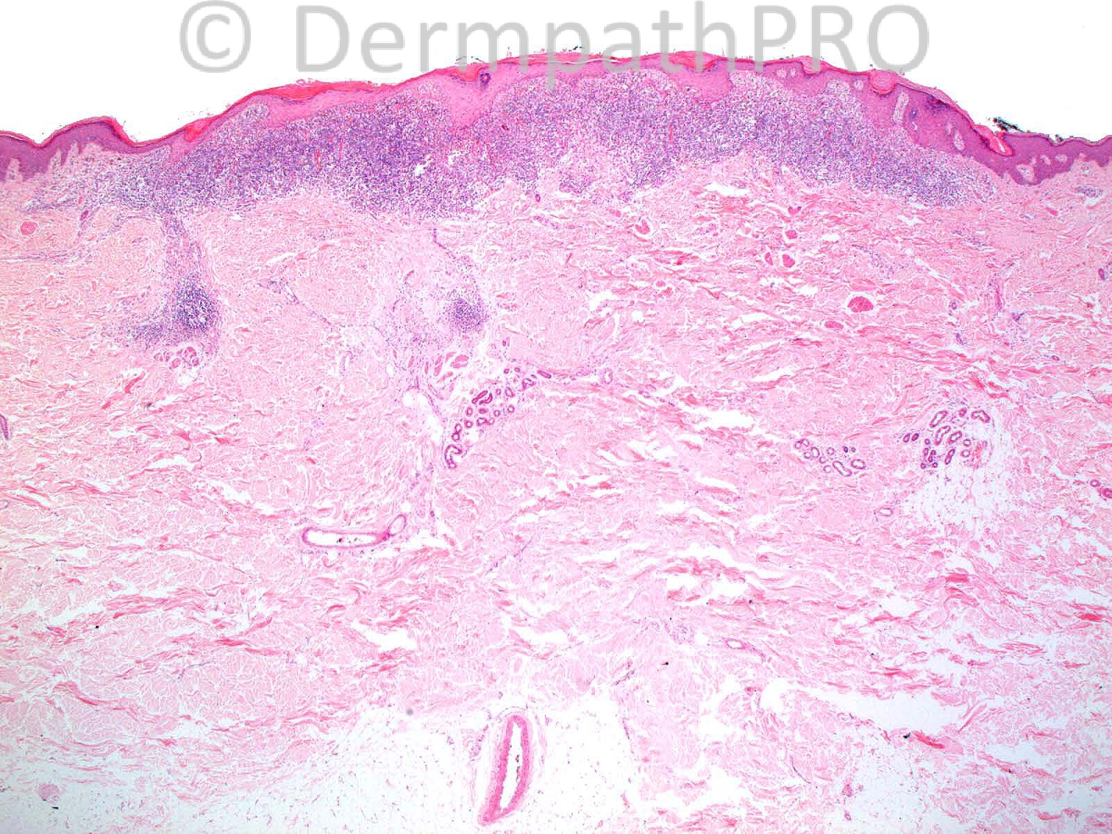
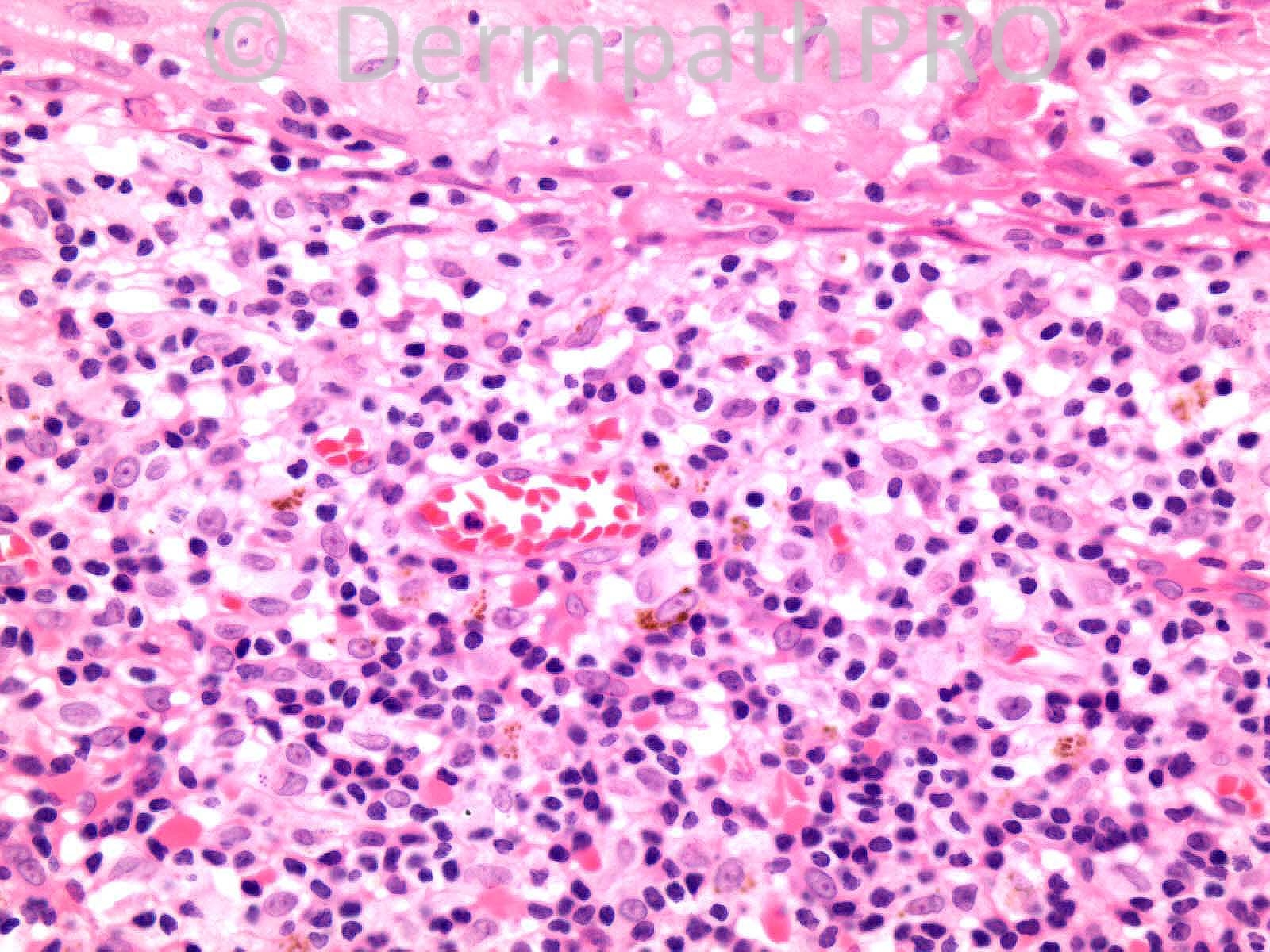
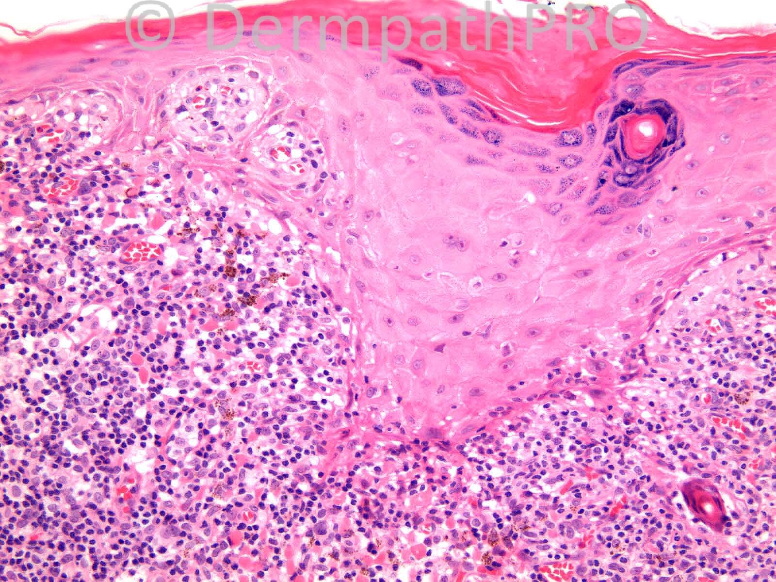
M38. Lesion right shoulder ??BCC 2 yrs in Cyprus and 2 years in Kenya.

Case posted by Dr. Richard Carr.

  Report Record

User Feedback


Eman El-Nabarawy

Posted

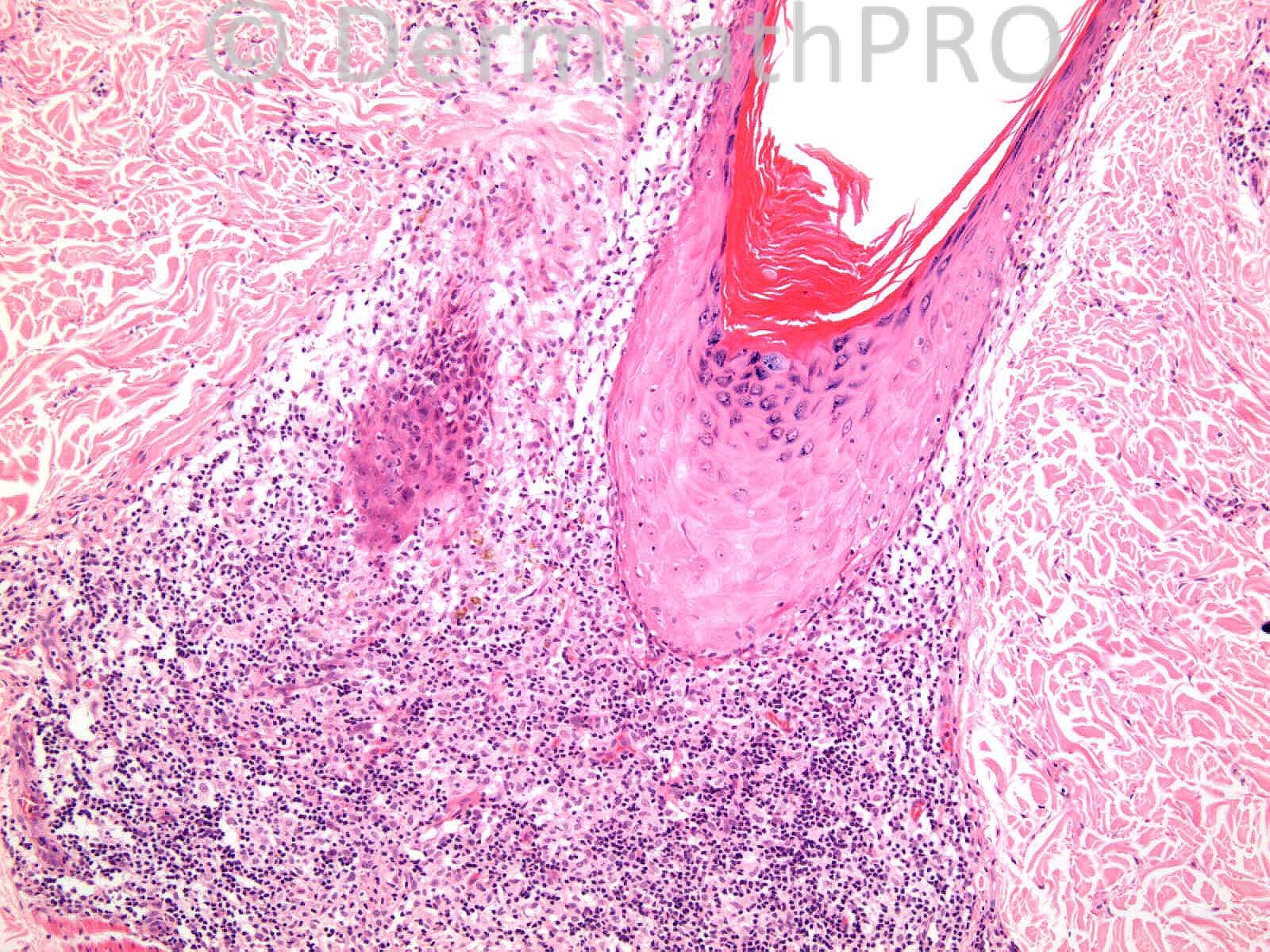
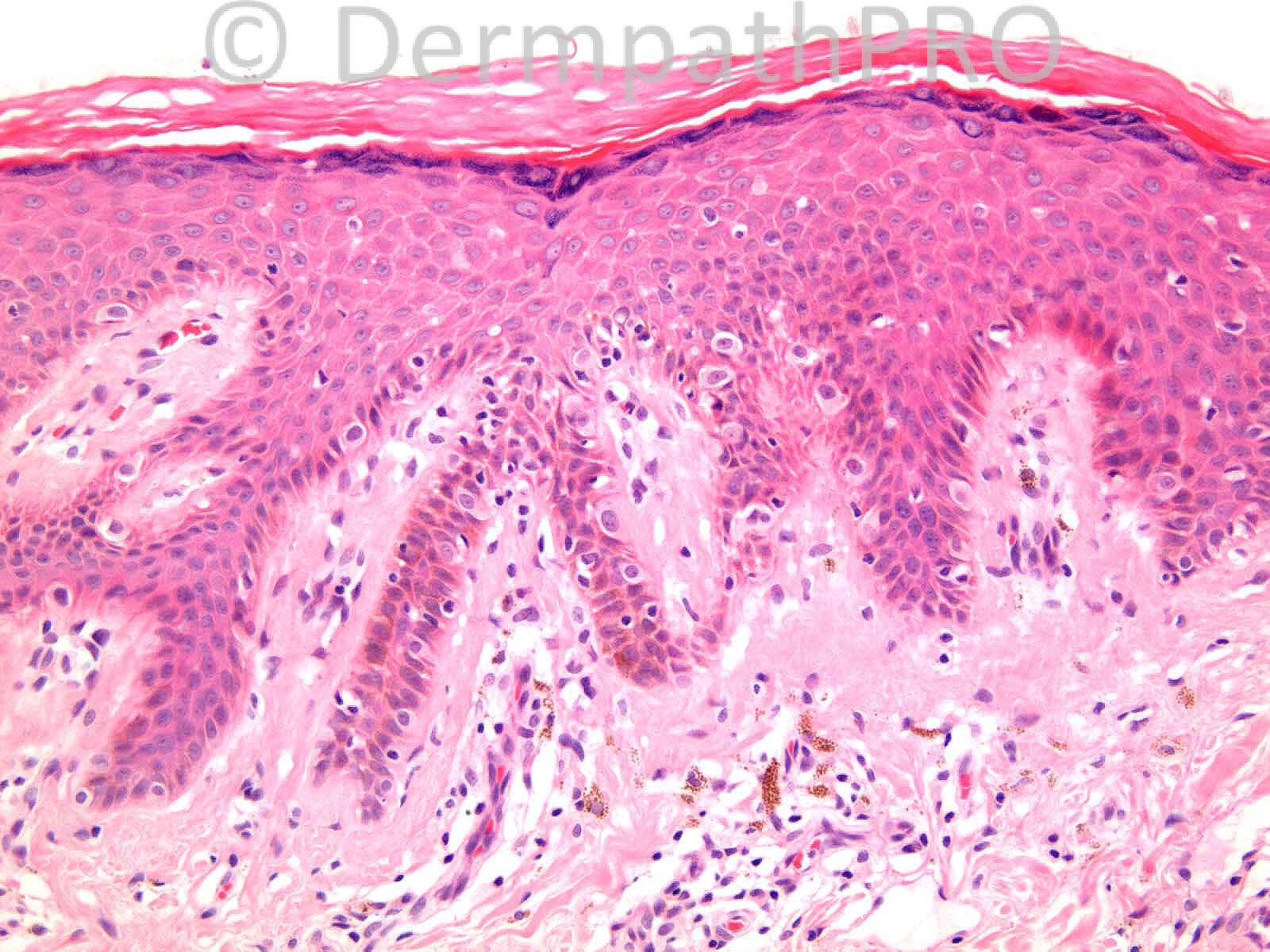
Lichen planus-like keratosis.

Share this comment


Link to comment
share_externally

Guest Lee Alexander

Posted

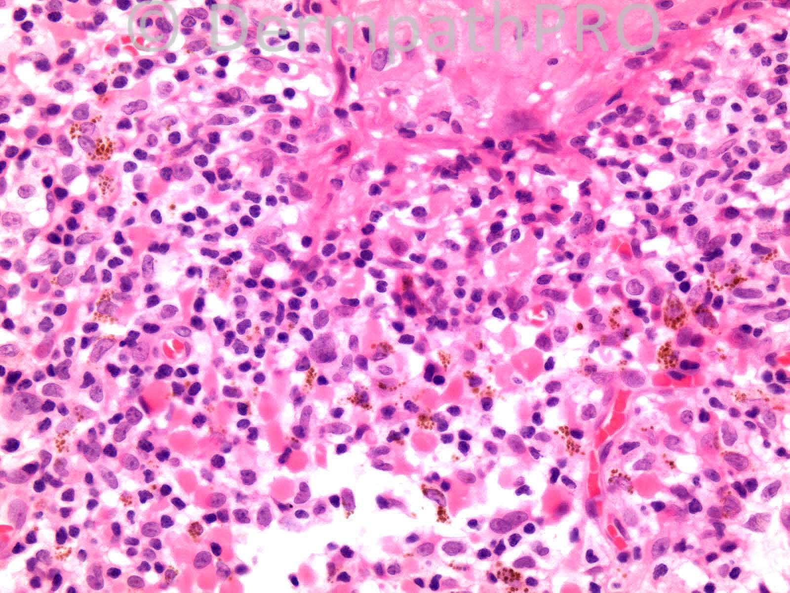
Lichen planus-like keratosis. Hemorrhage?

Share this comment


Link to comment
share_externally

Arti Bakshi

Posted

Agree with Lichenoid keratosis, if solitary lesion.

Share this comment


Link to comment
share_externally

Agree with the above, but would do melanocytic markers.

Share this comment


Link to comment
share_externally

Guest Romualdo

Posted

[quote name='I. Abdul-kadir' timestamp='1406283705']
Agree with the above, but would do melanocytic markers.
[/quote]

Share this comment


Link to comment
share_externally

Robledo F. Rocha

Posted

At first glance, this lesion looks lichenoid keratosis, but away from the lichenoid infiltrate there are lentiginous epithelioid melanocytes, albeit non-confluent. Bearing in mind this lesion is located on the back of a middle-aged man and a Dr. Carr’s comment in a [url="https://dermpathpro.com/spot-diagnosis-1/_/2013-spot-diagnoses/april-2013/case-734-9-apr-r757?st=0#comment_2859"]past spot diagnosis[/url], I think the brown pigment within the lichenoid infiltrate might raise suspicion of melanoma.

Share this comment


Link to comment
share_externally

Dr. Richard Carr

Posted

All agreed. I reported this as a lichen planus-like keratosis (adjacent solar lentigo) and reproduce here the link for those of you who only have the free access. Many thanks to Dr Rocha for reminding me of these comments.

Posted 10Apr13 in Discussion to Case 734:
[color=#1C2837][font=arial, verdana, tahoma, sans-serif][size=4]In answer to Dr Rocha's question "LPLK" is not a diagnosis it is a reaction pattern.[/size][/font][/color]
[color=#1C2837][font=arial, verdana, tahoma, sans-serif][size=4]Solar lentigo is possibly the most common association but it is basically a lichenoid reaction to a superficial lesion the nature of which almost by definition is uncertain. It could be seen in reaction to pre-existing actinic keratosis, thin seborrhoeic keratosis, thin SCC and indeend thin melanoma. Some pathologists recommend examination of multiple levels in all cases and I have heard some experts stating that a MelanA should be done on all cases. I am very wary about making the "diagnosis" of LPLK in a partial specimen e.g. punch biopsy and I emphasise it is a non-diagnosis / reaction pattern. Obviously if there is any clinical concern about a lesion it should be excised with clear margins in such cases. My comments may be conservative but unfortunately I have myself missed a thin melanoma (on punch biopsy due to brisk inflammatory reaction) that had very sparse histiocyte-like dermal cells and unfortunately the same lesion was punch biopsied a year latter and reported by another pathologist as LPLK (when on review there was more obvious SSMM). The final diagnosis of melanoma was delayed for two years in that particular case. All that said I don't personally automatically do MelanA myself on all cases I am very selective but I do automatically examine multiple levels in an attempt to make a specific diagnosis.[/size][/font][/color]

Share this comment


Link to comment
share_externally


Join the conversation

You can post now and register later. If you have an account, sign in now to post with your account.

Guest
Add a comment...

×   Pasted as rich text.   Paste as plain text instead

  Only 75 emoji are allowed.

×   Your link has been automatically embedded.   Display as a link instead

×   Your previous content has been restored.   Clear editor

×   You cannot paste images directly. Upload or insert images from URL.

Loading...

×
×
  • Create New...