Jump to content
In this section we have spot diagnoses posted on a daily basis since June 2010, now over 4000! You can review the archived cases and read the suggested diagnoses by users and the final comment by the contributors.
Case are uploaded each week day by 10 am UK time with the correct diagnosis will generally be posted at 8 pm UK time. Why not view the most recent spot diagnosis and proffer a diagnosis?

Case Number : Case 1068 - 28th July Posted By: Guest

Please read the clinical history and view the images by clicking on them before you proffer your diagnosis.
Submitted Date :
   (0 reviews)

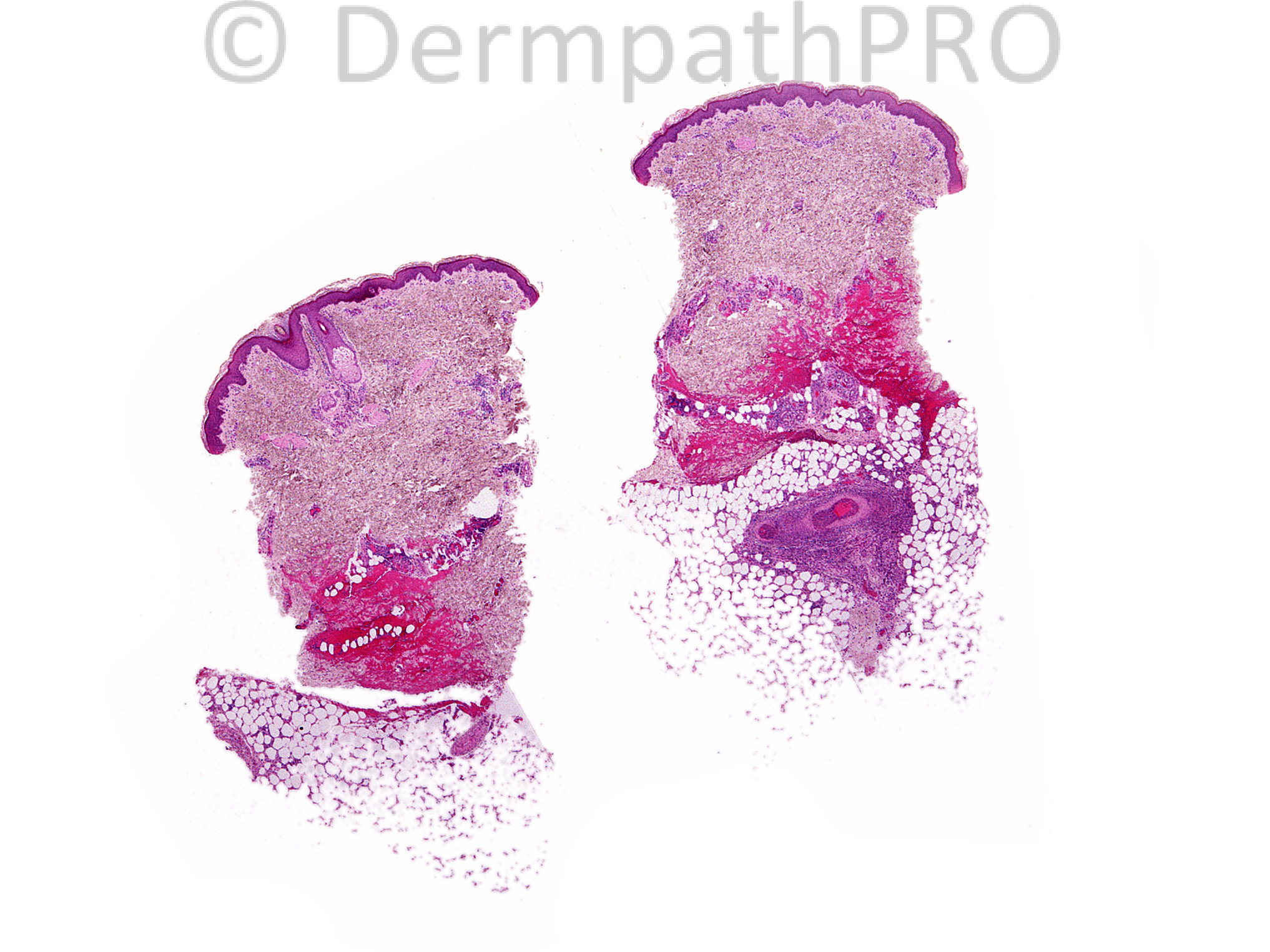
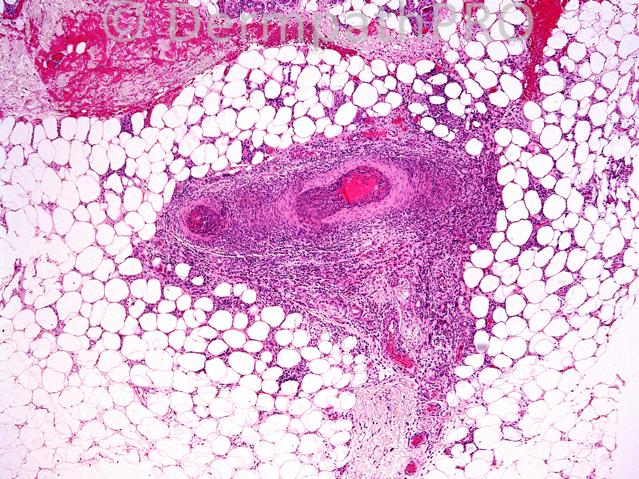
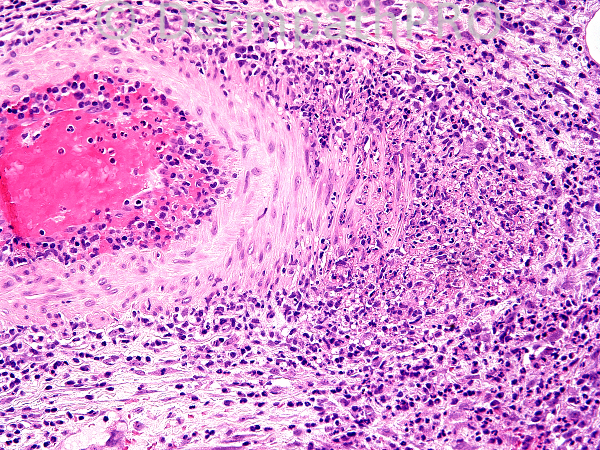
16 year old female with erythematous papules and nodules on her lower extremities. The biopsy is from a nodule on the left lower leg.

Case posted by Dr. Uma Sundram.


  Report Record

User Feedback


Superficial thrombophlebitis vs. polyarteritis nodosa.

Share this comment


Link to comment
share_externally

Guest Romualdo

Posted

Cutaneous polyarteritis nodosa.

Share this comment


Link to comment
share_externally

Robledo F. Rocha

Posted

Since the infiltrate is confined to the artery wall and to the subcutaneous fat immediately around it, I favor polyarteritis nodosa over nodular vasculitis.

Share this comment


Link to comment
share_externally

Robledo F. Rocha

Posted

Happy birthday, Romualdo!

Share this comment


Link to comment
share_externally

The lumen of the vessel is obstructed by trombosis and the infiltrate is compromising only focally the vessel wall thickness, so I believe this is a case of Superficial thrombophlebitis. Special stain with Orcein or other elastic tissue stain is necessary to better differentiate artery and vein. Most cases of PAN has the lumen patent.

Share this comment


Link to comment
share_externally

Uma Sundram

Posted

I called this PAN (polyarteritis nodosa). The differential diagnosis includes superficial thrombophlebitis and nodular vasculitis. The distinction between PAN and superficial thrombophlebitis can be very difficult. Elastin stains can be helpful as arteries have internal elastic lamina and veins do not. The assessment of smooth muscle bundle patterns can be very helpful; in veins the smooth muscle bundles are interrupted by collagen fibers, and in arteries the smooth muscle bundles are continuous, as they are in this case. I too favored nodular vasculitis less as the inflammation seems pretty confined to the vessels rather than showing significant involvement of the panniculus.

Share this comment


Link to comment
share_externally


Join the conversation

You can post now and register later. If you have an account, sign in now to post with your account.

Guest
Add a comment...

×   Pasted as rich text.   Paste as plain text instead

  Only 75 emoji are allowed.

×   Your link has been automatically embedded.   Display as a link instead

×   Your previous content has been restored.   Clear editor

×   You cannot paste images directly. Upload or insert images from URL.

Loading...

×
×
  • Create New...