Jump to content
In this section we have spot diagnoses posted on a daily basis since June 2010, now over 4000! You can review the archived cases and read the suggested diagnoses by users and the final comment by the contributors.
Case are uploaded each week day by 10 am UK time with the correct diagnosis will generally be posted at 8 pm UK time. Why not view the most recent spot diagnosis and proffer a diagnosis?

Case Number : Case 1586 - 25 July Posted By: Guest

Please read the clinical history and view the images by clicking on them before you proffer your diagnosis.
Submitted Date :
   (0 reviews)

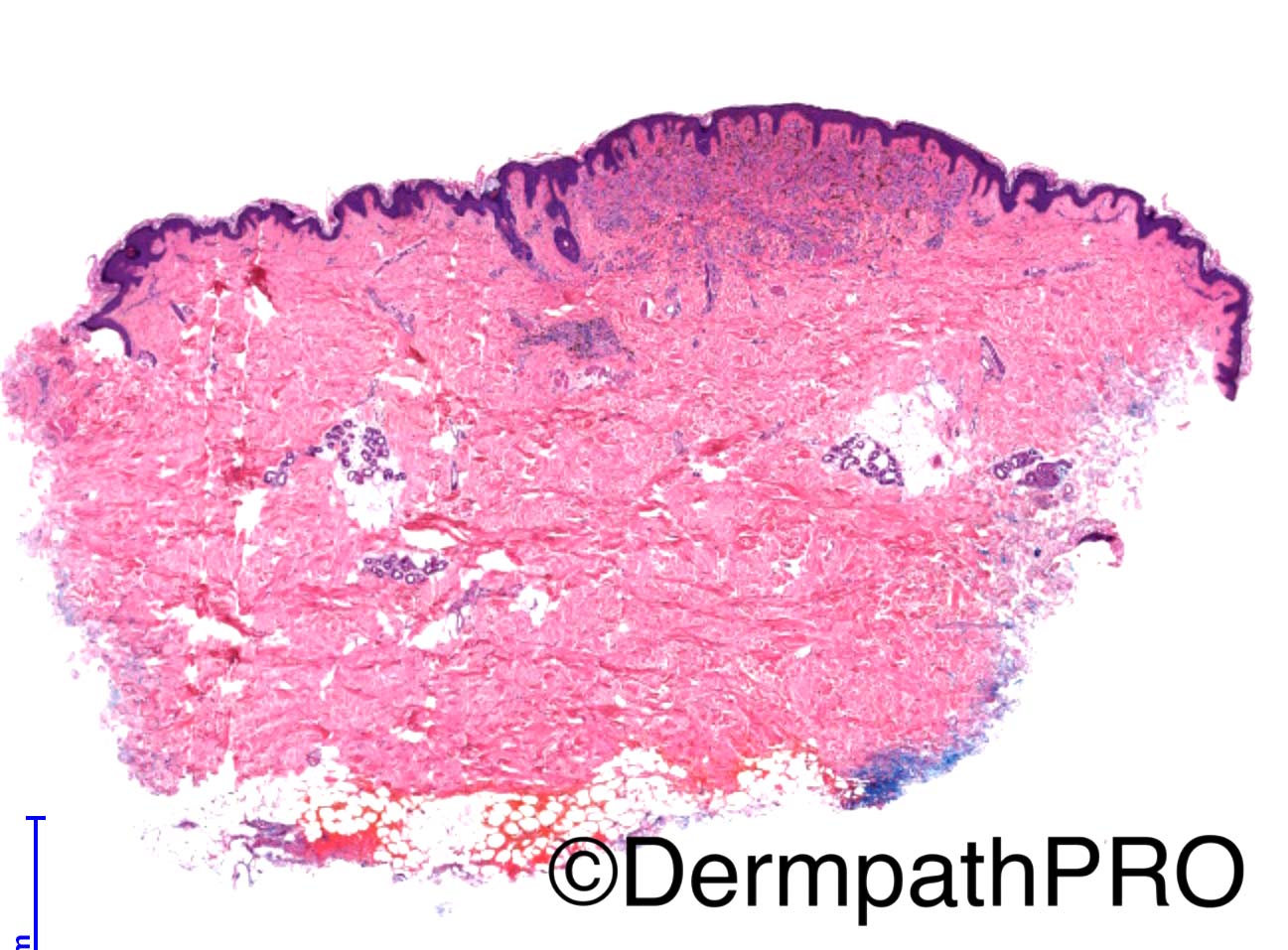
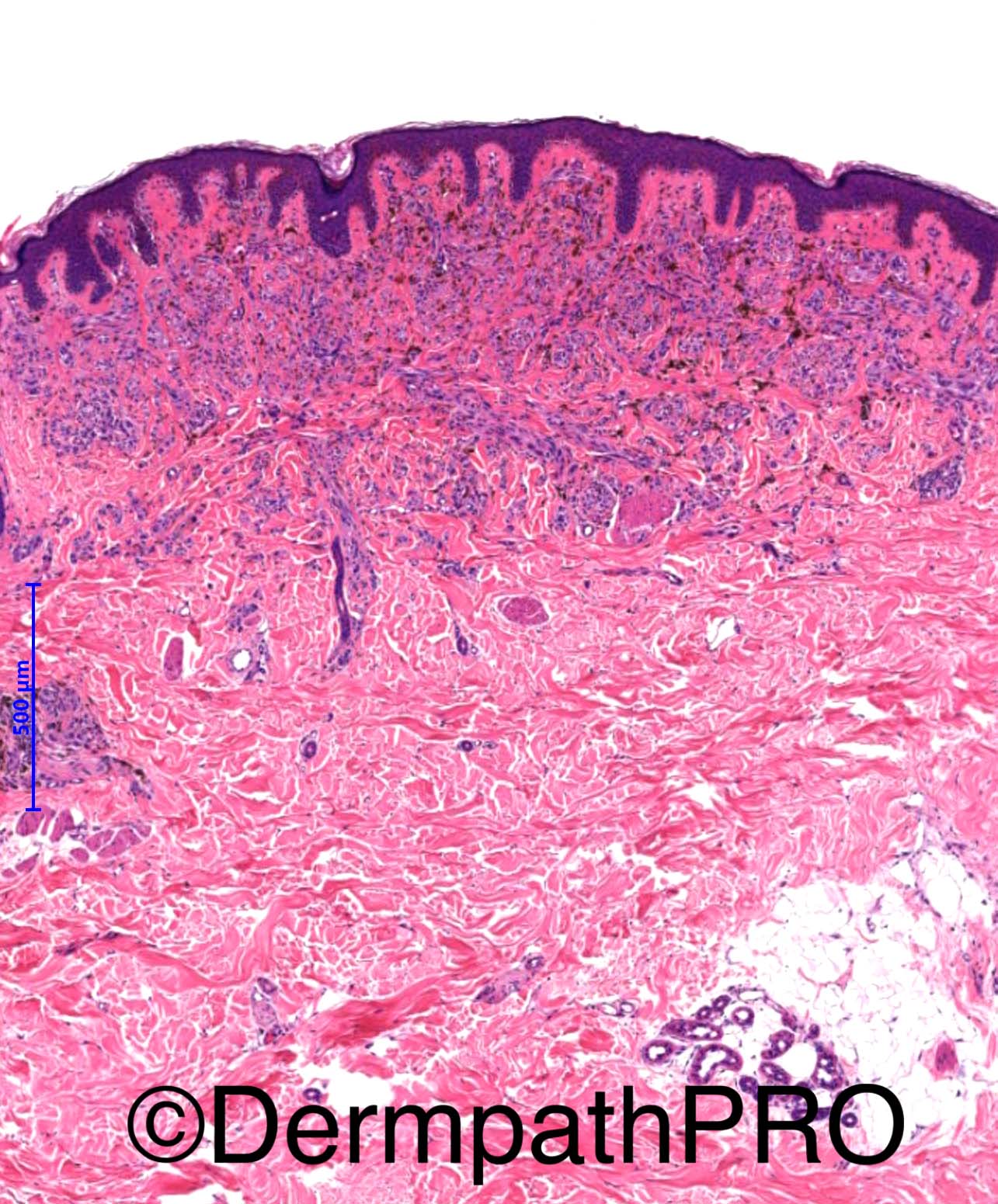
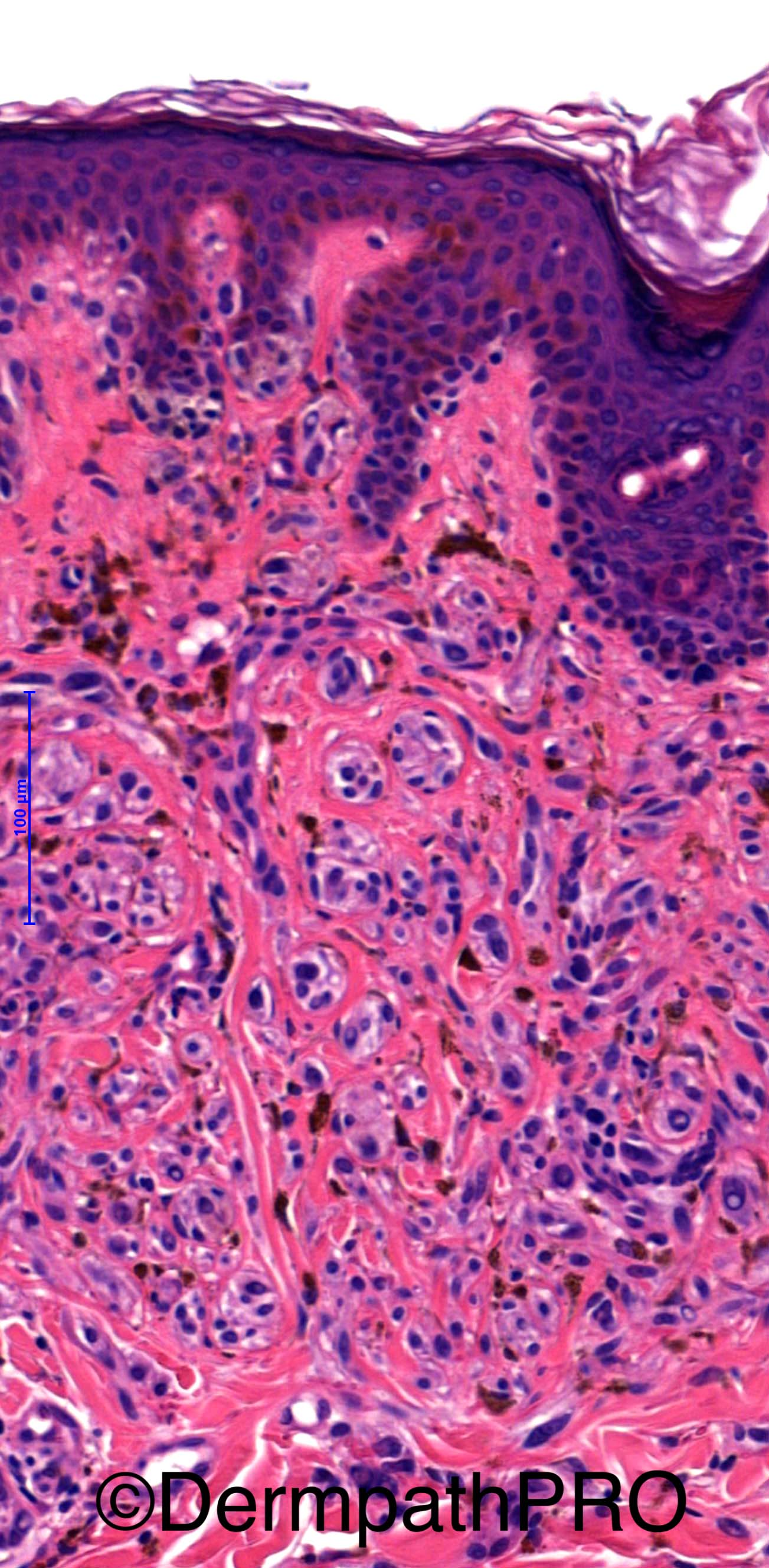
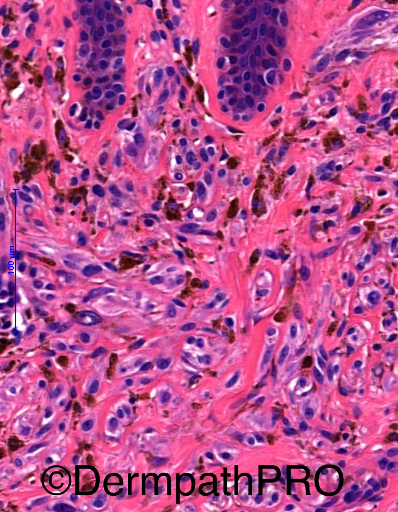
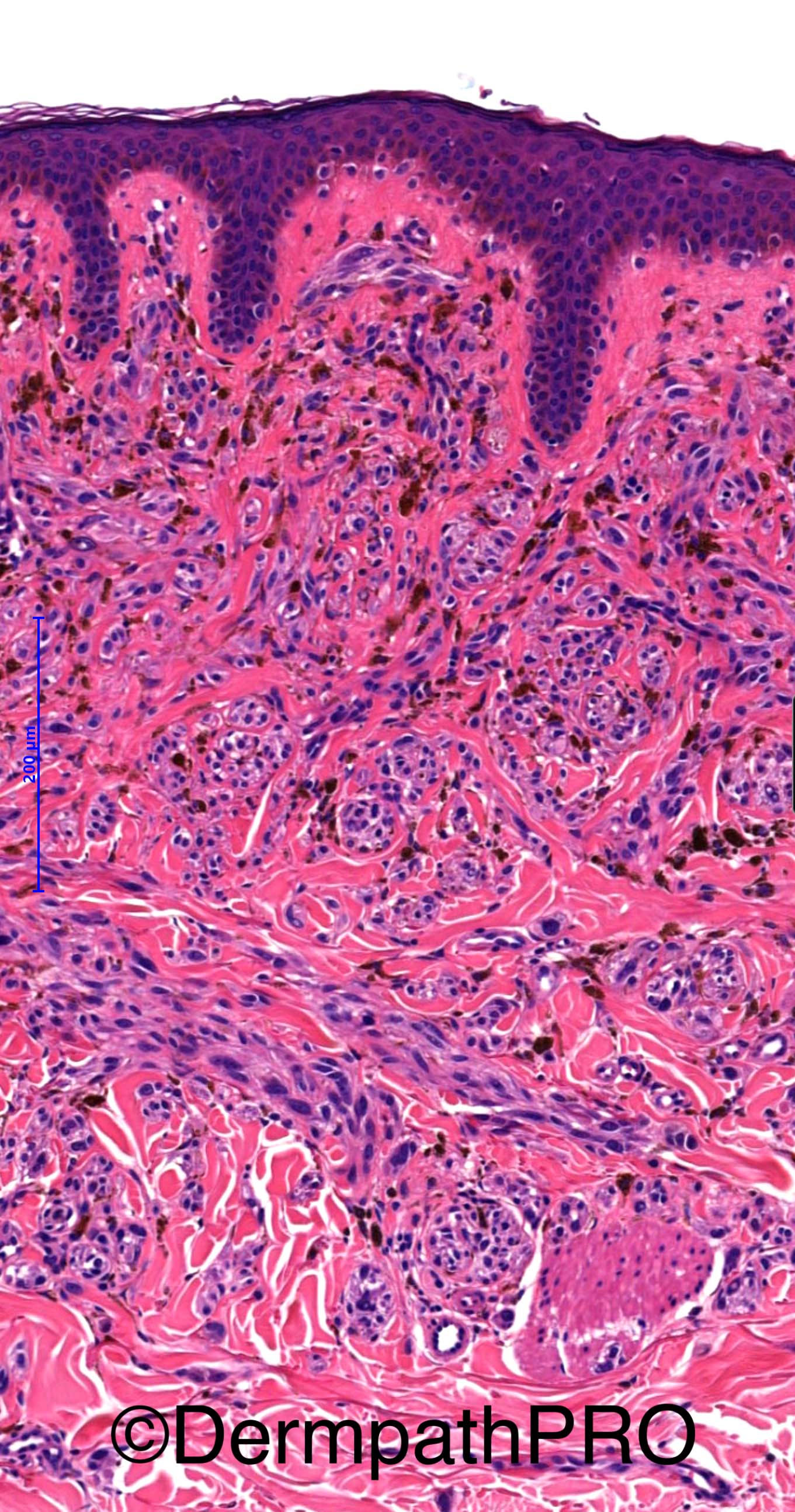
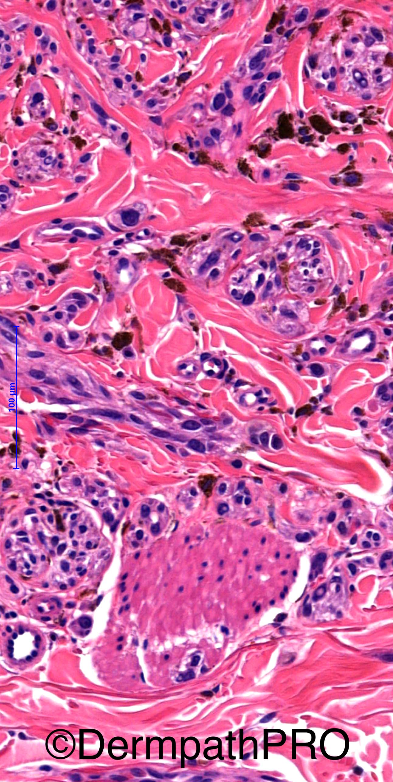
35 year old male with pigmented lesion on arm ?

Spot Diagnosis provided by Iskander Chaudhry


  Report Record

User Feedback


Guest Romualdo

Posted

Wiesner nevus, to IHQ confirmation.

Share this comment


Link to comment
share_externally

Arti Bakshi

Posted

Deep penetrating naevus.

The cytomorphology of cells with plump oval to spindle cells with heavily pigmented macrophages would fit with this entity. Can see a few intranuclear inclusions too.These lesions may not always have deep extension.

Lack of maturation and some degree of cytological atypia is acceptable. Havent seen any mitoses and presuming there are none.

Share this comment


Link to comment
share_externally

Raul Perret

Posted

This is a compound benign, superficial, wedge shaped and symmetrical lesion. It has a dermal component that tends to be arranged in a plexiform fashion. On my eyes there is a dual cytological component: dendritic cells (last image) and banal nevocytes that in areas surround adnexae. I agree with Arti, it fits in the blue nevus category: Superficial deep penetrating nevus, I think we could also discuss a combined nevus (phenotypic heterogeneity) with banal and blue nevus components. However this discussion would be pointless as there is no difference in prognosis and the entities share many morphological features.

I did not think of Wiesner nevus, as this is frequently a dermal based lesion (although probably this is not an exclusive parameter) and to my knowledge the presence of a component of dendritic melanocytes is not a feature.  

Share this comment


Link to comment
share_externally

vincenzo polizzi

Posted

It's a nevus!!! Not a BAP1-negative cells nevus. Not an atypical Spitz Nevus. Not a melanoma!!! Nevus it is, so I agree with all of you.

Share this comment


Link to comment
share_externally

Nitin Khirwadkar

Posted

Deep penetrating naevus/naevus with phenotypic heterogeneity. Favour DPN.

Share this comment


Link to comment
share_externally

vincenzo polizzi

Posted

I have review more carefully the images, and agree with Romualdo... 

Share this comment


Link to comment
share_externally

Iskander H. Chaudhry

Posted

Thank you all! 

 
It was diagnosed as a deep penetrating naevus. 

Share this comment


Link to comment
share_externally


Join the conversation

You can post now and register later. If you have an account, sign in now to post with your account.

Guest
Add a comment...

×   Pasted as rich text.   Paste as plain text instead

  Only 75 emoji are allowed.

×   Your link has been automatically embedded.   Display as a link instead

×   Your previous content has been restored.   Clear editor

×   You cannot paste images directly. Upload or insert images from URL.

Loading...

×
×
  • Create New...