Jump to content
In this section we have spot diagnoses posted on a daily basis since June 2010, now over 4000! You can review the archived cases and read the suggested diagnoses by users and the final comment by the contributors.
Case are uploaded each week day by 10 am UK time with the correct diagnosis will generally be posted at 8 pm UK time. Why not view the most recent spot diagnosis and proffer a diagnosis?

Case Number : Case 1028 - 2nd June Posted By: Guest

Please read the clinical history and view the images by clicking on them before you proffer your diagnosis.
Submitted Date :
   (0 reviews)

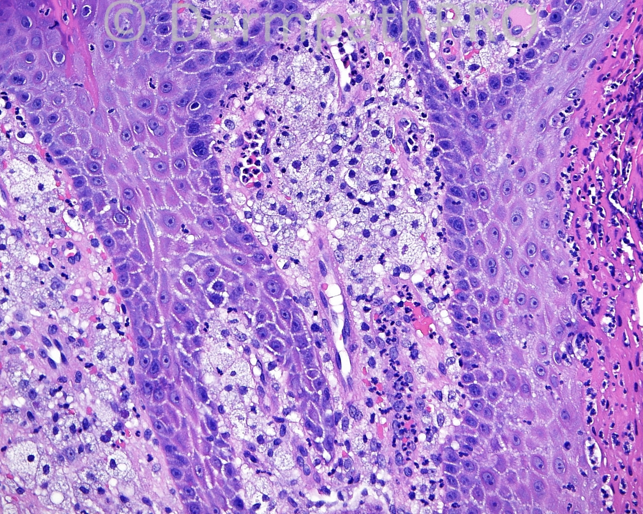
The patient is a 74 year old man with a shave biopsy of a lesion on the right side of the scrotum.

Case posted by Dr. Mark Hurt.


  Report Record

User Feedback


nick turnbull

Posted

Hmm..Heavily inflamed and spongiotic fbroepithelial polyp, with markerd parakeratosis with numerous neutrophils, reactive pagetoid dyskeratosis and verrucous xanthoma. I would wonder about trauma, infection (didn't see any fungi or bacteria), infestation (scabies), and contact/irritant dermatitis. Way off?

Share this comment


Link to comment
share_externally

Eman El-Nabarawy

Posted

Verruciform xanthoma.

Share this comment


Link to comment
share_externally

Arti Bakshi

Posted

Agree with Verruciform xanthoma.
There is evidence of keratinocyte necrosis in image 2 and 3, which is thought to be pathogenic in causing macrophage influx and xanthomatisation.

Share this comment


Link to comment
share_externally

Dr. Mona Abdel-Halim

Posted

Verruciform xanthoma

Share this comment


Link to comment
share_externally

Robledo F. Rocha

Posted

Verruciform xanthoma.

Share this comment


Link to comment
share_externally

Guest Tiberiu Tebeica

Posted

Verruciform xanthoma, which is fairly common on the scrotum.

Share this comment


Link to comment
share_externally

Guest Omobolade Obadofin

Posted

Classical Veruciform xanthoma. Nice case.

Share this comment


Link to comment
share_externally

Robledo F. Rocha

Posted

[quote name='arti bakshi' timestamp='1401709557']
Agree with Verruciform xanthoma.
There is evidence of keratinocyte necrosis in image 2 and 3, which is thought to be pathogenic in causing macrophage influx and xanthomatisation.
[/quote]
Very interesting this pathogenic hypothesis, Dr. Bakshi. I remember an ultrastructural study performed by [url="http://www.jaad.org/article/0190-9622(91)70117-K/abstract"]Balus et cols.[/url] which have suggested a transferation of lipid droplets from the cytoplasm of the melanocytes in the basal layer of the epidermis to the unknown-lineage foam cells that house in the in the dermis between rete pegs.

Share this comment


Link to comment
share_externally


Join the conversation

You can post now and register later. If you have an account, sign in now to post with your account.

Guest
Add a comment...

×   Pasted as rich text.   Paste as plain text instead

  Only 75 emoji are allowed.

×   Your link has been automatically embedded.   Display as a link instead

×   Your previous content has been restored.   Clear editor

×   You cannot paste images directly. Upload or insert images from URL.

Loading...

×
×
  • Create New...