Jump to content
In this section we have spot diagnoses posted on a daily basis since June 2010, now over 4000! You can review the archived cases and read the suggested diagnoses by users and the final comment by the contributors.
Case are uploaded each week day by 10 am UK time with the correct diagnosis will generally be posted at 8 pm UK time. Why not view the most recent spot diagnosis and proffer a diagnosis?

Case Number : Case 1029 - 3rd June Posted By: Guest

Please read the clinical history and view the images by clicking on them before you proffer your diagnosis.
Submitted Date :
   (0 reviews)

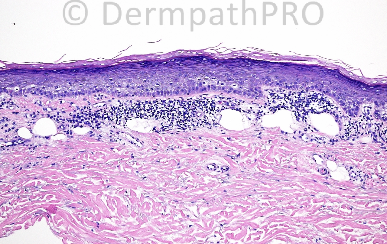
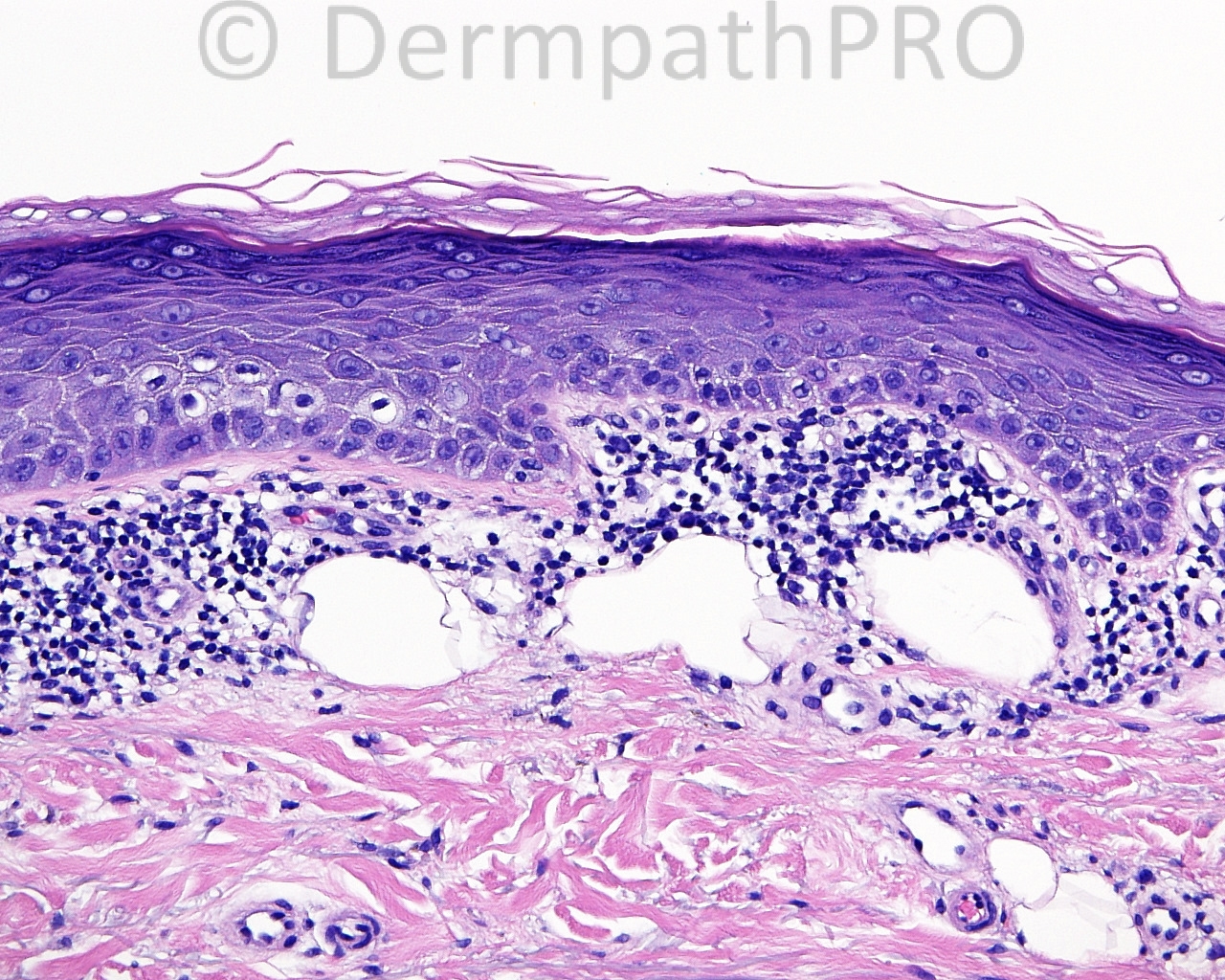
The patient is a 64 year old white woman with shave biopsy a lesion on the right posterior thigh.

Case posted by Dr. Mark Hurt.


  Report Record

User Feedback


Dr. Mona Abdel-Halim

Posted

? Atypical melanocyte proliferation within basal layer , ? Focal supra basal ascent... Or ? basal keratinocyte atypia? I think I will do Melan A first for this lesion...

Share this comment


Link to comment
share_externally

IgorSC

Posted

This is not an easy case. I´m thinking there is some atypical keratinocytes at the basal layer with palisade Deep sections could show us a superficial CBC

Share this comment


Link to comment
share_externally

Dr. Mona Abdel-Halim

Posted

Inspired by the comment of Dr Igor, may be the empty spaces are clue of BCC, mucinous artefacts !!!!!!

Share this comment


Link to comment
share_externally

Arti Bakshi

Posted

Difficult...not sure if the epidermal changes are significant or dermal!
Wondering about the nature of the superficial dermal vacuoles ?lymphatics ?pseudolipomatosis cutis

Share this comment


Link to comment
share_externally

Robledo F. Rocha

Posted

My first impression is superficial lymphatic malformation, aka lymphangioma circunscriptum, but the typical clinical presentation of grouped vesicle-like papules doesn’t fit on an impression of squamous cell carcinoma. I’m not sure about relevant epidermal abnormalities. Looking forward for new insights.

Share this comment


Link to comment
share_externally

Guest Jim Davie MD

Posted

[size=4]Agree this is a challenge. I'd consider an unusual presentation of lichen aureus / pigmented purpura, or MF. As Igor suggested, the edge of an epidermal neoplasm such as BCC or SCC in situ, although the epidermis looks more reactive than neoplastic to me.[/size]

[size=4]The centrally atrophic epidermis looks like benign reactive inflammatory changes, with sparse lymphocyte exocytosis, spongiosis, hypergranulosis, hydropic changes, and compact hyperkeratosis. There is a lichenoid chronic inflammatory infiltrate (focally hyperchromatic lymphocytes, and more peripheral aggregates of plasma cells or lymphoplasmacytoid cells) with scattered clear, slightly refractile, oil-droplet-like, focally compartmented spaces with what appears to be foamy histiocyte (or adipocyte-like) vesiculated large pale nuclei at the periphery; these agree with this might represent adipose tissue, foreign-body oil, pseudolipomatosis vs.lymphangioma. There is subtle sparse brown pigment in the dermis, which may be non-specific melanin from inflammation, vs. hemosiderin [last photo]. [/size]

Share this comment


Link to comment
share_externally

dermpath1

Posted

Mycosis fungoides.a typical variant.

Share this comment


Link to comment
share_externally

Guest Tiberiu Tebeica

Posted

I think this is superficial lymphangioma (lymphangioma circumscriptum). Sometimes, the epidermis overlying these lesions looks warty, which is true in our case and also explains the clinical presentation.

Share this comment


Link to comment
share_externally

Eman El-Nabarawy

Posted

What about lichen planus like keratosis?

Share this comment


Link to comment
share_externally

Dr. Mona Abdel-Halim

Posted

I was all the time worried about the basal layer !!!!

Share this comment


Link to comment
share_externally

Dr. Richard Carr

Posted

I think we saw similar fat cells high in the dermis (anatomically located on the calf) last week in a "lichenoid keratosis" or regression reaction in a superficial BCC. Not sure I would put to much emphasis on them but fun to speculate.

Share this comment


Link to comment
share_externally


Join the conversation

You can post now and register later. If you have an account, sign in now to post with your account.

Guest
Add a comment...

×   Pasted as rich text.   Paste as plain text instead

  Only 75 emoji are allowed.

×   Your link has been automatically embedded.   Display as a link instead

×   Your previous content has been restored.   Clear editor

×   You cannot paste images directly. Upload or insert images from URL.

Loading...

×
×
  • Create New...