Jump to content
In this section we have spot diagnoses posted on a daily basis since June 2010, now over 4000! You can review the archived cases and read the suggested diagnoses by users and the final comment by the contributors.
Case are uploaded each week day by 10 am UK time with the correct diagnosis will generally be posted at 8 pm UK time. Why not view the most recent spot diagnosis and proffer a diagnosis?

Case Number : Case 1032 - 6th June Posted By: Guest

Please read the clinical history and view the images by clicking on them before you proffer your diagnosis.
Submitted Date :
   (0 reviews)

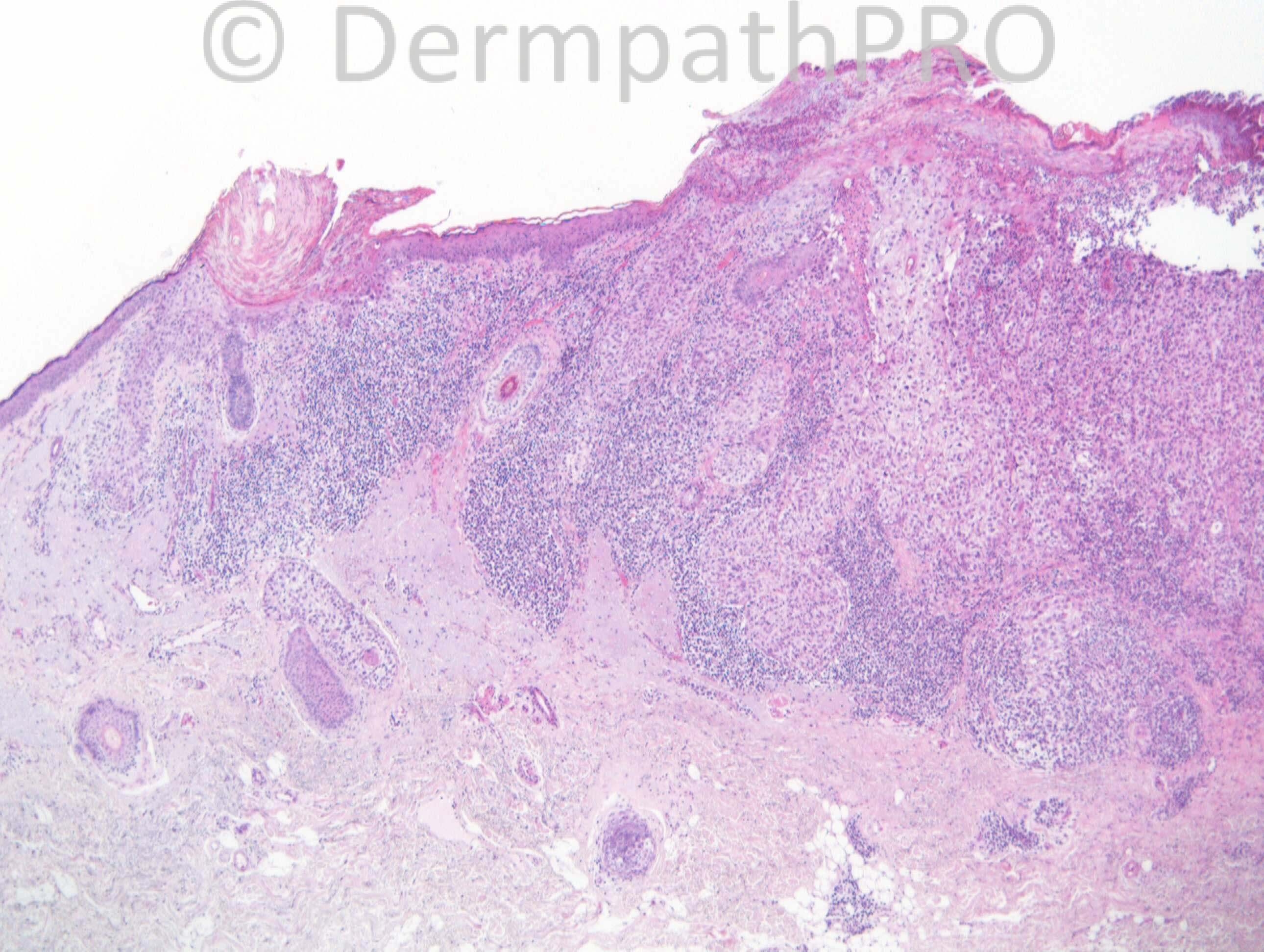
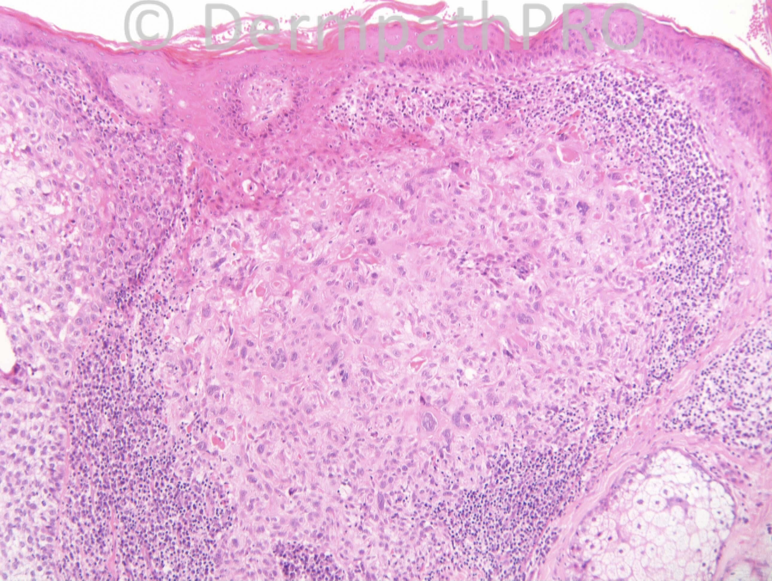
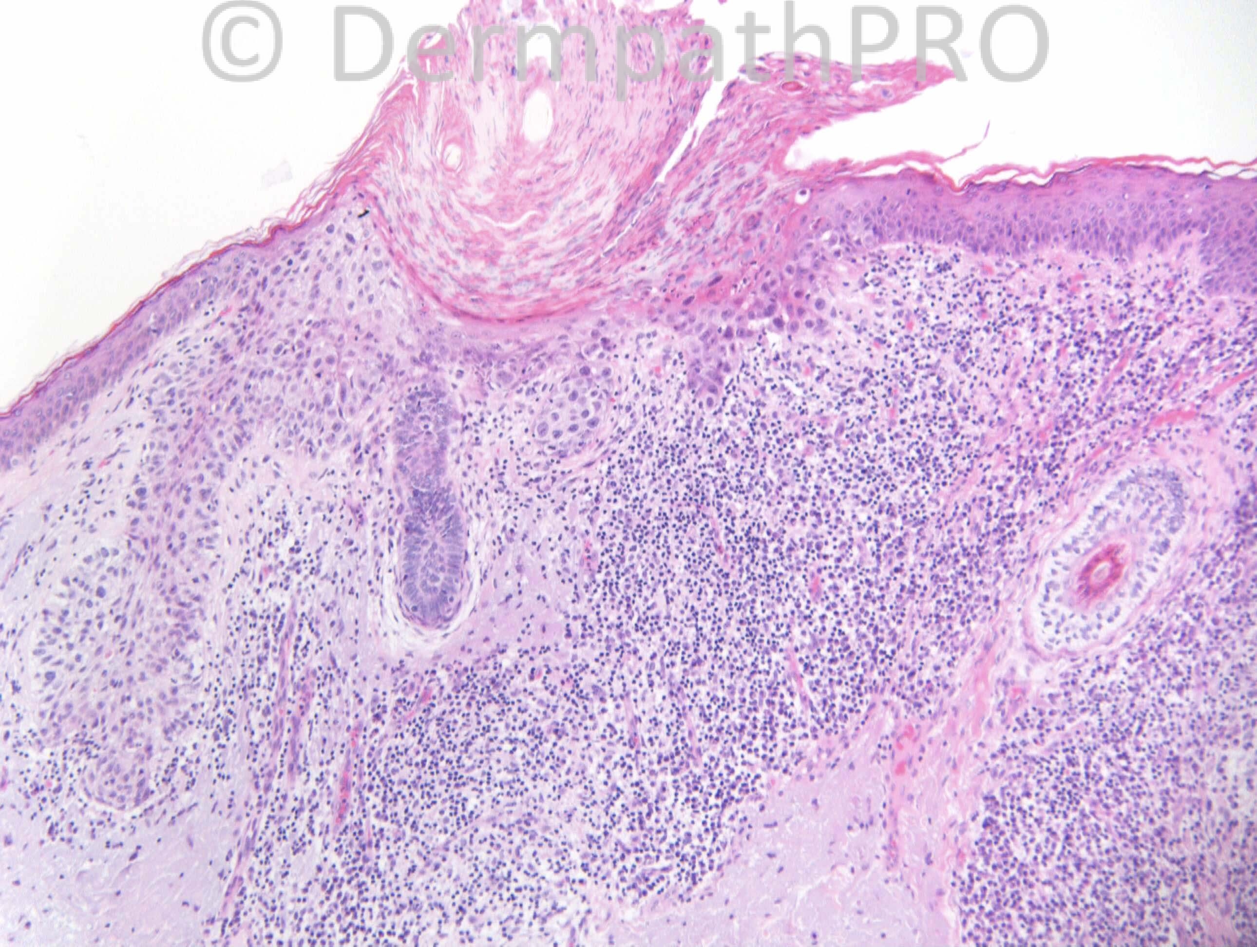
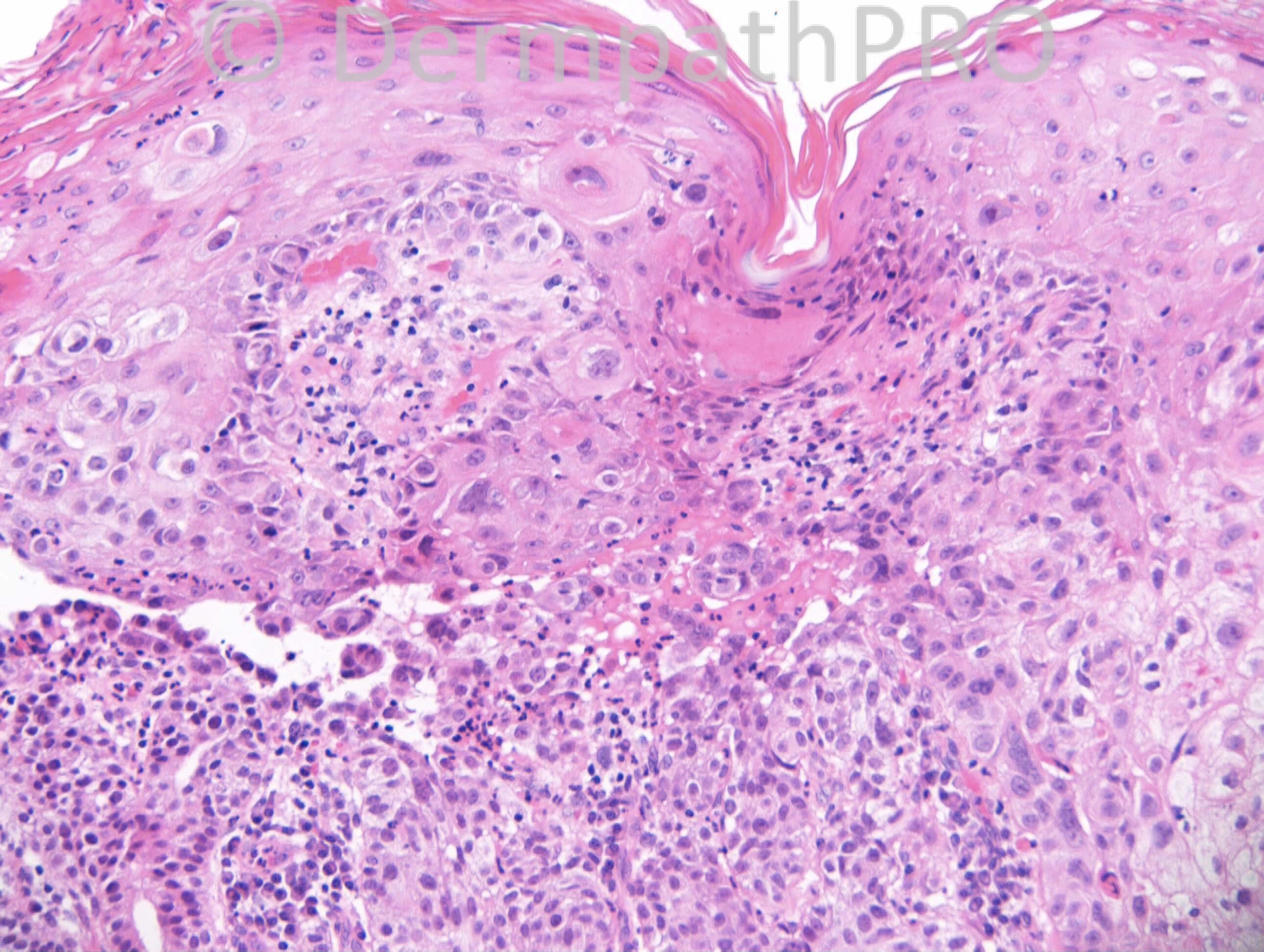
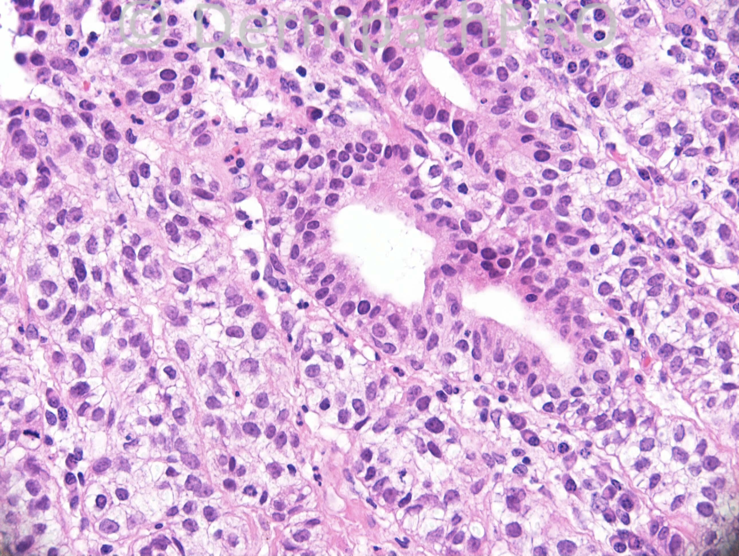
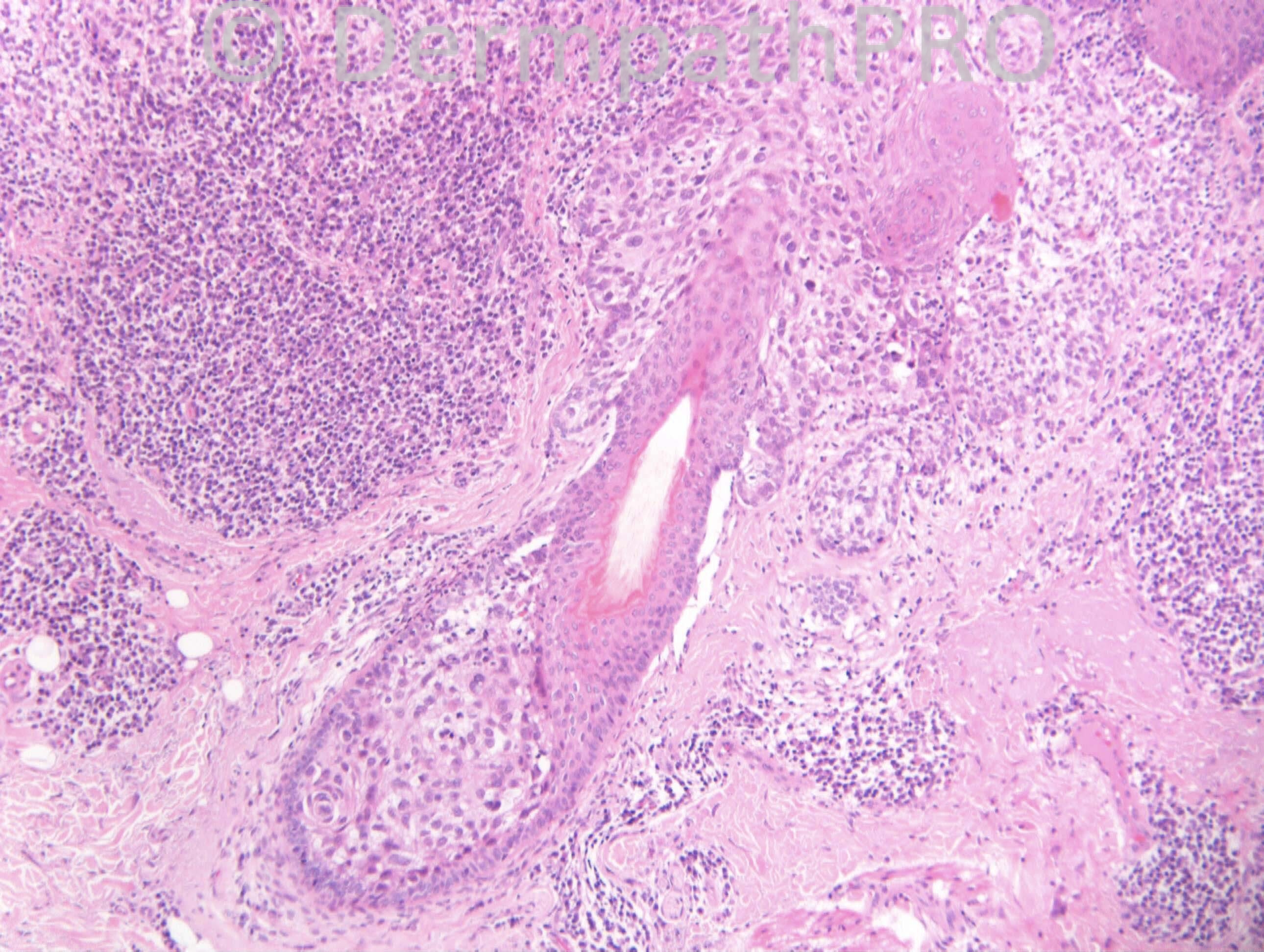
M72. Right temple, ?AK, ?BCC

Case posted by Dr. Richard Carr.

  Report Record

User Feedback


Dr. Mona Abdel-Halim

Posted

SCC with clear cell and acantholytic changes.

Share this comment


Link to comment
share_externally

Arti Bakshi

Posted

Agree with SCC
There are some features of follicular SCC ( well circumscribed, evidence of follicular origin in image 6 and clear cell change); but also overlying dysplasia; so may represent a hybrid (conventional and follicular )SCC

Share this comment


Link to comment
share_externally

Guest Romualdo

Posted

I think there is true glandular differentiaton so my vote is for adenosquamous carcinoma.

Share this comment


Link to comment
share_externally

Dr. Mona Abdel-Halim

Posted

I think in adenosquamous carcinoma there r two distinct areas, one of a moderate to undifferentiated SCC and the other of a true mucin secreting glandular component mimicking adenocarcinoma. I could not perceive the areas in the last image as true glandular component. I am in favor of them being pseudoglandular... May be a mucin stain or CEA can help.

Share this comment


Link to comment
share_externally

IgorSC

Posted

Clear cell SCC. Dif. diagnosis with SCC associated with Melanoma. Immunostains with SOX-10 and 34BE12 are usefull.

Share this comment


Link to comment
share_externally

Dr. Richard Carr

Posted

[img]https://dermpathpro.com/uploads/spot_diagnosis_comment_img/RAC4152x10_CD34_Label.jpg[/img]

[img]https://dermpathpro.com/uploads/spot_diagnosis_comment_img/RAC4152x10_p53_Label.jpg[/img]

[img]https://dermpathpro.com/uploads/spot_diagnosis_comment_img/RAC4152x20_PAS_Label.jpg[/img]

[img]https://dermpathpro.com/uploads/spot_diagnosis_comment_img/RAC4152x20_PASD_Label.jpg[/img]

[img]https://dermpathpro.com/uploads/spot_diagnosis_comment_img/RAC4152x2_p16_Label.jpg[/img]

[img]https://dermpathpro.com/uploads/spot_diagnosis_comment_img/RAC4152x5_BerEP4_Label.jpg[/img]

[img]https://dermpathpro.com/uploads/spot_diagnosis_comment_img/RAC4152x5_EMA_Label.jpg[/img]

Share this comment


Link to comment
share_externally

Robledo F. Rocha

Posted

One thing that struck me is the epidermis, which is virtually undisturbed, except for some reactive changes due to pagetoid spread from the underlying tumor. So, my first impression was of this tumor didn’t arise from the above epidermis, but it could be an epidermotropic metastatic carcinoma or a squamous cell carcinoma developed in the upper part of hair follicle.

Share this comment


Link to comment
share_externally

Dr. Richard Carr

Posted

I reported this as a follicular (infundibular-tricholemmal) SCC with focal glandular/ductal differentiation. This lesion has many of our diagnostic criteria for follicular SCC i.e. abrupt / circumscript connection with the epidermis (best seen at the lateral borders), clear cut focal follicular infundibular involvement and absence of bowenoid dsyplasia in the adjacent epidermis. This particular case is unusual being predominantly tricholemmal (lower outer root sheath) clear cell pattern, so much so that the existence of the tumour as a distinct (tricholemmal carcinoma) entity has even been challenged. This is the first case I have seen personally with pagetoid spread but it is noted in some classical tricholemmal carcinoma in the literature. The features of lower outer root sheath are here nicely demonstrated including prominent glycogen, distinct cell membranes in the clear cells, distinct basement membrane. In contrast most follicular SCC show differentiation towards the upper (in addition to infundibular and lower) outer root sheath including lobulated profile, subtle palisading of pale or even basaloid cells and central squamous morules with abundant pale cytoplasm and abrupt bright orange keratinisation with inconspicuous or absent keratohyaline granules. Remember that around 60% have central acantholytic mucin pools (follicular mucin) that seems to correlate with the infundibular differentiation also apparent in most FSCC (but also seen in tricholemmoma and inverted follicular keratosis/seborrhoeic keratosis). In my experience this is the first case of FSCC that has had diffuse p16 (normally typical of bowenoid actinic keratosis and classical bowen's). However a null p53 is at least as common in FSCC as diffuse positivity. Finding ductal / glandular differentiation is not unexpected as we know tumours of the follicular-sebaceous-apocrine unit (including BCC) may show mixed differentiation although this is the first example I have reported in more than 200 cases of FSCC. Finally I wanted you to know in our recent audit of SCC we found the follicular subtype was by far the most common (58% of our excisions for SCC although about 50% of them were entirely circumscript and for practical purposes in situ). We hope to submit a series (>100) with clinical follow-up in the very near future. There is a sub-group of crateriform FSCC that do metastasise and could, in a superficial biopsy, be confused with KA (more anon). Tricholemmoma are almost invariably focally CD34+ (in contrast FSCC including classical tricholemmal carcinomas of clear cell type that are rarely CD34+) and would not have diffuse p16 or null p53 and would not have pagetoid spread. I tend to limit adenosquamous carcinomas to a waste basket of high grade tumours that lack areas distinctive of other entities such as typical porocarcinoma, follicular SCC, hidradenocarcinoma etc.
Regards to all and enjoy your weekends.

Share this comment


Link to comment
share_externally

Arti Bakshi

Posted

Great case, Richard and thanks for your very educational comments! Having learnt the concept of follicular SCC from you, I do report these more often now, but sometimes struggle with the decision of insitu vs invasive in this group of lesions. Did you regard this case as in situ or invasive?

Share this comment


Link to comment
share_externally

Dr. Richard Carr

Posted

Difficult to assess I agree. Probably at worst pushing invasion only. It was an old case reported at St Elsewhere.

Share this comment


Link to comment
share_externally


Join the conversation

You can post now and register later. If you have an account, sign in now to post with your account.

Guest
Add a comment...

×   Pasted as rich text.   Paste as plain text instead

  Only 75 emoji are allowed.

×   Your link has been automatically embedded.   Display as a link instead

×   Your previous content has been restored.   Clear editor

×   You cannot paste images directly. Upload or insert images from URL.

Loading...

×
×
  • Create New...