Jump to content
In this section we have spot diagnoses posted on a daily basis since June 2010, now over 4000! You can review the archived cases and read the suggested diagnoses by users and the final comment by the contributors.
Case are uploaded each week day by 10 am UK time with the correct diagnosis will generally be posted at 8 pm UK time. Why not view the most recent spot diagnosis and proffer a diagnosis?

Case Number : Case 1033 - 9th June Posted By: Guest

Please read the clinical history and view the images by clicking on them before you proffer your diagnosis.
Submitted Date :
   (0 reviews)

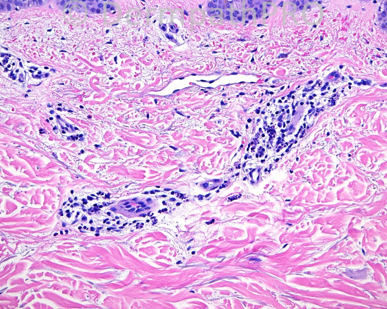
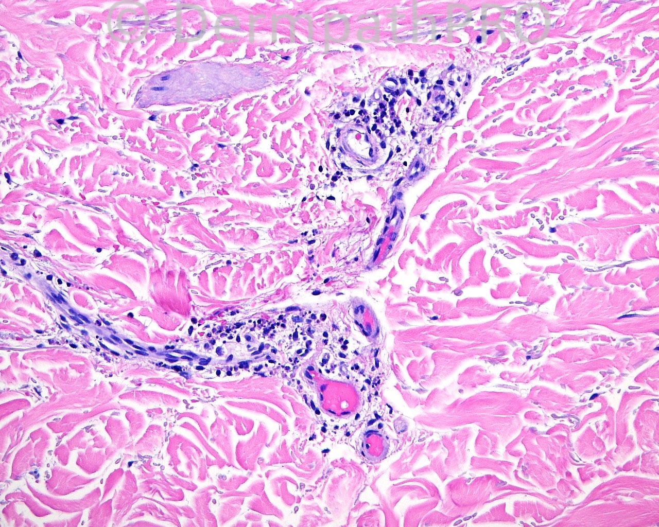
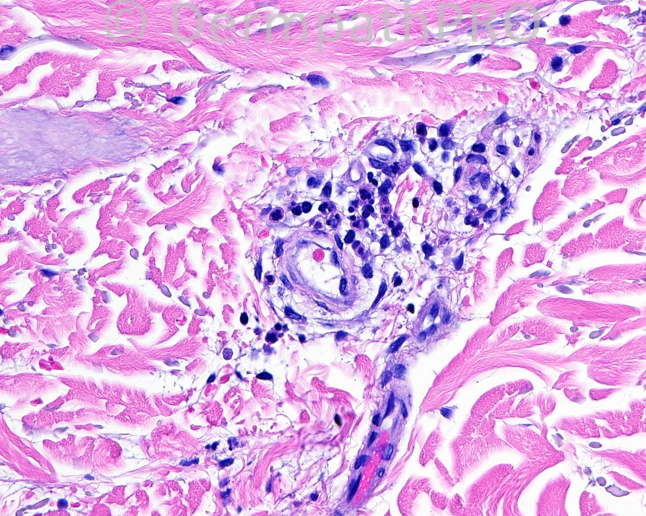
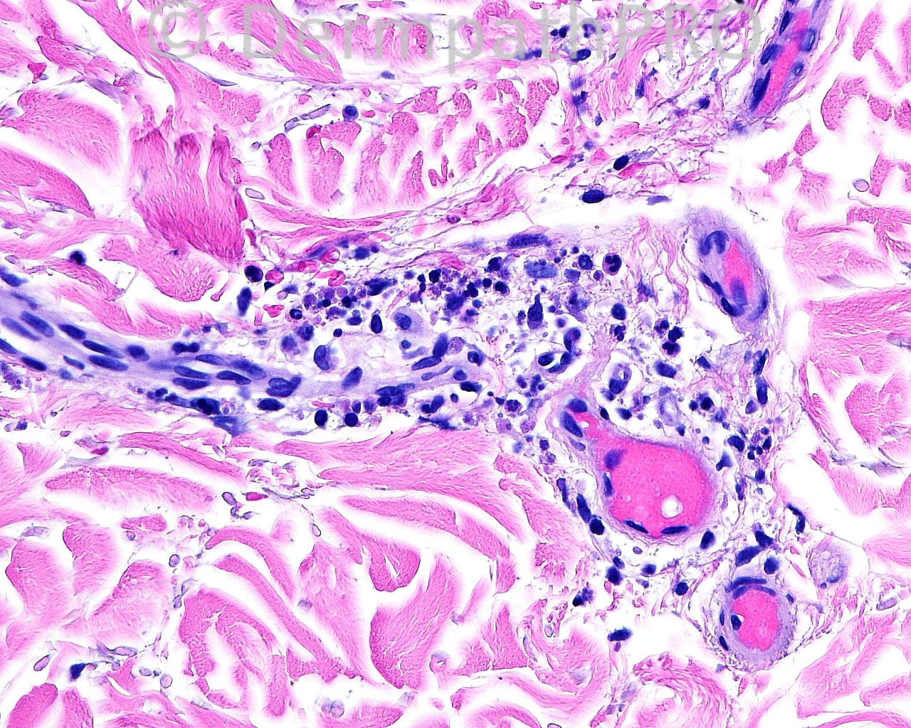
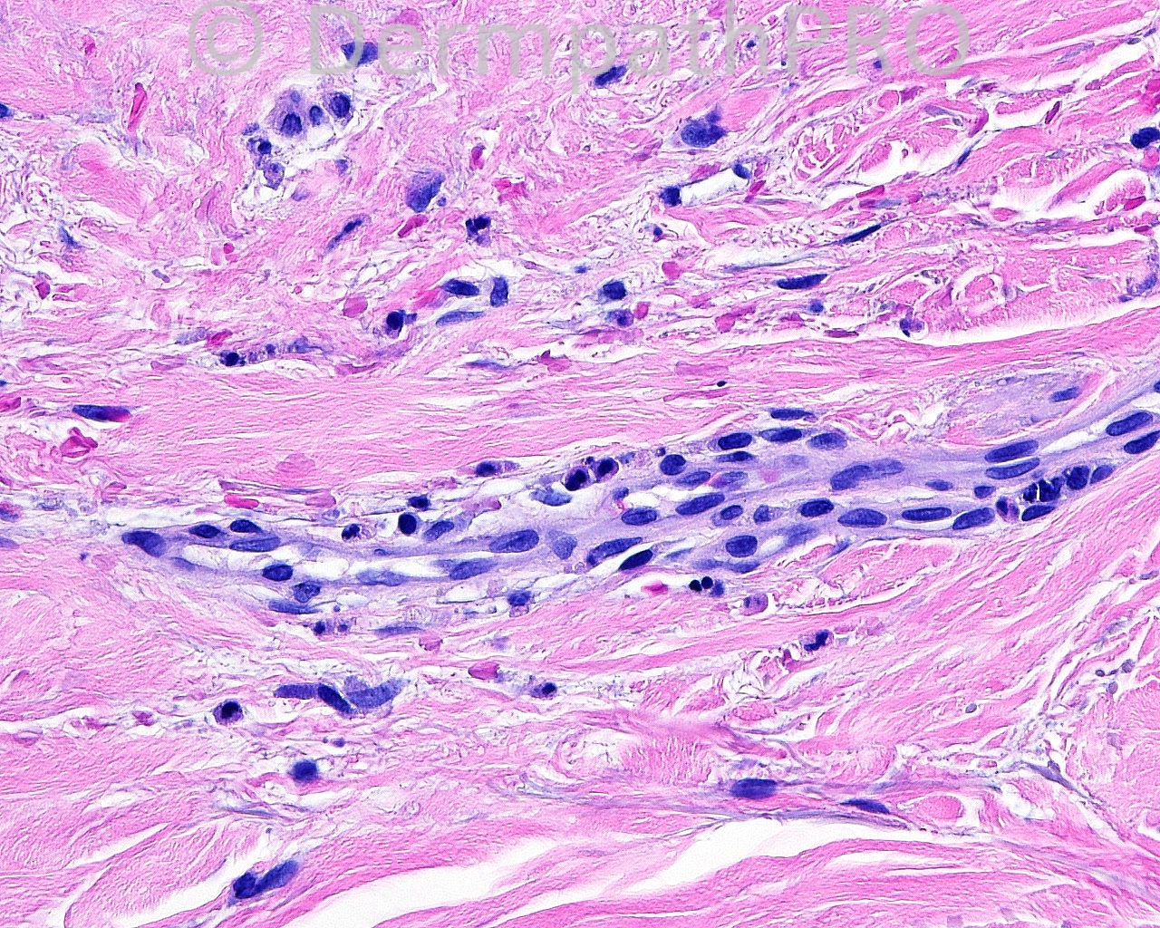
The patient is a 65 year old white man with a biopsy taken from the right ulnar mid volar forearm for direct immunofluorescence.

Case posted by Dr. Mark Hurt.


  Report Record

User Feedback


dermpath1

Posted

Urticarial vasculitis.
Other things to be mentioed are other causes of vasculopathy such as DIC , cryoglobulinemia......etc.

Share this comment


Link to comment
share_externally

Eman El-Nabarawy

Posted

Indeterminate leprosy.

Share this comment


Link to comment
share_externally

Arti Bakshi

Posted

Agree that histology is certainly suggestive of urticarial vasculitis. Ofcourse, needs correlation with clinical presentation[b].[/b]

Share this comment


Link to comment
share_externally

Urticaria-like lesion. I do not see vasculitis.

Share this comment


Link to comment
share_externally

Dr. Richard Carr

Posted

c/w late-stage leucocytoclastic vasculitis (LCV). The prominent orange pink deposits raise cryoglobulins or other coagulopathy in the differential. Cryoglobulins can cause LCV.

Share this comment


Link to comment
share_externally

Guest Jim Davie MD

Posted

[quote name='Dr. Richard Carr' timestamp='1402325960']
c/w late-stage leucocytoclastic vasculitis (LCV). The prominent orange pink deposits raise cryoglobulins or other coagulopathy in the differential. Cryoglobulins can cause LCV.
[/quote]

Agree with Richard...LCV vs. cryoglobulinemia. This neutrophil-rich sparse perivascular lymphocytic infiltrate has variably degraded neutrophils and leukocytoclastic debris, with extravasated red cells. Vessels show equivocal fibrinoid changes with some apparently plugged with eosinophilic material (which may be either packed RBCs vs. cryoglobulin on these images). Eosinophils are notable for their absence. No epidermal changes.
I would imagine the DIF was ordered for a differential of vasculitis vs. urticaria.

Share this comment


Link to comment
share_externally

Robledo F. Rocha

Posted

The presence of sparse perivascular infiltrate with neutrophilic karyorrhexis, small number of extravasated erythrocytes and slight dermal edema, and the lack of both fibrin in vessel walls and thrombi within lumina, make me think of early leukocytoclastic vasculitis. Granular deposits of IgA in vessels suggest Henoch-Schönlein purpura in a rare clinical presentation in the upper extremity of an old adult.
The so-called urticarial vasculitis is nothing more than an early leukocytoclastic vasculitis which expresses itself clinically as edematous urticarial papules due to paucity of extravasated erythrocytes.

Share this comment


Link to comment
share_externally

Dr. Mona Abdel-Halim

Posted

Old lesion of LCV vasculitis, IgA mediated. Strange age for Henoch Schonlein as Robledo said.

Share this comment


Link to comment
share_externally

I agree with HSP, based on the IMF.

Share this comment


Link to comment
share_externally


Join the conversation

You can post now and register later. If you have an account, sign in now to post with your account.

Guest
Add a comment...

×   Pasted as rich text.   Paste as plain text instead

  Only 75 emoji are allowed.

×   Your link has been automatically embedded.   Display as a link instead

×   Your previous content has been restored.   Clear editor

×   You cannot paste images directly. Upload or insert images from URL.

Loading...

×
×
  • Create New...