Jump to content
In this section we have spot diagnoses posted on a daily basis since June 2010, now over 4000! You can review the archived cases and read the suggested diagnoses by users and the final comment by the contributors.
Case are uploaded each week day by 10 am UK time with the correct diagnosis will generally be posted at 8 pm UK time. Why not view the most recent spot diagnosis and proffer a diagnosis?

Case Number : Case 1038 - 16th June Posted By: Guest

Please read the clinical history and view the images by clicking on them before you proffer your diagnosis.
Submitted Date :
   (0 reviews)

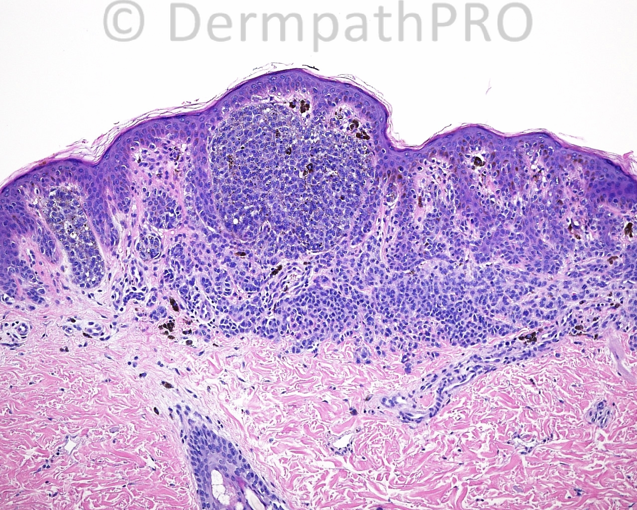
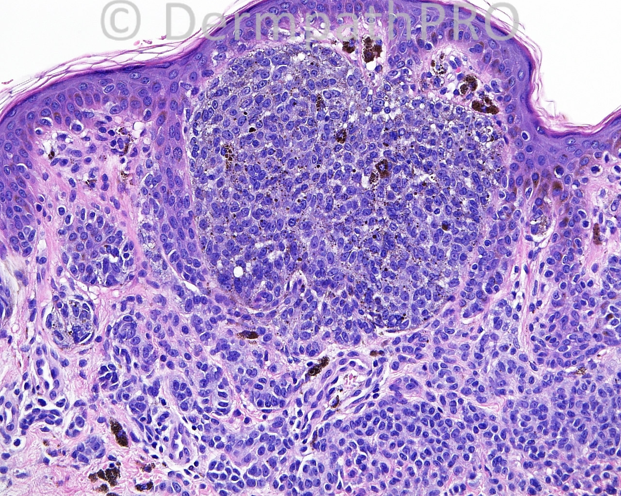
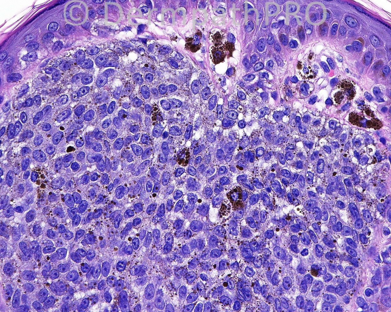
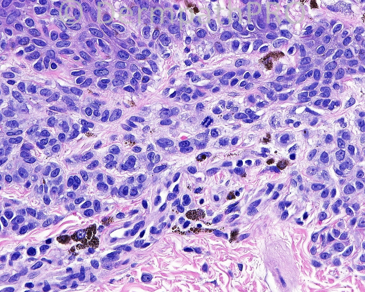
The patient is a 13 year old boy with excisions with margin exam of darkening lesions, present six months, taken from the left, upper aspect of the back.

Case posted by Dr. Mark Hurt.


  Report Record

User Feedback


amiratawdy

Posted

is it possible to be a minimal deviation melanoma? ki67 and HMB45 are needed

Share this comment


Link to comment
share_externally

Dr. Mona Abdel-Halim

Posted

This is a somehow difficult lesion. Fairly circumscribed, yet asymmetrical. The lesion appear nicely maturing. However, I am worried about this expansile nodule that is sharply delineated from the rest of the nevus in the background. It reminded me of proliferation nodules in congenital nevi. From the history, I understood that the patient presents with darkening lesions of 6 months. Does this mean multiple lesions? Are the darkening lesions arising de novo or on top of another lesion in the background, ? Congenital nevus? and has been present for 6months? Cytologically, the cells of this nodule do not look like typical epithelioid melanoma cells, yet they show prominent nucleoli and are different than the background dermal nevocytic cells, but they show no mitotic activity. It does not fit in clonal or (inverted type A) nevus. There is an isolated typical mitosis in the background dermal component, I do not think it is worrisome because the patient is a child (13 years) and I think this is expected in such age.
What is worrying me is this dermal nodule!!!!!
If these are lesions developing within a giant congenital nevus, I will consider it proliferation nodules.
If they are arising de novo, then may be it is better to describe this lesion as MELTUMP and do further work including IHC and genetic studies?? Waiting to read expert opinions...

Share this comment


Link to comment
share_externally

Robledo F. Rocha

Posted

A late proliferative nodule arising within a congenital melanocytic nevus.

Share this comment


Link to comment
share_externally

I don´t think this is a melanoma. I agree with Robledo, proliferative nodule arising in a congenital nevus.

Share this comment


Link to comment
share_externally

Guest Romualdo

Posted

Agree with Dr. Richard Carr: clonal nevus.

Share this comment


Link to comment
share_externally

Eman El-Nabarawy

Posted

Agree with Dr Abdul-Kadir: Nevoid melanoma.

Share this comment


Link to comment
share_externally

Dr. Mona Abdel-Halim

Posted

Interesting case that highlights the value of genetic studies in problematic melanocytic lesions...

Share this comment


Link to comment
share_externally

Dr. Richard Carr

Posted

I think we will find all clonal naevi are separated from their background lesion by an additional mutation / translocation etc. Not sure this proves a fully capable malignant melanoma though. I suspect this lesion will be of low risk (all be it uncertain potential).

Share this comment


Link to comment
share_externally


Join the conversation

You can post now and register later. If you have an account, sign in now to post with your account.

Guest
Add a comment...

×   Pasted as rich text.   Paste as plain text instead

  Only 75 emoji are allowed.

×   Your link has been automatically embedded.   Display as a link instead

×   Your previous content has been restored.   Clear editor

×   You cannot paste images directly. Upload or insert images from URL.

Loading...

×
×
  • Create New...