Jump to content
In this section we have spot diagnoses posted on a daily basis since June 2010, now over 4000! You can review the archived cases and read the suggested diagnoses by users and the final comment by the contributors.
Case are uploaded each week day by 10 am UK time with the correct diagnosis will generally be posted at 8 pm UK time. Why not view the most recent spot diagnosis and proffer a diagnosis?

Case Number : Case 1044 - 24th June Posted By: Guest

Please read the clinical history and view the images by clicking on them before you proffer your diagnosis.
Submitted Date :
   (0 reviews)

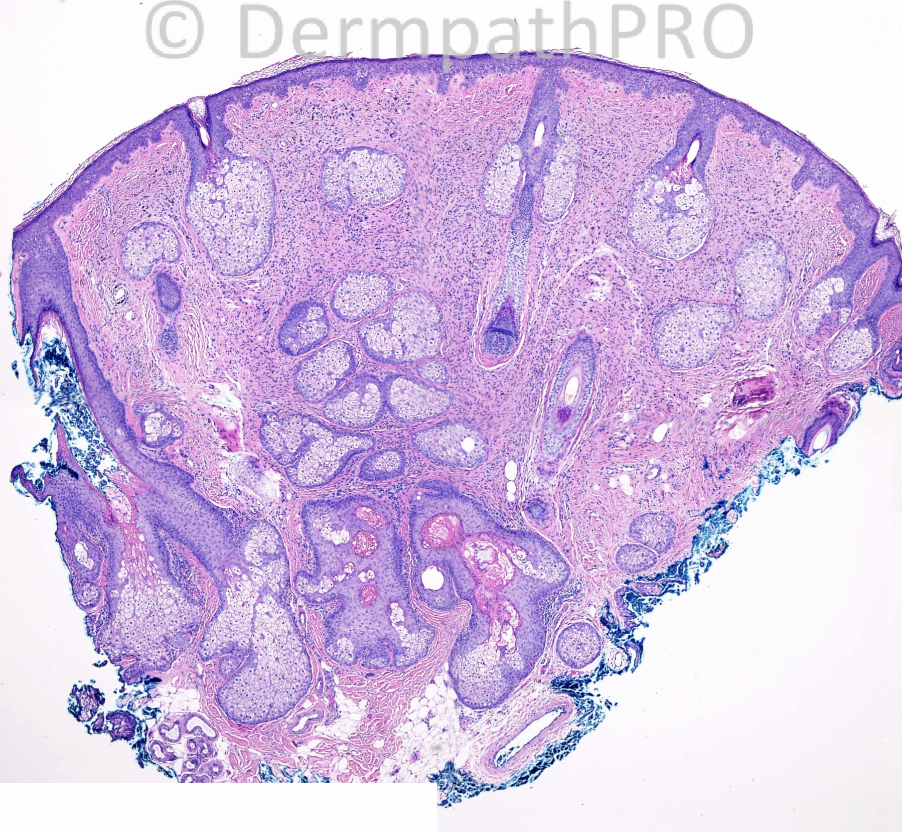
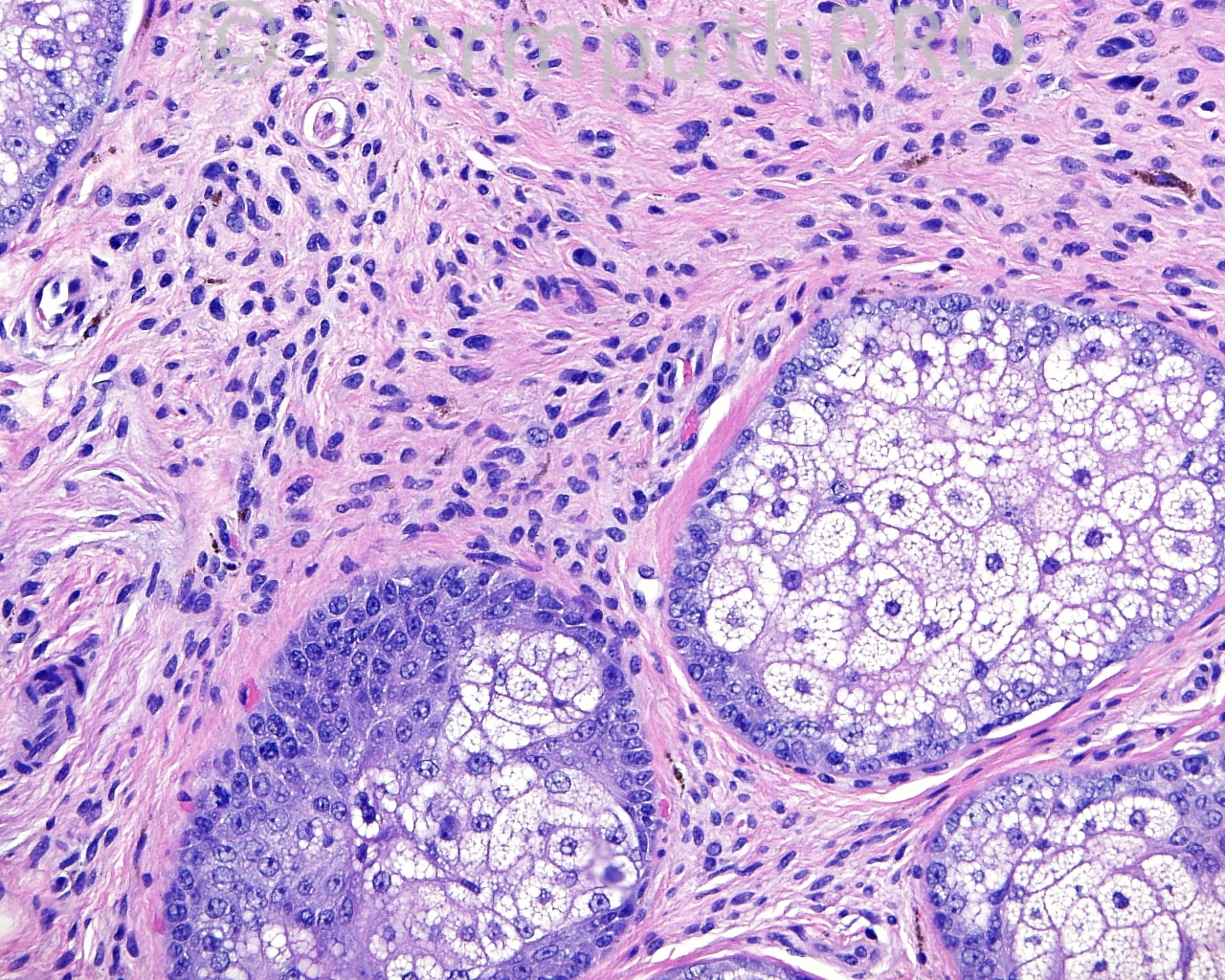
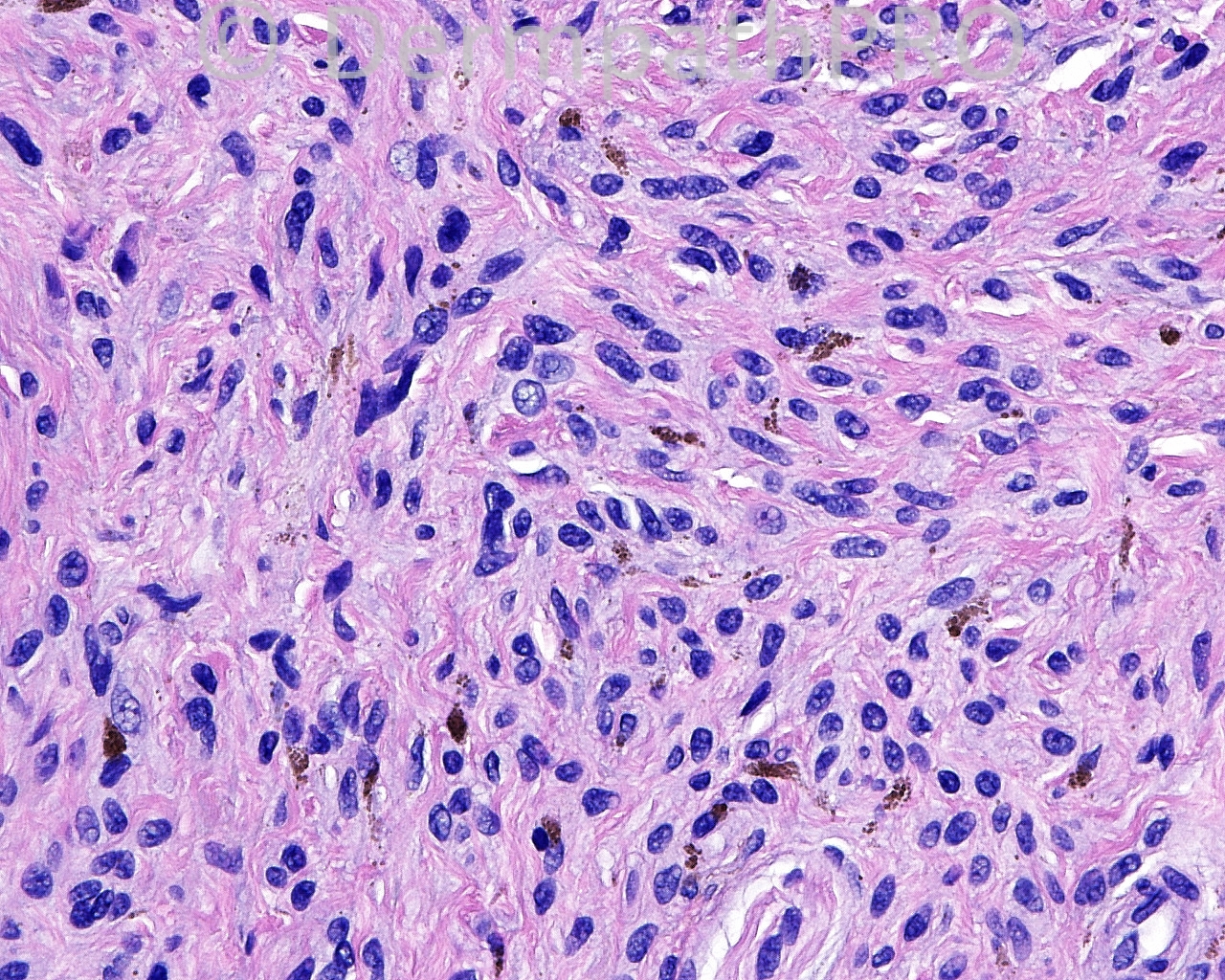
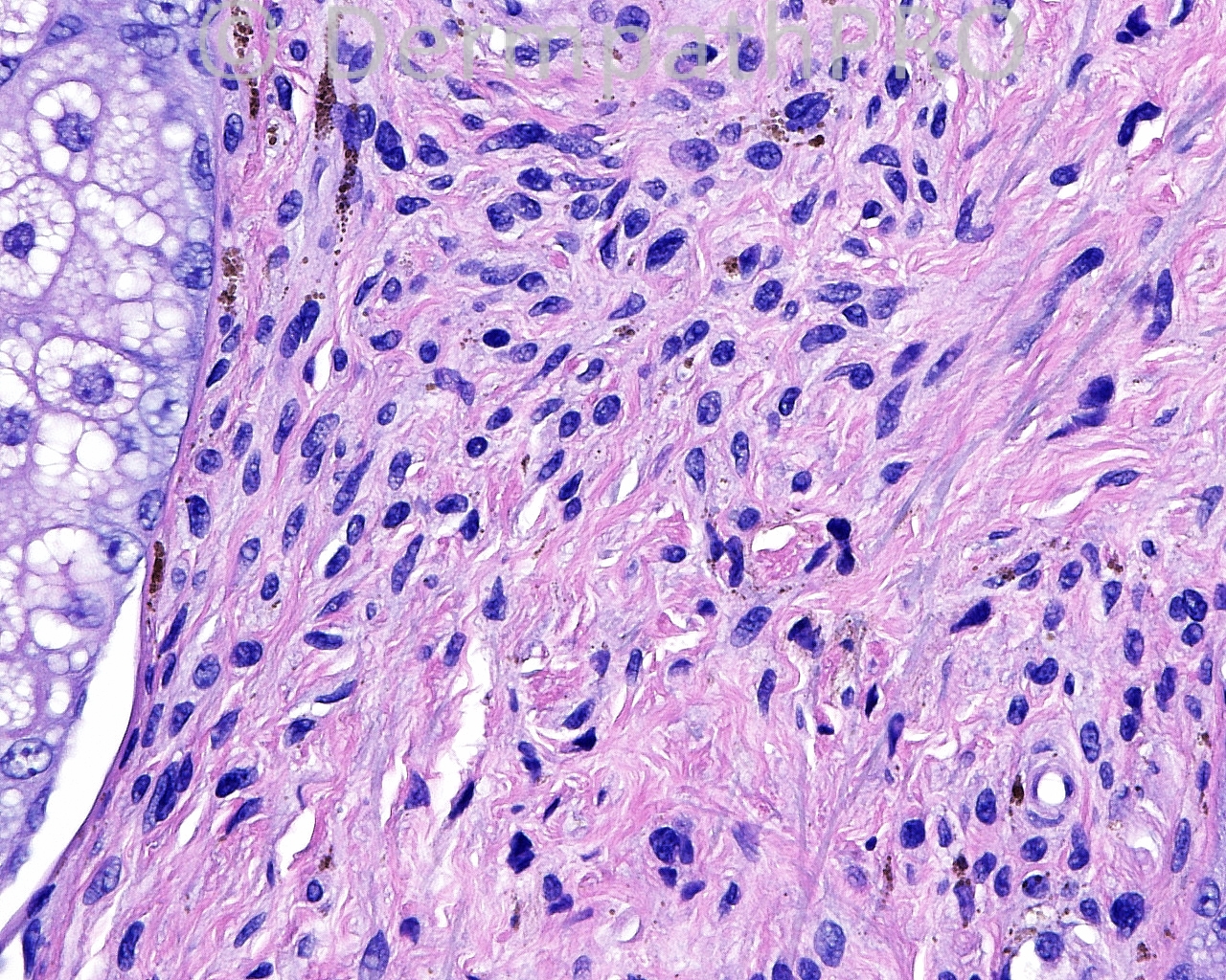
The patient is a 38-year-old white woman with an excision of a pigmented lesion on the nasal tip.

Case posted by Dr. Mark Hurt.


  Report Record

User Feedback


Eman El-Nabarawy

Posted

Amelanotic cellular blue nevus.

Share this comment


Link to comment
share_externally

Arti Bakshi

Posted

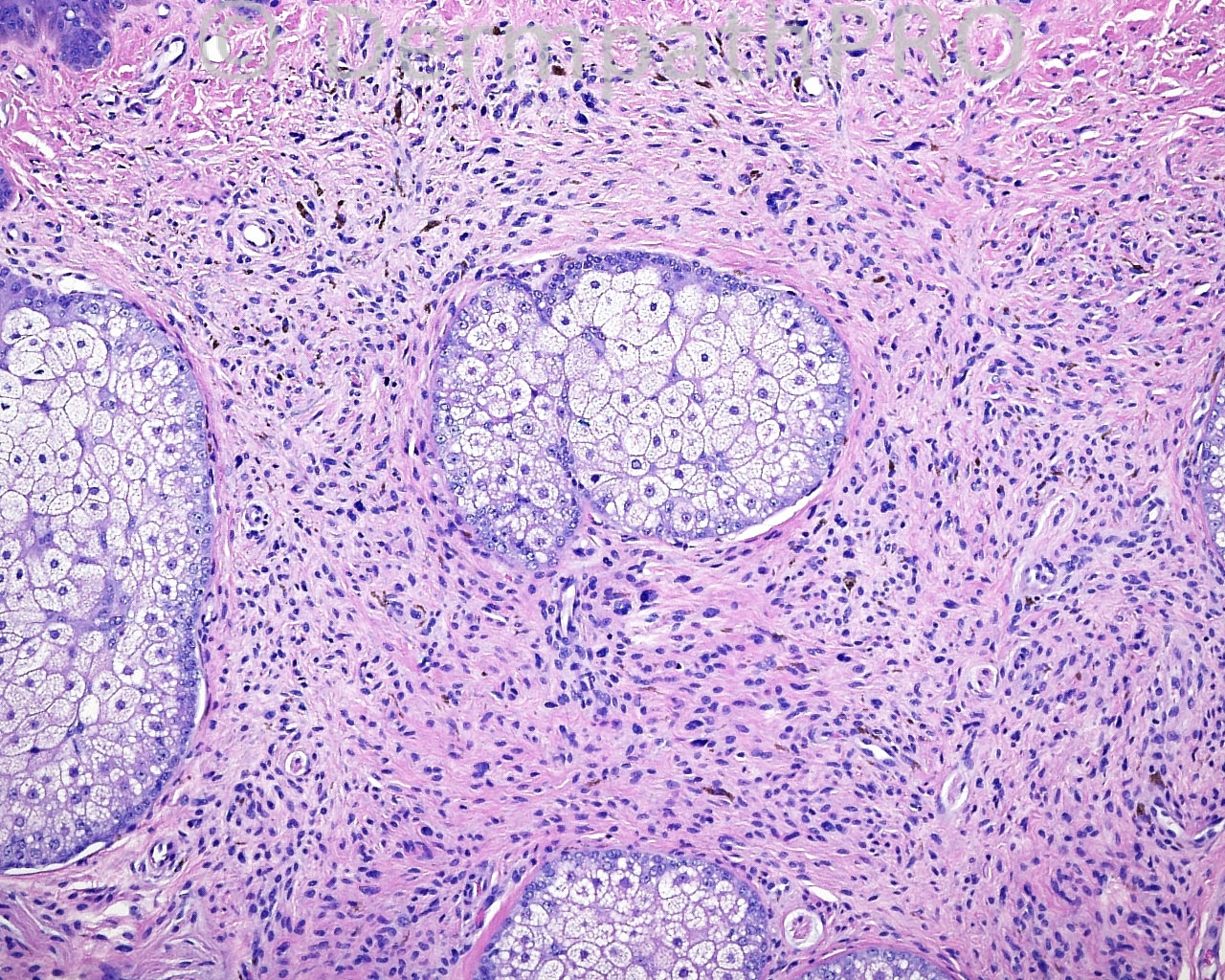
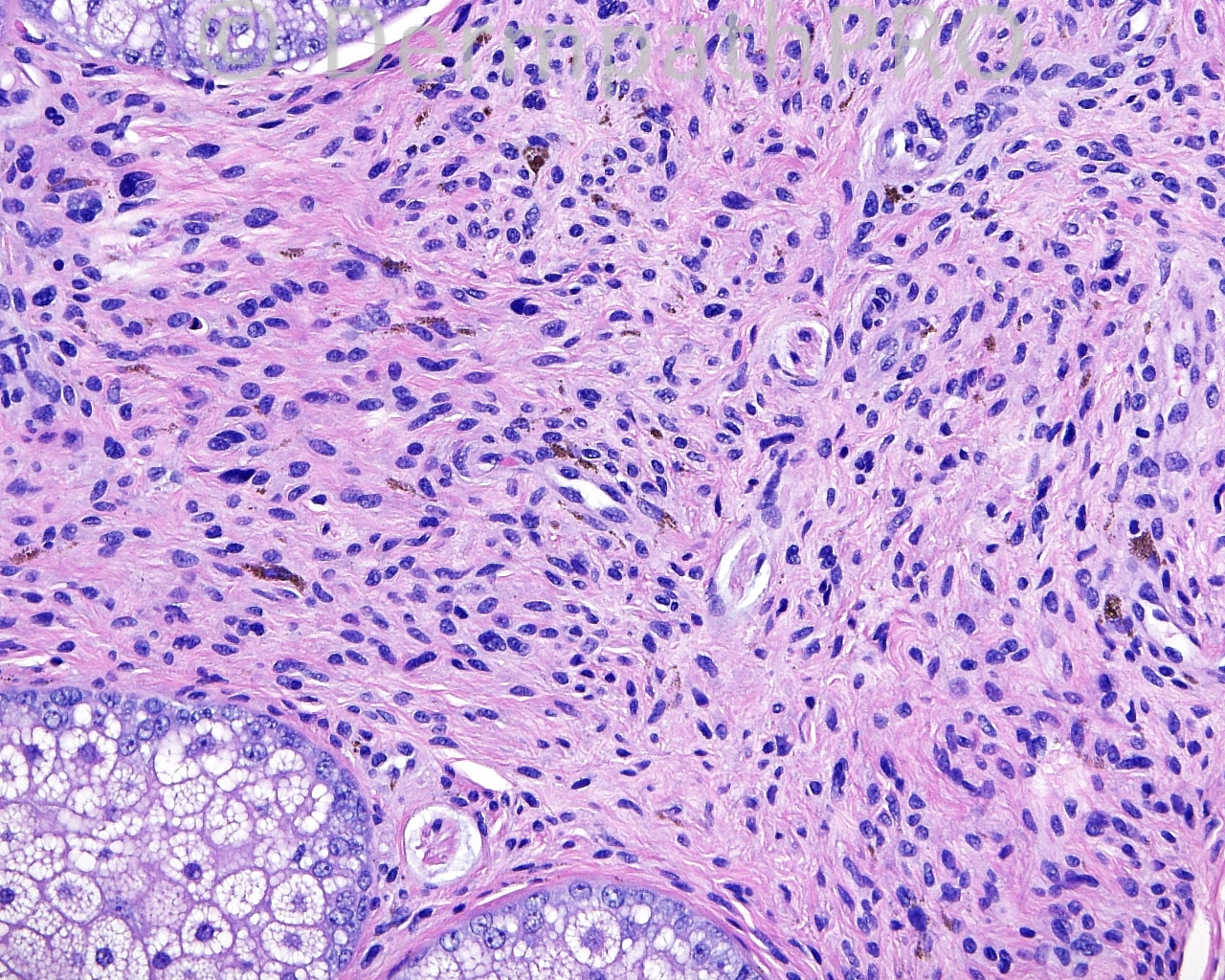
Wedge shaped intradermal melanocytic proliferation with an admixture of spindle and dendritic cells. There is variable pleomorphism, but no mitoses.
Benign, probably a variant of blue naevus.

Share this comment


Link to comment
share_externally

nick turnbull

Posted

Agree, hypopigmented blue nevus

Share this comment


Link to comment
share_externally

Dr. Mona Abdel-Halim

Posted

Hypopigmented blue nevus

Share this comment


Link to comment
share_externally

Robledo F. Rocha

Posted

My first impression was a benign fibrous tumor, but the presence of a few pigmented cells with bipolar or dendritic processes drove me to the diagnosis of hypopigmented blue nevus.

Share this comment


Link to comment
share_externally

Guest Dr Gonzalo de Toro

Posted

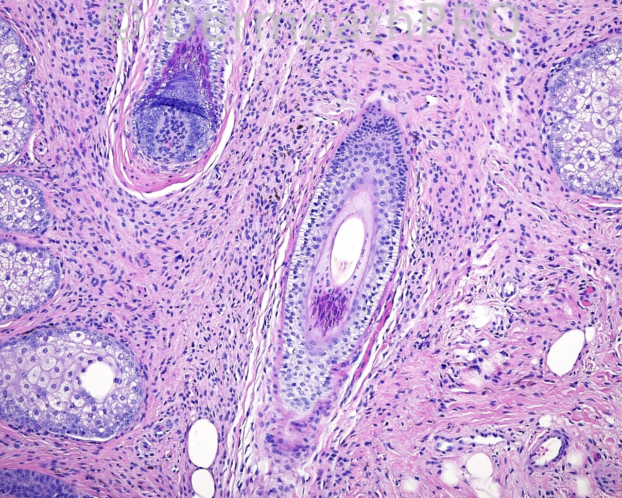
The lesión is wedge shaped, with some dendritic melanocytes with elongated and neurofibroma-like featues. I favor a hypopigmented blue nevus.

Share this comment


Link to comment
share_externally

Dr. Richard Carr

Posted

Agree with hypopigmented blue naevus. Most of the lesional cells are S100 negative as expected. Nice internal control in Langerhan's cells and nerves.

Share this comment


Link to comment
share_externally

Guest Jim Davie MD

Posted

Agree with hypomelanotic blue nevus (possibly congenital).
The bland histology (slightly hyperchromatic senescent nuclei with small to inconspicuous nucleoli), non-expansile architecture (that surrounds and does not significantly disturb/displace arrangement of adnexa), sparse evenly distributed interspersed melanophages, absence of mitotic activity, and absence of inflammatory reaction help support the diagnosis; the immunostains are similarly congruent (no Ki67 or PHH3 detectable proliferative / mitotic activity, and expected positive staining for Melan-A and S100.)

Share this comment


Link to comment
share_externally


Join the conversation

You can post now and register later. If you have an account, sign in now to post with your account.

Guest
Add a comment...

×   Pasted as rich text.   Paste as plain text instead

  Only 75 emoji are allowed.

×   Your link has been automatically embedded.   Display as a link instead

×   Your previous content has been restored.   Clear editor

×   You cannot paste images directly. Upload or insert images from URL.

Loading...

×
×
  • Create New...