Jump to content
In this section we have spot diagnoses posted on a daily basis since June 2010, now over 4000! You can review the archived cases and read the suggested diagnoses by users and the final comment by the contributors.
Case are uploaded each week day by 10 am UK time with the correct diagnosis will generally be posted at 8 pm UK time. Why not view the most recent spot diagnosis and proffer a diagnosis?

Case Number : Case 1048 - 30th June Posted By: Guest

Please read the clinical history and view the images by clicking on them before you proffer your diagnosis.
Submitted Date :
   (0 reviews)

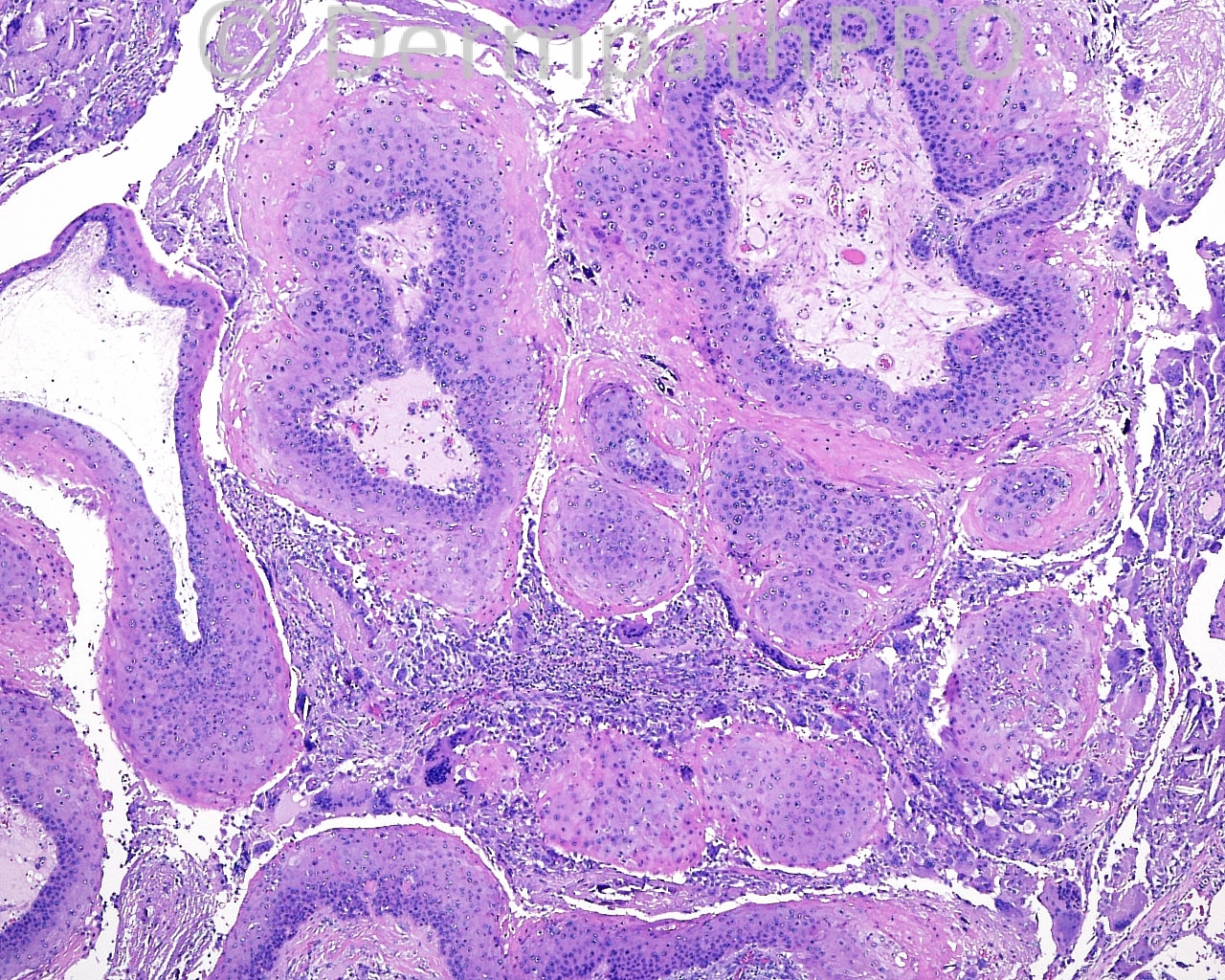
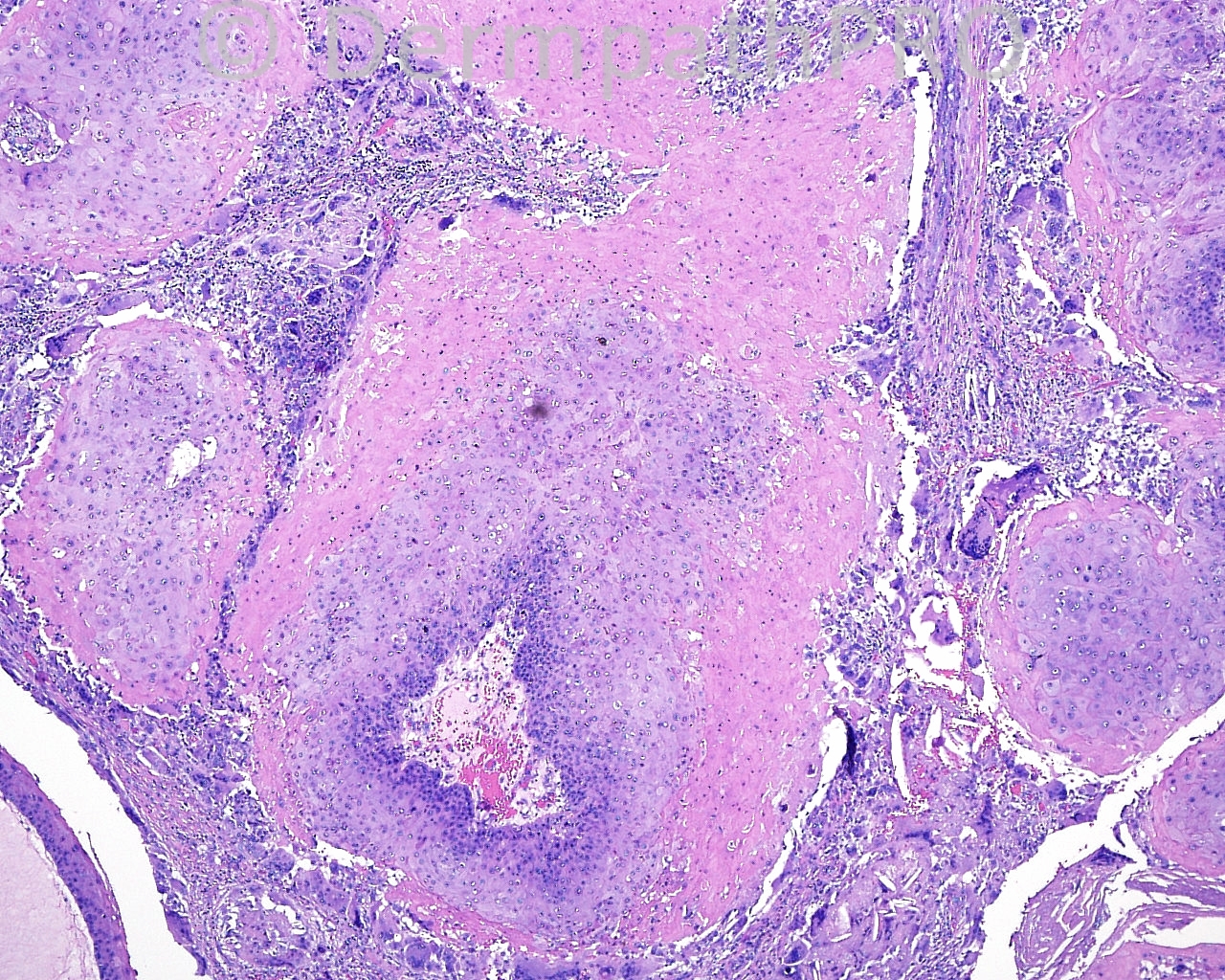
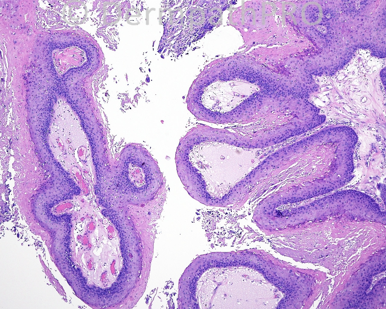
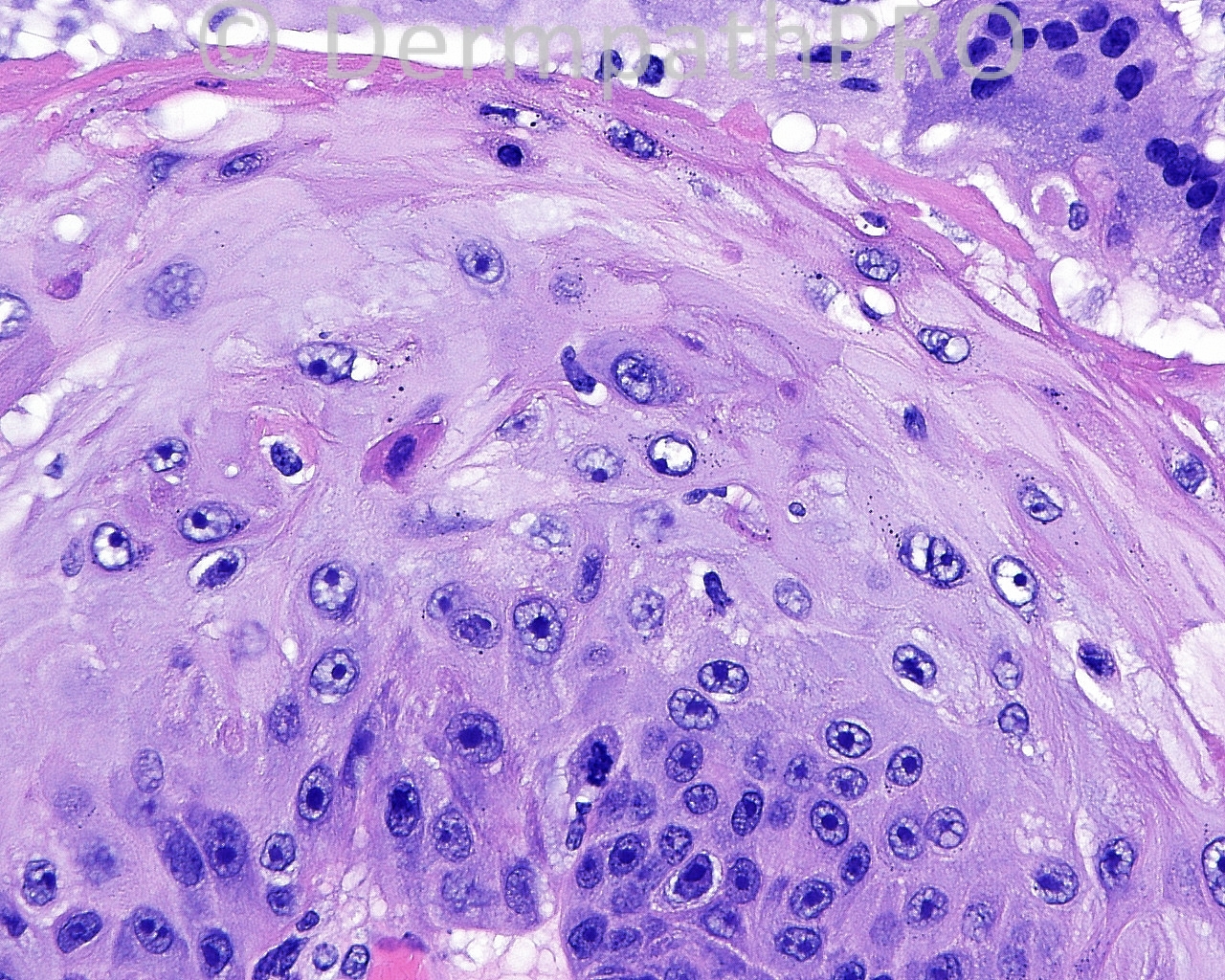
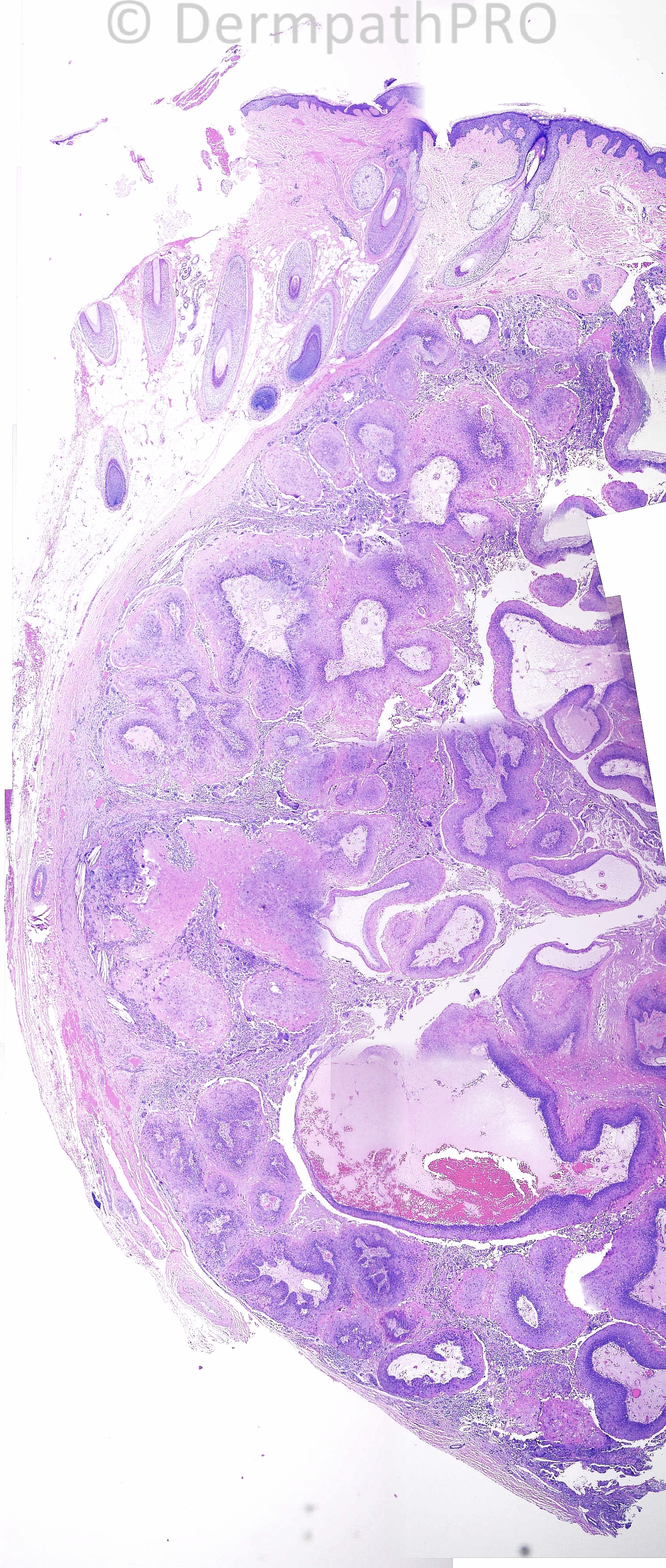
The patient is a 56 year old woman with an excision with margin examination (if malignant), of a lesion taken from the scalp.

Case posted by Dr. Mark Hurt


  Report Record

User Feedback


Arti Bakshi

Posted

Proliferating trichilemmal cyst (pilar tumour of scalp)

Share this comment


Link to comment
share_externally

Squamous cell carcinoma with trichilemmal keratinisation (malignant proliferating trichilemmal cyst).

Share this comment


Link to comment
share_externally

Robledo F. Rocha

Posted

Proliferating tricholemmal tumor. I don’t believe this could be considered a variant of squamous cell carcinoma because these two tumors differ in the cytokeratin profile and in behavior. Proliferating tricholemmal tumors express [url="http://journals.lww.com/amjdermatopathology/pages/articleviewer.aspx?year=2002&issue=08000&article=00011&type=abstract"]fetal and tricholemmal hair follicle phenotypes[/url], suggesting differentiation from hair stem cells.

Share this comment


Link to comment
share_externally

Dr. Mona Abdel-Halim

Posted

Proliferating trichilemmal cyst (pilar tumor)

Share this comment


Link to comment
share_externally

Proliferating trichilemmal tumor. This kind of lesion can evolve to malignancy very rarelly, generally occuring after many relapses. Other histological features that are generally seen in Malignant proliferating trichilemmal tumor area infiltrating borders, high mitotic count and high nuclear grande that we don´t see in this case.
I recently recieved 2 opinion cases of Proliferating trichilemmal tumor that were diagnosed before as SCC.

Share this comment


Link to comment
share_externally

Guest Romualdo

Posted

Proliferating trichilemmal (pilar) cyst (tumor).

Share this comment


Link to comment
share_externally

Guest Jim Davie MD

Posted

Agree with Proliferating pilar (trichilemmal) cyst.

Architecture is spherical/expansile with partial pseudocapsule, gentle displacement/distortion of adnexa, and absence of definitive infiltrative edges that I'd expect in the carcinomatous variant. The prominent foreign body type granulomatous reaction (featuring very large, "ancient type" foreign-body type giant cells with numerous nuclei), suggests a very long-standing lesion with indolent growth. There is sparse mitotic activity with balancing apoptotic activity within the atypical keratinocytes.

Share this comment


Link to comment
share_externally

Dr. Richard Carr

Posted

Looks nicely circumscript so would have gone with majority (benign diagnosis) here I think with a comment on the possibility of local recurrence. Obviously tumours that have been interferred with by the surgical knife can be implanted in to lymphatics so spread to local nodes would not prove malignancy beyond all reasonable doubt. Hopefully in the future we will get the molecular tools to help us.

Share this comment


Link to comment
share_externally


Join the conversation

You can post now and register later. If you have an account, sign in now to post with your account.

Guest
Add a comment...

×   Pasted as rich text.   Paste as plain text instead

  Only 75 emoji are allowed.

×   Your link has been automatically embedded.   Display as a link instead

×   Your previous content has been restored.   Clear editor

×   You cannot paste images directly. Upload or insert images from URL.

Loading...

×
×
  • Create New...