Jump to content
In this section we have spot diagnoses posted on a daily basis since June 2010, now over 4000! You can review the archived cases and read the suggested diagnoses by users and the final comment by the contributors.
Case are uploaded each week day by 10 am UK time with the correct diagnosis will generally be posted at 8 pm UK time. Why not view the most recent spot diagnosis and proffer a diagnosis?

Case Number : Case 1552 - 07 June Posted By: Guest

Please read the clinical history and view the images by clicking on them before you proffer your diagnosis.
Submitted Date :
   (0 reviews)

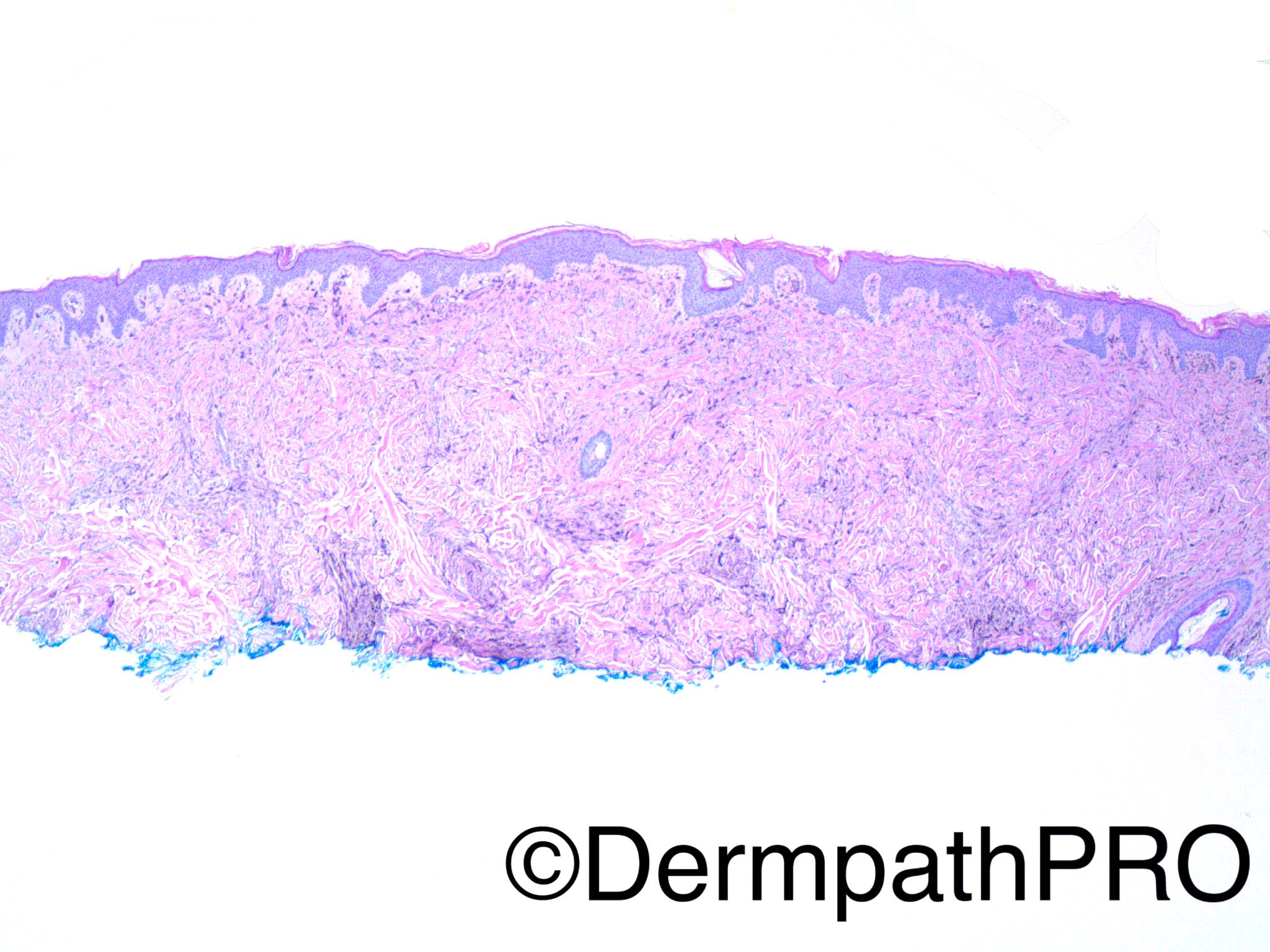
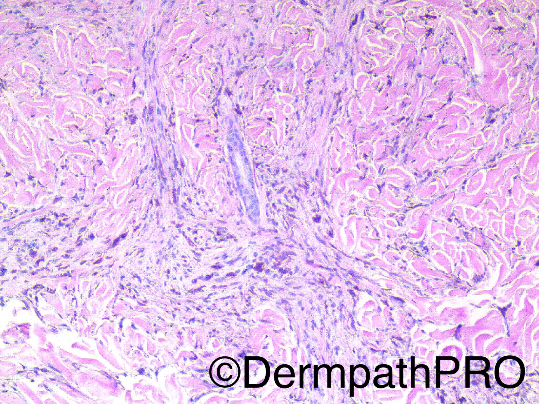
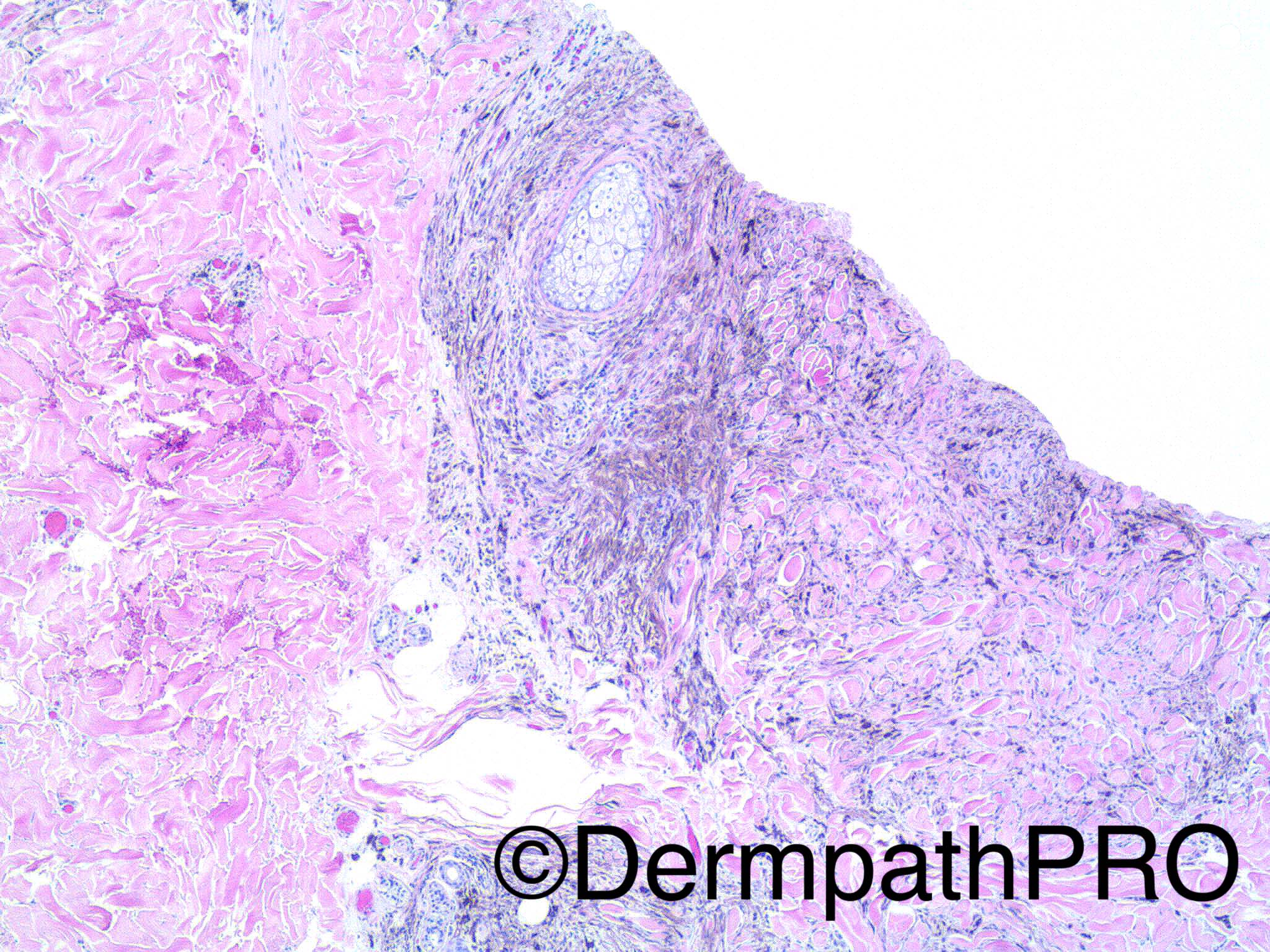
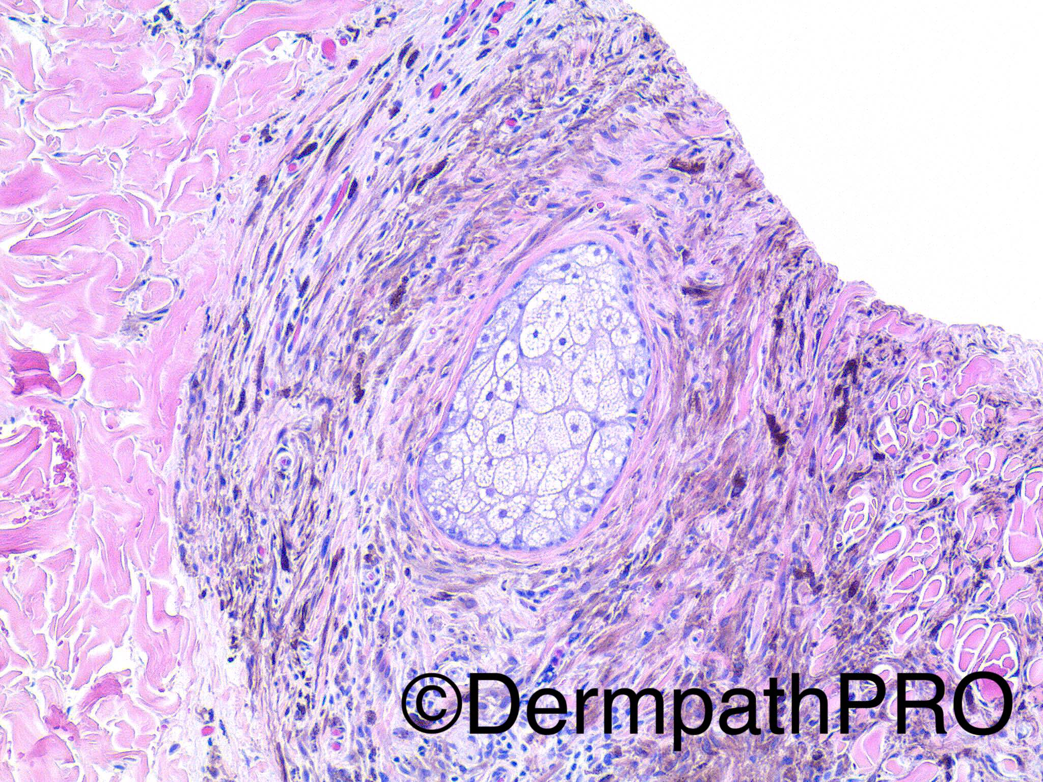
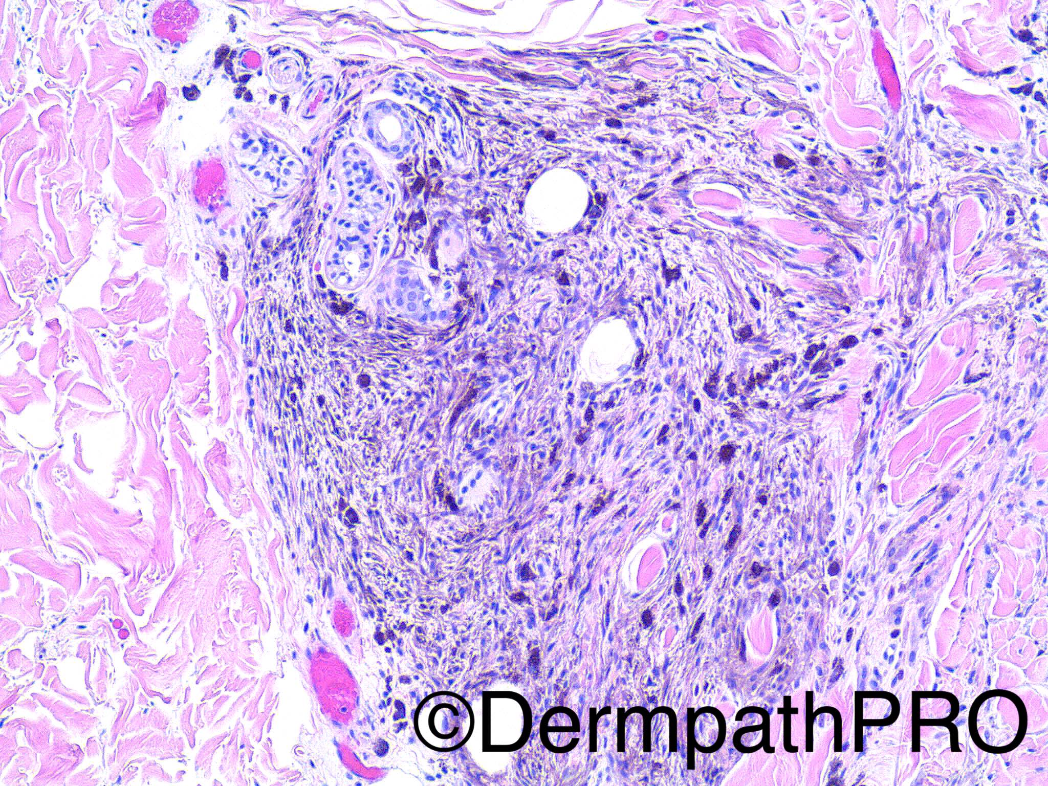
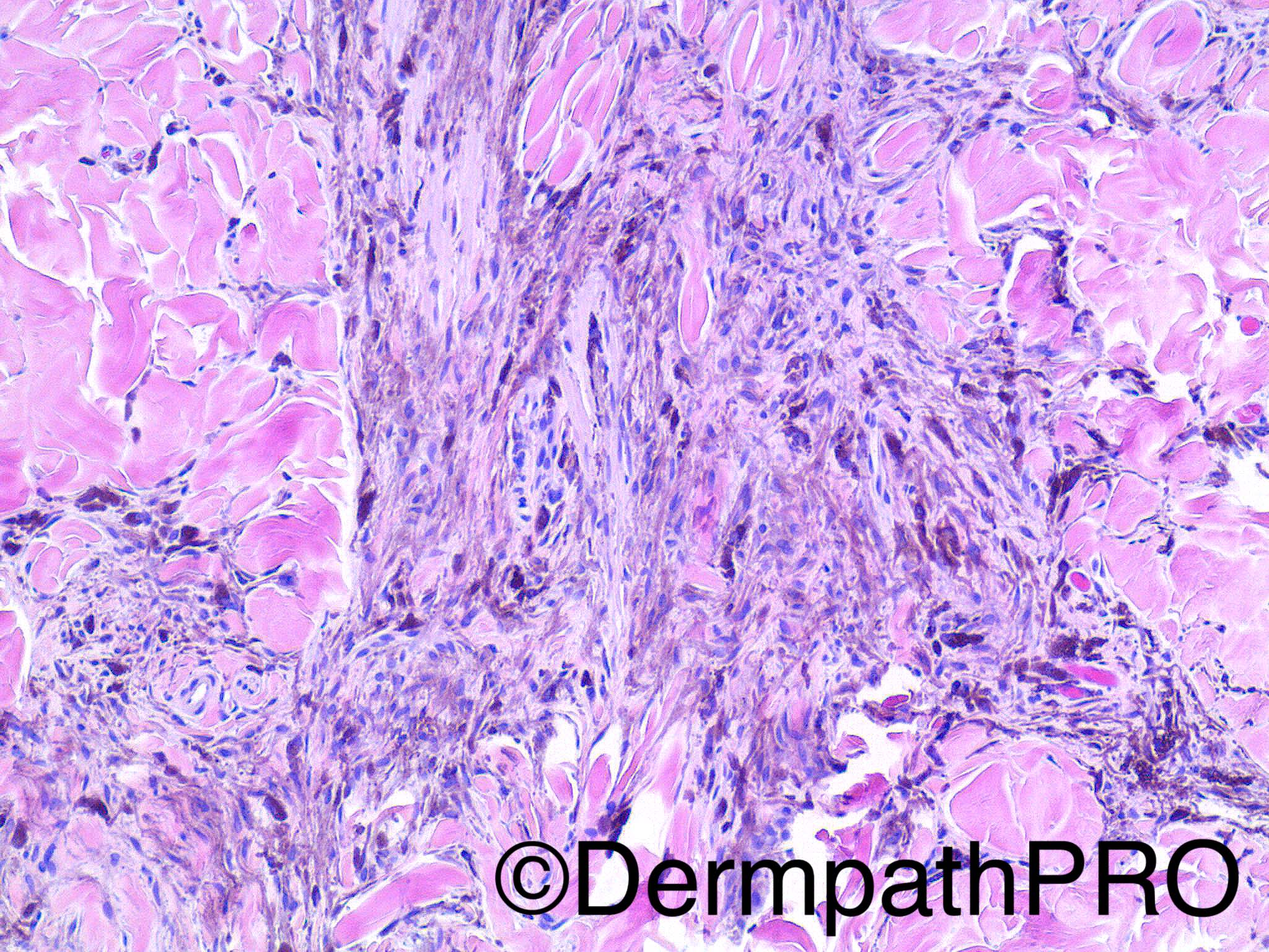
29 year old woman with 5 mm pigmented lesion on the left buttocks.

Dr Uma Sundram.


  Report Record

User Feedback


Raul Perret

Posted

Looks like blue nevus but the limits are compromised so should state it to get a full excision (for complete evaluation), these lesions can recur and be quite deep, particularly in that location

Share this comment


Link to comment
share_externally

Cellular blue naevus, which would correlate with the site. 

Share this comment


Link to comment
share_externally

vincenzo polizzi

Posted

Deep penetrating nevus.

Share this comment


Link to comment
share_externally

Nitin Khirwadkar

Posted

Would favour a cellular blue naevus, typical site.

Share this comment


Link to comment
share_externally

vincenzo polizzi

Posted

Let me explain why favor a DPN. -the superficial derma is totally busy while the deepest one shows a focal free zone, that makes me think about some wedge shaped pattern deeply ( the biopsy catched only the superficial part of the lesion ). -there are many periadnexial growth patterns deeply. -I don't see any poorly pigmented nest together the deeply pigmented fascicles, so typical of cellular blue nevus.

Share this comment


Link to comment
share_externally

Arti Bakshi

Posted

Cellular blue naevus.

There is clearly a spindle/dendritic melanocytic component, which puts the lesion more in the blue naevus category than DPN…I think.

Periadnexal spread is a feature of both.

Share this comment


Link to comment
share_externally

vincenzo polizzi

Posted

Ok. I'm finally learning the dd between BN and DPN. Thanks Arti!

Share this comment


Link to comment
share_externally

Guest Charlie Keen

Posted

Incomplete shave biopsy of BN.

Share this comment


Link to comment
share_externally

Uma Sundram

Posted

I called this a cellular blue nevus and asked for a complete excision. This lesion addresses the difficulty of separating blue nevi that are on the cellular end of things and the so-called cellular blue nevus. I have had some issues with clinicians misinterpreting these lesions as benign (with a local recurrence rate of zero), so have called these cellular blue nevi to facilitate a re excision.  Dermal dendritic melanocytes characterize these lesions, but plenty of overlap among CBN, DPN and pigmented Spitz nevi that are not as epithelioid.

Share this comment


Link to comment
share_externally

Dr. Richard Carr

Posted

Uma,  I'm slightly confused.  Cellular blue naevi are benign and any benign tumour incompletely excised may recur locally (but not always). What is the reason for recommending re-excision - are you concerned for example the lesion may have a risk for malignant transformation or that the diagnosis is not 100% certain and you would rather see it completely excised with clear margins for that reason - analogous to Spitz tumours?

Share this comment


Link to comment
share_externally


Join the conversation

You can post now and register later. If you have an account, sign in now to post with your account.

Guest
Add a comment...

×   Pasted as rich text.   Paste as plain text instead

  Only 75 emoji are allowed.

×   Your link has been automatically embedded.   Display as a link instead

×   Your previous content has been restored.   Clear editor

×   You cannot paste images directly. Upload or insert images from URL.

Loading...

×
×
  • Create New...