Jump to content
In this section we have spot diagnoses posted on a daily basis since June 2010, now over 4000! You can review the archived cases and read the suggested diagnoses by users and the final comment by the contributors.
Case are uploaded each week day by 10 am UK time with the correct diagnosis will generally be posted at 8 pm UK time. Why not view the most recent spot diagnosis and proffer a diagnosis?

Case Number : Case 1556 - 13 June Posted By: Guest

Please read the clinical history and view the images by clicking on them before you proffer your diagnosis.
Submitted Date :
   (0 reviews)

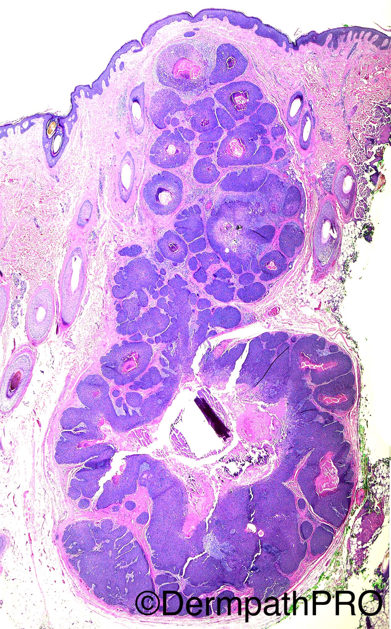
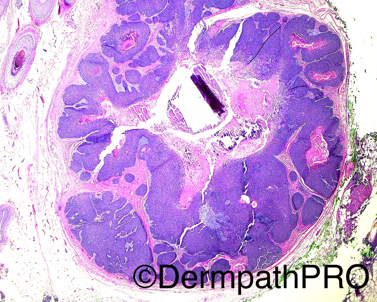
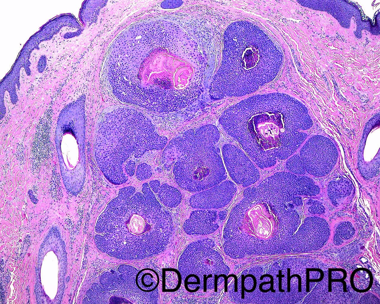
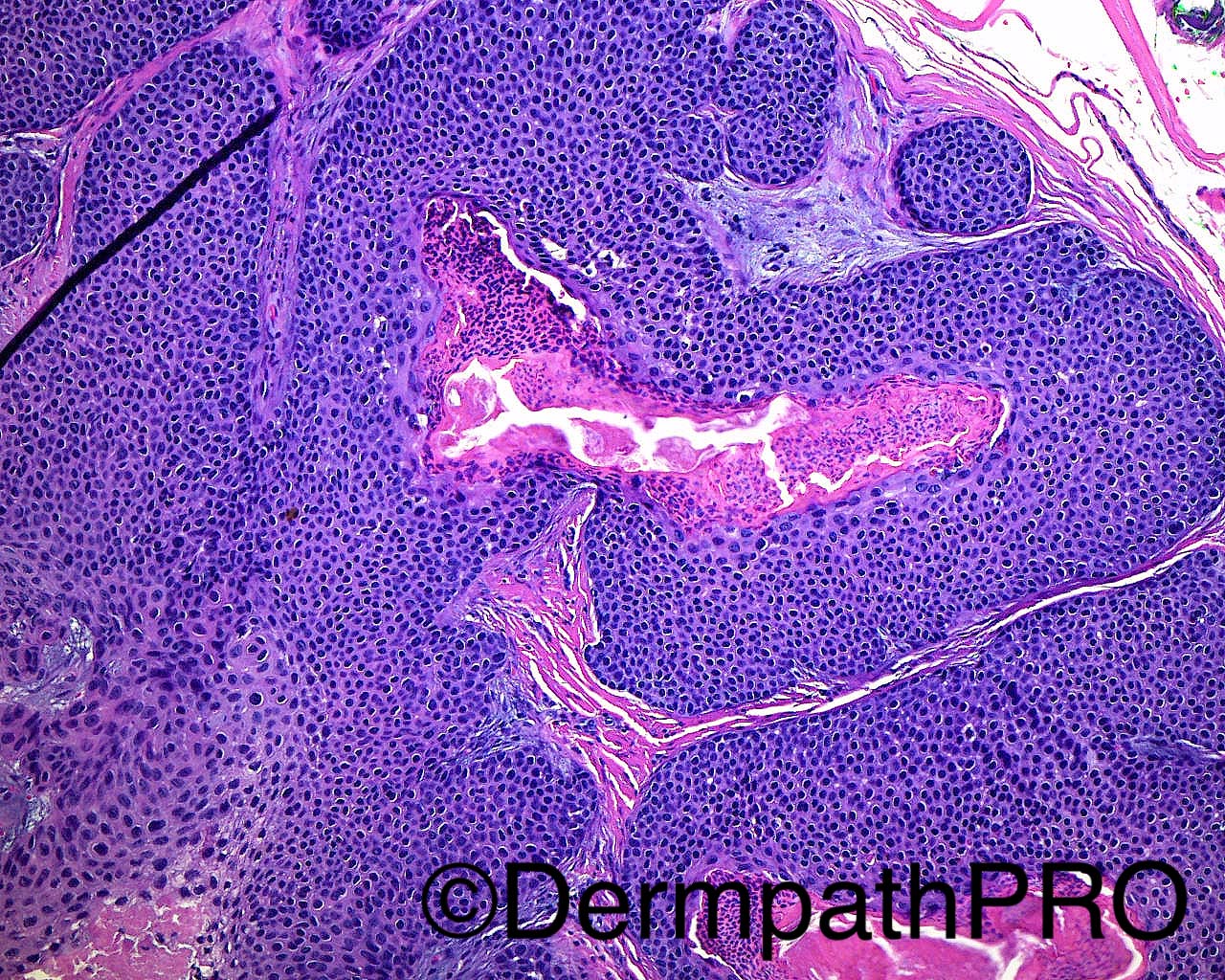
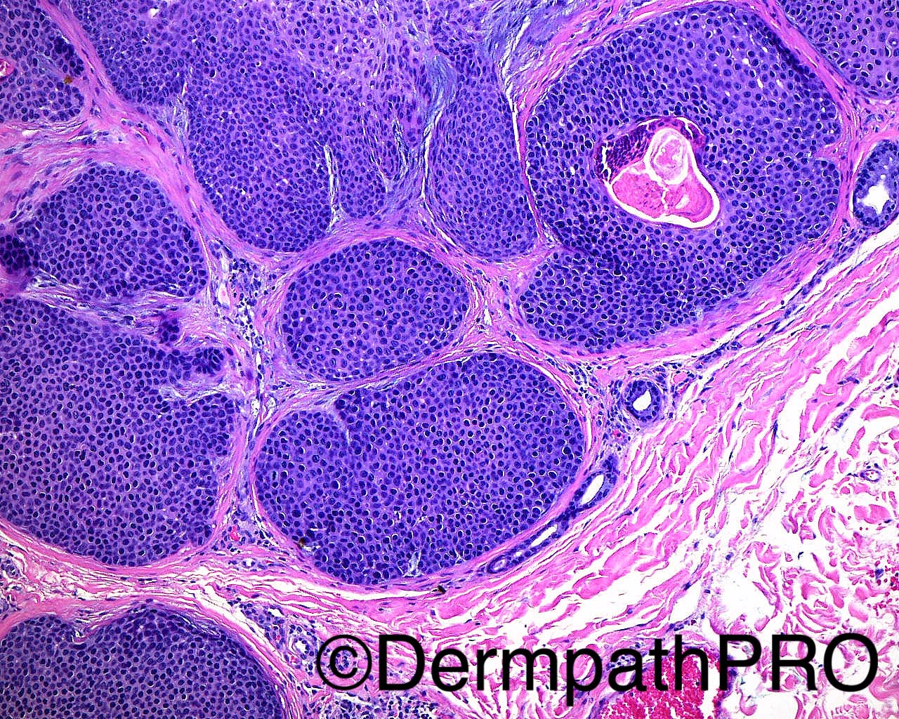
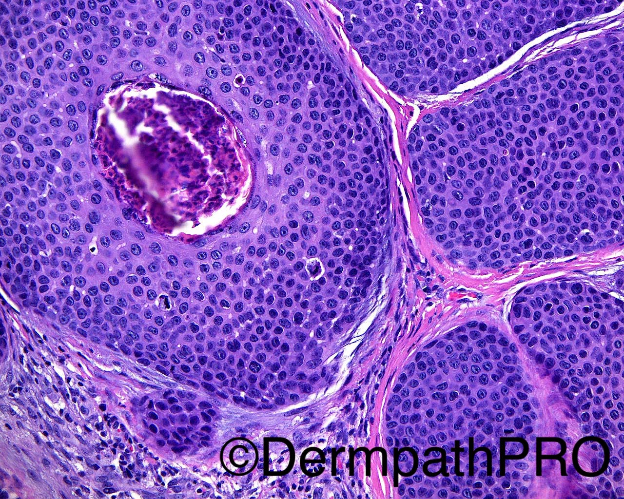
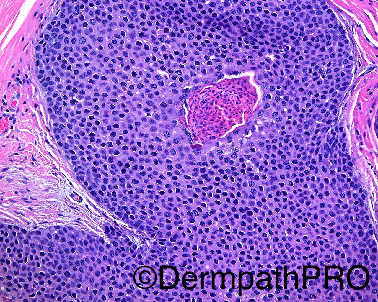
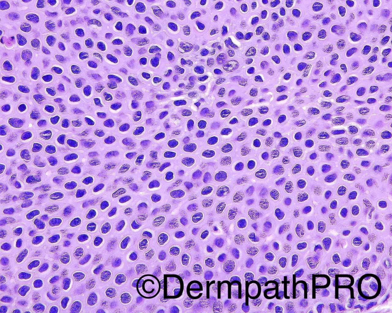
The patient is a 48-year-old woman with a biopsy of a scalp mass. This was a consult case with the following history: "The clinical impression is basal cell carcinoma, I do not know if there was a previous biopsy. As you can see by my provisional report, I am confused by the growth pattern of this lesion and am considering proliferating trichilemmal tumor and squamous cell carcinoma."
Dr Mark Hurt


  • Like 1
  Report Record

User Feedback


I agree with dermal duct tumour, as the cells look poroid, on the condition that ducts are demonstrated by immunohistochemistry. 

Share this comment


Link to comment
share_externally

Nitin Khirwadkar

Posted

Agree. Can't see ducts on H&E, but very poroid looking cells.

Share this comment


Link to comment
share_externally

Arti Bakshi

Posted

I think better categorised as a poroid hidradenoma, in view of lack of connection to surface and absence of obvious ductal differentiation.

Share this comment


Link to comment
share_externally

Dr. Richard Carr

Posted

I find this lesion very odd. No ducts although agree poroid-like.  I see foci of subtle bowenoid dysplasia (including individual cell necrosis and atypical mitotic figures) developing into confluent parakeratosis. Profile is reminiscent of pilar sheath acanthoma. Could it be bowenoid changes within pilar sheath acanthoma? On second thoughts a low-grade hidradenocarcinoma is also a possibility.

Share this comment


Link to comment
share_externally

Raul Perret

Posted

To be honest my first impression was that of a neoplasm with predominant follicular differentiation. We have small cells poroid like with abrupt keratinization and calcification, individual apoptosis, some mitotic figures. There are some pictures where the cells look with a bit more cytoplasm and clearer. I actually thought that the suggestion  of the referring pathologist was quite good I think this is a lesion in the spectrum of proliferating trichilemmal tumor.

Share this comment


Link to comment
share_externally

vincenzo polizzi

Posted

I think there isn't any follicular differentiation, but a very ductal/poroid differentiation! May be i'm wrong but in Fig5/6/7/8 i see a ductal-cystic differentiation and the cells are too poroid...

Share this comment


Link to comment
share_externally

Raul Perret

Posted

I think there isn't any follicular differentiation, but a very ductal/poroid differentiation! May be i'm wrong but in Fig5/6/7/8 i see a ductal-cystic differentiation and the cells are too poroid...

Look at the last picture, the cells look ORS like, there is individual cell apoptosis, and on the left side of the picture there is abrupt keratinization and there are some granular layer like vestiges (variable size keratohyaline granules). I dont know it really looks like the neoplasm has follicular differentiation to my eyes

Share this comment


Link to comment
share_externally


Join the conversation

You can post now and register later. If you have an account, sign in now to post with your account.

Guest
Add a comment...

×   Pasted as rich text.   Paste as plain text instead

  Only 75 emoji are allowed.

×   Your link has been automatically embedded.   Display as a link instead

×   Your previous content has been restored.   Clear editor

×   You cannot paste images directly. Upload or insert images from URL.

Loading...

×
×
  • Create New...