Jump to content
In this section we have spot diagnoses posted on a daily basis since June 2010, now over 4000! You can review the archived cases and read the suggested diagnoses by users and the final comment by the contributors.
Case are uploaded each week day by 10 am UK time with the correct diagnosis will generally be posted at 8 pm UK time. Why not view the most recent spot diagnosis and proffer a diagnosis?

Case Number : Case 972 - 14th March Posted By: Guest

Please read the clinical history and view the images by clicking on them before you proffer your diagnosis.
Submitted Date :
   (0 reviews)

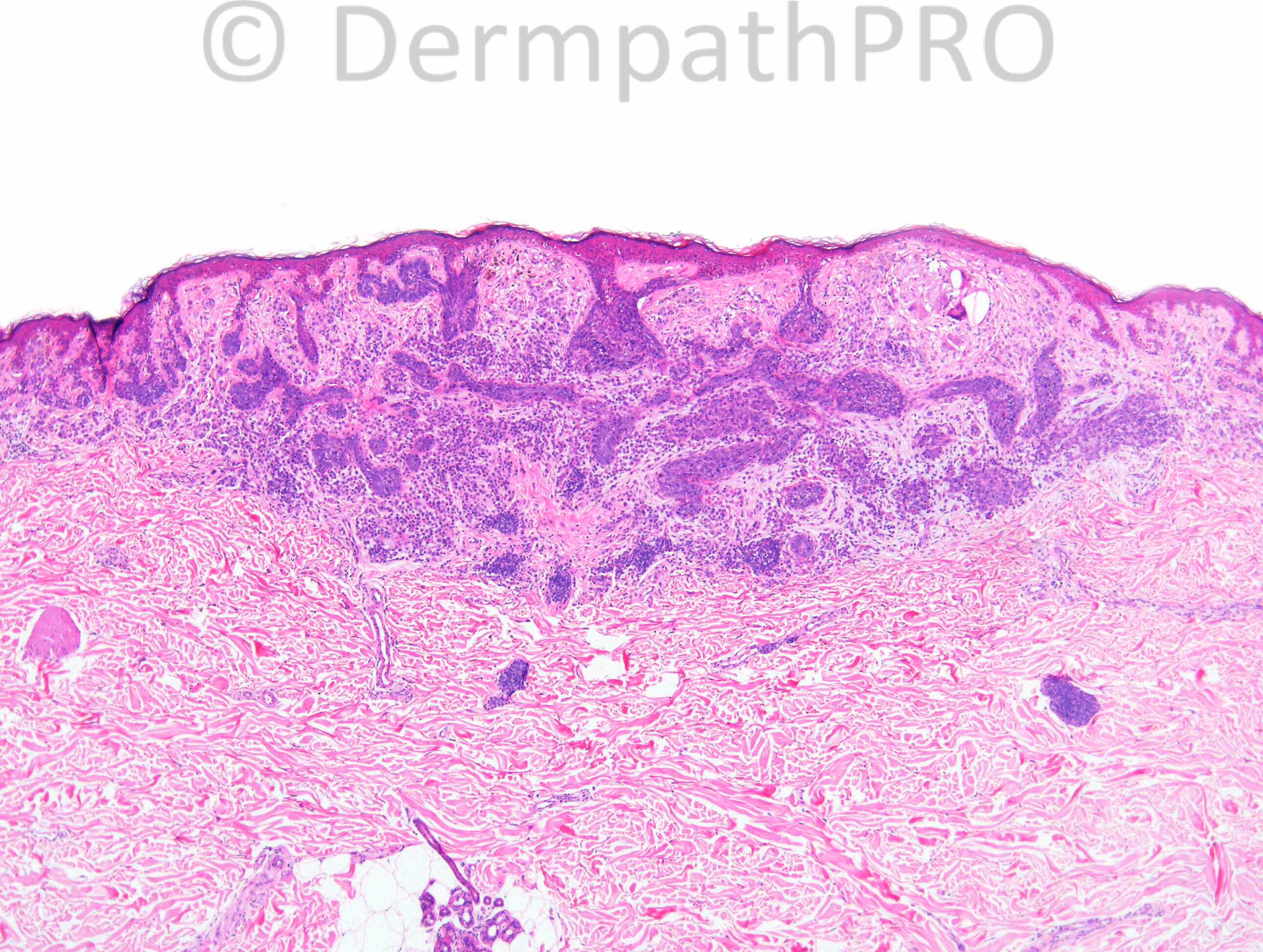
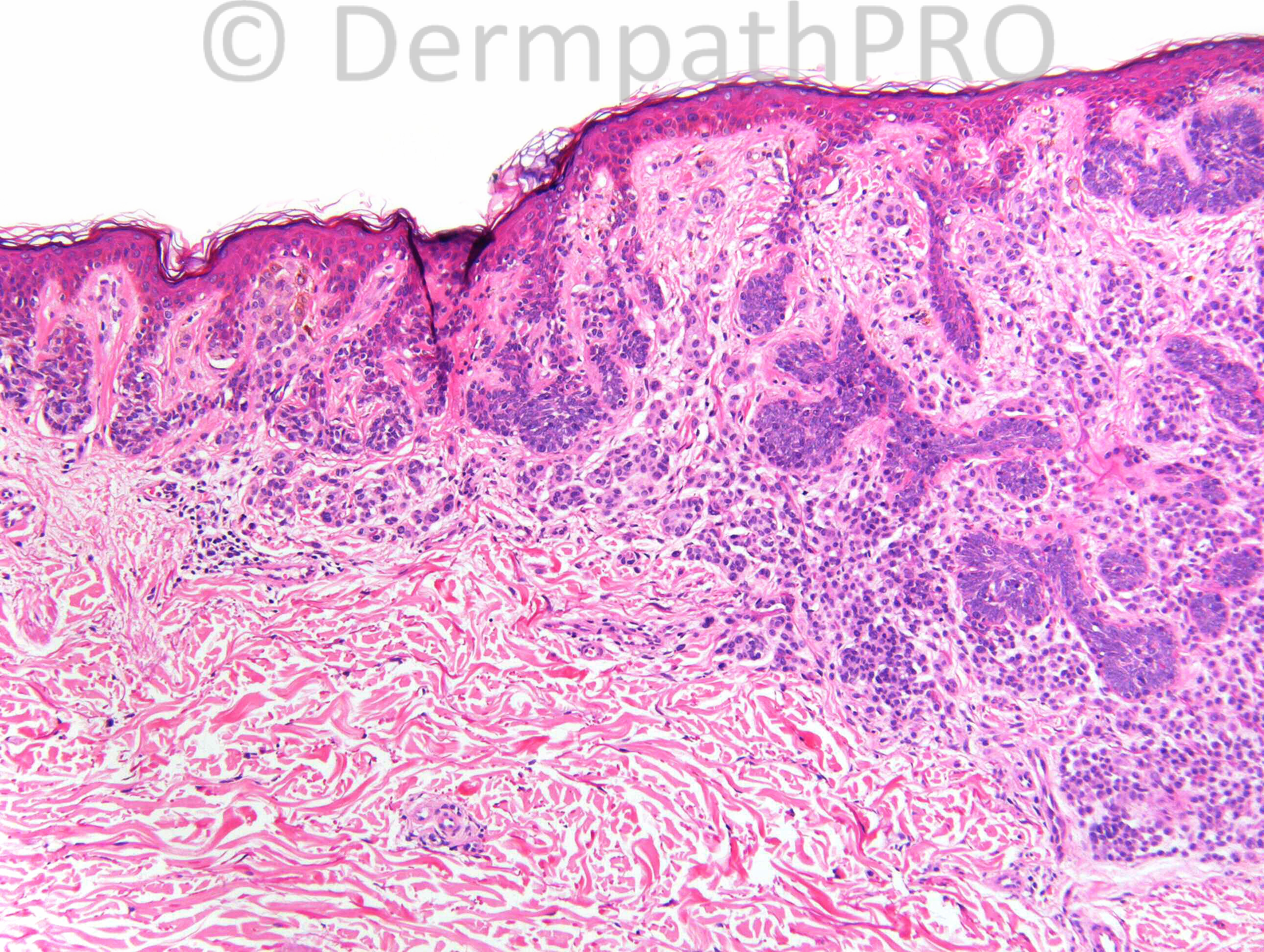
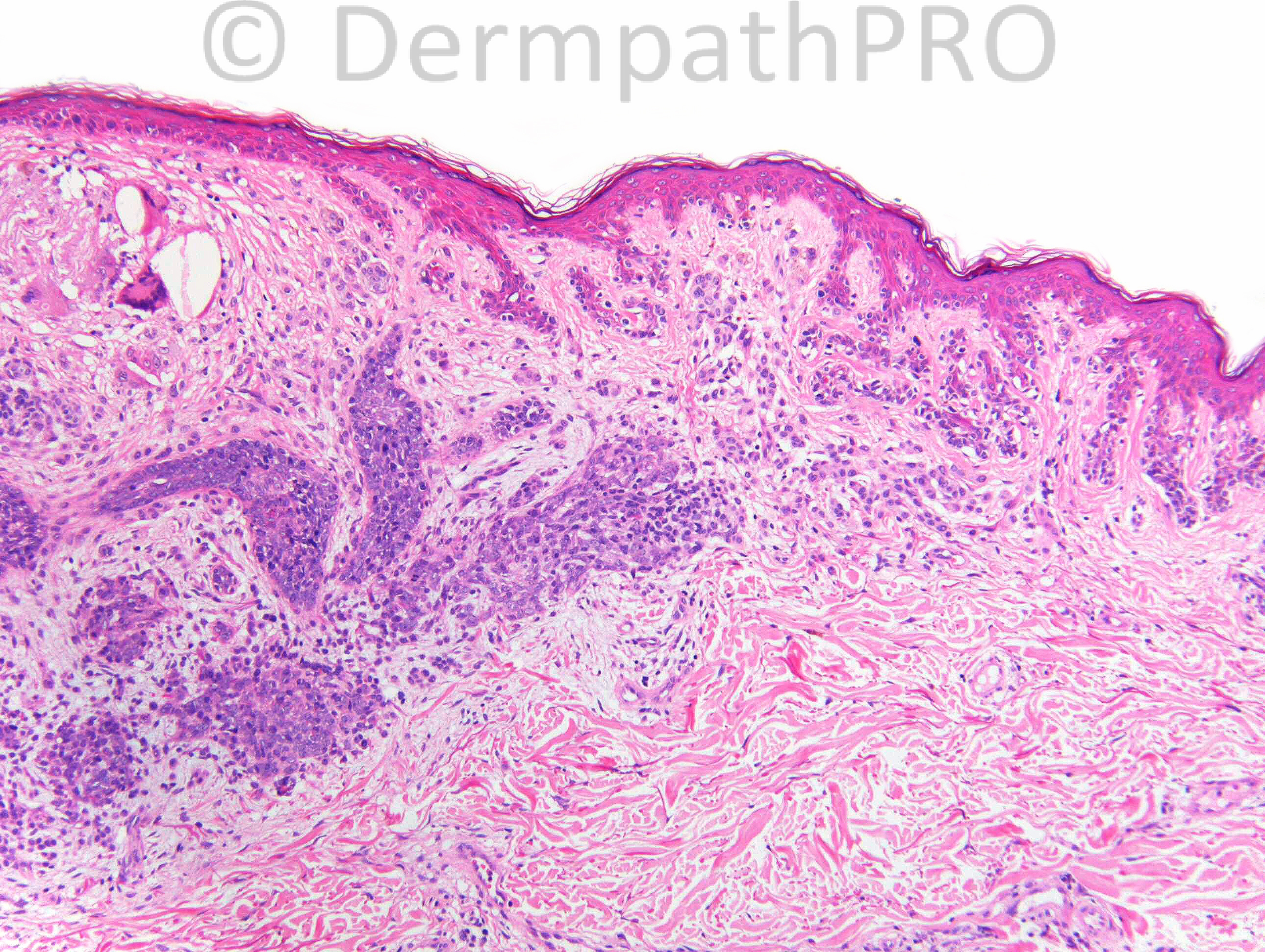
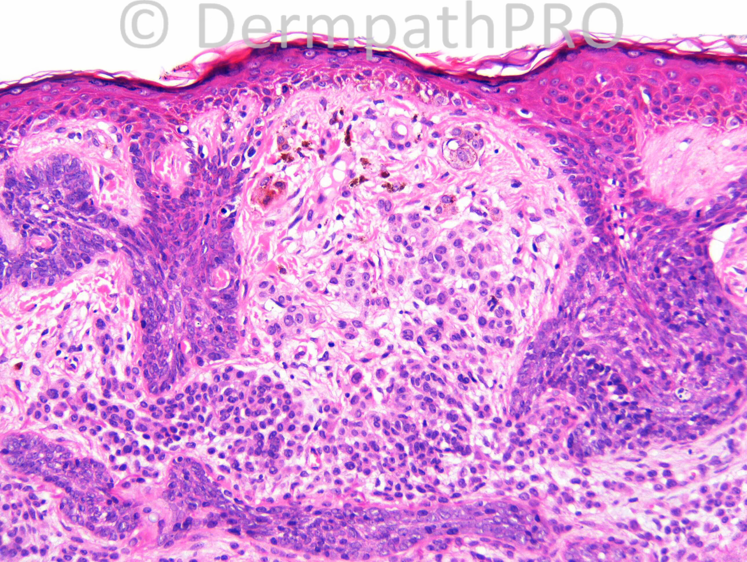
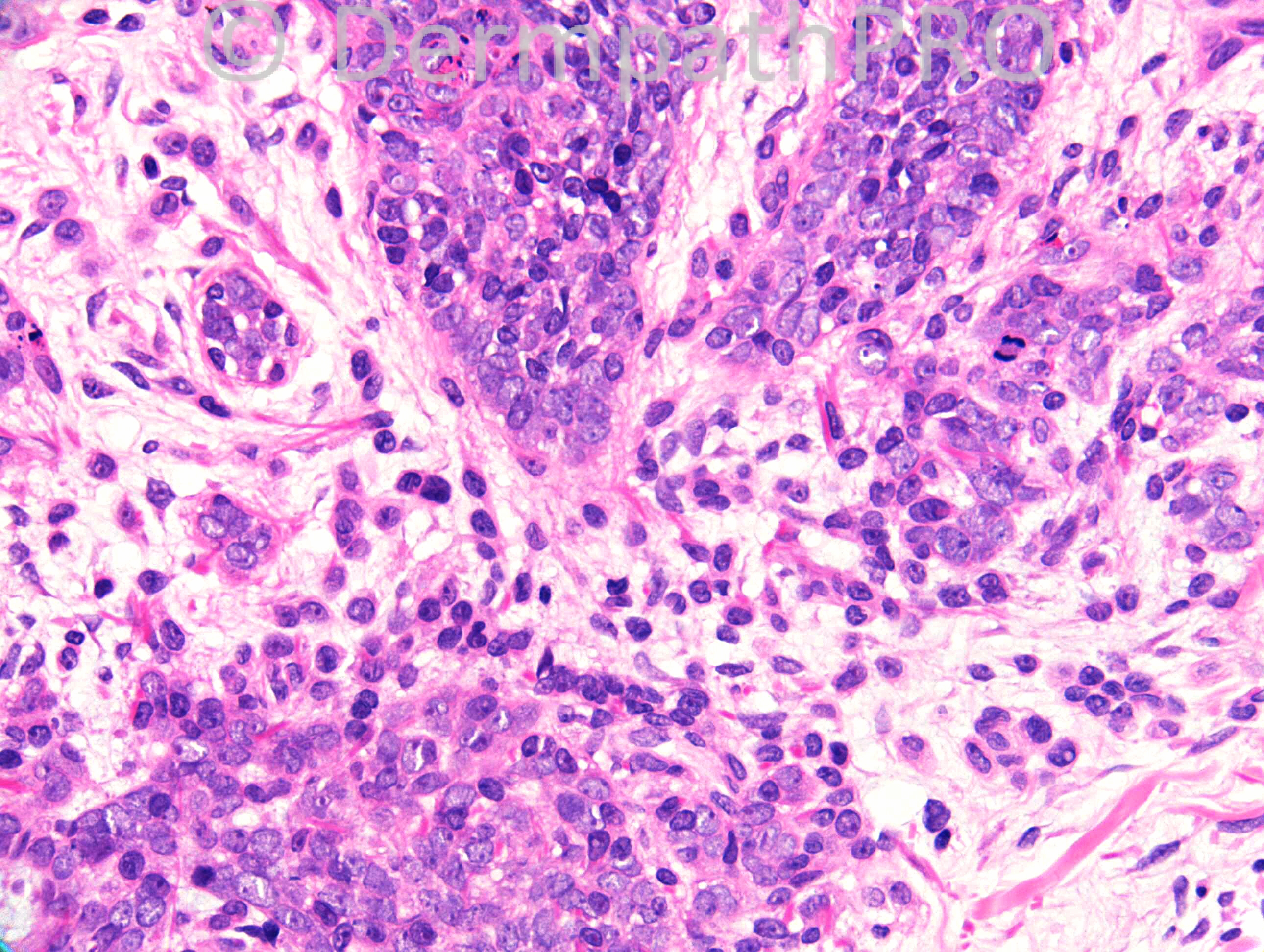
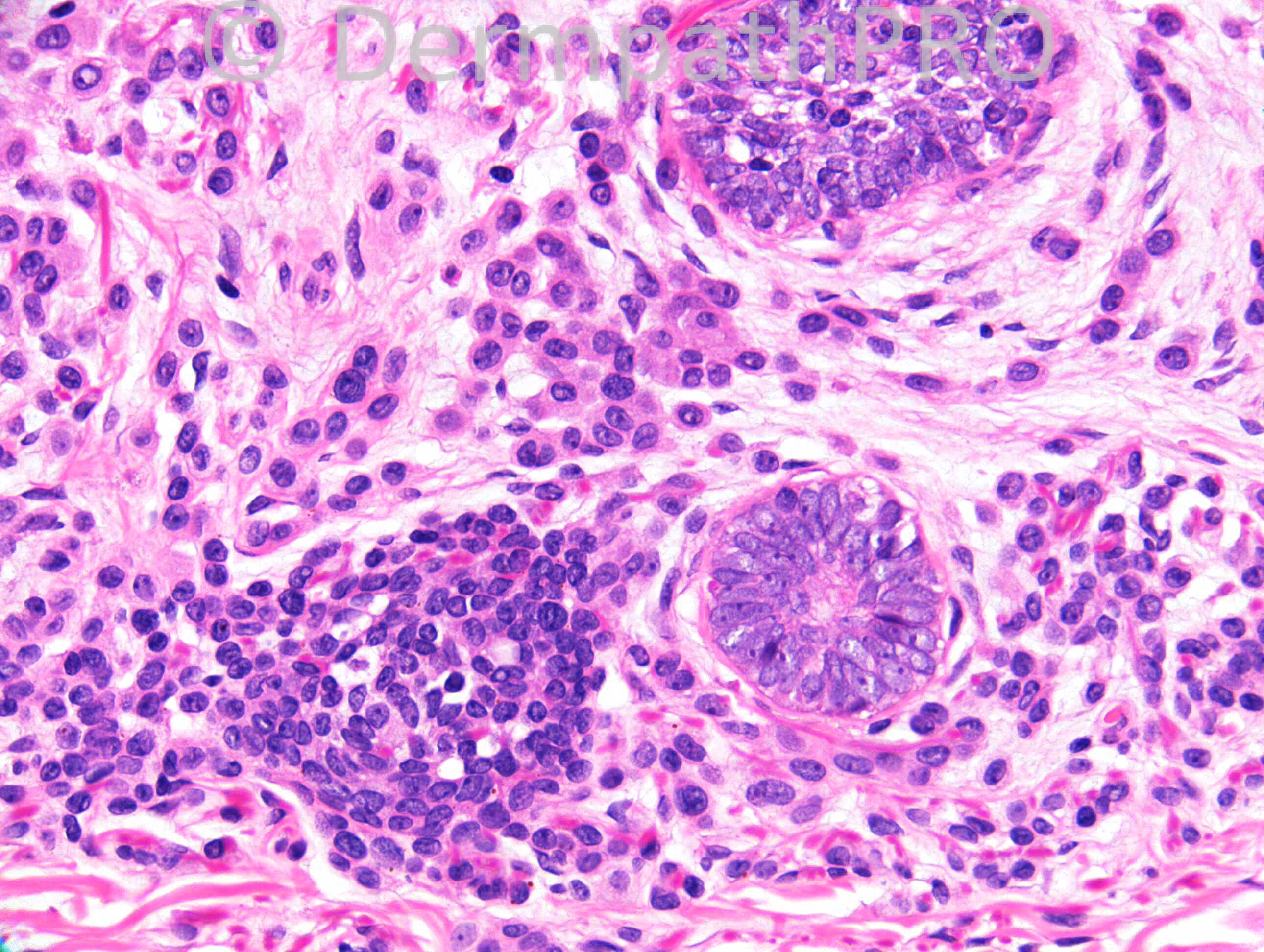
F51. Atypical naevus syndrome, 8 x 6 mm irregularly pigmented naevus R
chest ?dysplastic.

Case posted by Dr. Richard Carr.


  Report Record

User Feedback


Composite basal cell carcinoma and dysplastic naevus.

Share this comment


Link to comment
share_externally

Dr. Mona Abdel-Halim

Posted

Collision between BCC and dysplastic nevus.

Share this comment


Link to comment
share_externally

Eman El-Nabarawy

Posted

METRO( MElanocytic tumor+TRichObastoma). Collision of dysplastic nevus with trichobastoma.

Share this comment


Link to comment
share_externally

Guest Tiberiu Tebeica

Posted

melanocytic nevus associated with trichoepithelioma

Share this comment


Link to comment
share_externally

Dr. Richard Carr

Posted

I agree with nearly half of you on nearly half the diagnoses! IHC to follow hopefully from Dermpathpro colleagues.

Share this comment


Link to comment
share_externally

Dr. Mona Abdel-Halim

Posted

Before the immunos, I would like to throw another suggestion: basomelanocytic tumor. I have been looking at the images all the day and I think melanocytes are colonizing the basaloid islands in many areas. I think this is more appropriate than a collision lesion.

Share this comment


Link to comment
share_externally

Dr. Richard Carr

Posted

[img]https://dermpathpro.com/uploads/spot_diagnosis_comment_img/Case972_BerEp4.jpg[/img]

[img]https://dermpathpro.com/uploads/spot_diagnosis_comment_img/Case972_CD10.jpg[/img]

[img]https://dermpathpro.com/uploads/spot_diagnosis_comment_img/Case972_S100.jpg[/img]

Share this comment


Link to comment
share_externally

Robledo F. Rocha

Posted

Combination of lentiginous compound nevus and small nodular basal cell carcinoma with follicular differentiation.

Share this comment


Link to comment
share_externally

Composite basal cell carcinoma and melanoma.

Share this comment


Link to comment
share_externally

amiratawdy

Posted

i like the suggestion of dear Mona of basomelanocytic tumor

Share this comment


Link to comment
share_externally

Dr. Richard Carr

Posted

Sorry for delay.

This was a [b]basal cell carcinoma arising in a pre-existing (mildly dysplastic) melanocytic naevus[/b]. H&E features were typical of BCC. CD10 confirmed absence of papillary mesenchymal cells and typical epithelial staining pattern of BCC that we have found pretty reliable in distinguishing BCC from TE (Pham et al 2006, Turnbull et al 2013). Two of your made the correct diagnosis at the outset than changed your minds. Well done Robledo!

My view of baso- squamo- melanocytic tumours are a mixed group of tumours that according to Satter et al 2009) include:
1) basaloid or squamous tumours with [b]reactive dendritic melanocytes[/b] (e.g. melanocytic matricoma, pigmenthed trichoblastoma, pigmented BCC),
2) simple [b]collision[/b] of two lesions that are clearly demarcated from each other e.g. BCC next to or abutting a naevus
3) basaloid or squamous tumours, more intimately, [b]colonised[/b] by other melanocytic neoplasms e.g. BCC colonised by lentigo maligna (the neoplastic cells of the melanoma colonising the epithelium of the epithelial neoplasm. Taibjee et al 2007) - I did not see melanocytic nests in the BCC in the case here.
4) [b]Combined[/b] tumour (as in this case) when the two lesions are closely intermingled but identifiably separate (intertwined) populations.
5) Very rare [b]bi-phenotypic[/b] tumours with mixed epithelial-melanocytic differentiation (really would have to confirm by electron microscopy or IHC showing dual immunophenotype in individual cells e.g. p63/melanA dual staining) and assumes a common pre-cursor / stem cell of origin.
Note: Re-edited!

[b]References[/b]
Pham TT, Selim MA, Burchette JL Jr, Madden J, Turner J, Herman C. CD10 expression in trichoepithelioma and basal cell carcinoma. J Cutan Pathol. 2006 Feb;33(2):123-8.

Taibjee SM, Gee BC, Sanders DS, Smith A, Carr RA. Lentigo maligna involving the tumour nests and stroma of a nodular basal cell carcinoma. Br J Dermatol. 2007 Jul;157(1):184-8.

Satter EK, Metcalf J, Lountzis N, Elston DM. Tumors composed of malignant epithelial and melanocytic populations: a case series and review of the literature. J Cutan Pathol. 2009 Feb;36(2):211-9.

Turnbull N, Ghumra W, Vella J, Sanders DSA, Taibjee SM, Carr RA. An immunohistochemical study of trichoepithelioma and trichoepithelioma-like basal cell carcinoma. Poster ISDP Meeting Florence 2013.

Share this comment


Link to comment
share_externally

Dear Dr Carr

Could you possibly elaborate more on this point. You said:

"CD10 confirmed absence of papillary mesenchymal cells and typical epithelial staining pattern of BCC that we have found pretty reliable in distinguishing BCC from TE . . ."

Immunohistochemically speaking, do rely solely on CD10 in separating BCCs and TEs?

Thank you.

Share this comment


Link to comment
share_externally

Dr. Richard Carr

Posted

Thanks Dr Abdul-Kadir for the further enquiry. The H&E is still the best stain but we do use IHC to hold our hands in difficult cases (often small biopsies). CD10 is generally pretty good at highlighting papillary mesencymal cells (PMC, the histological hallmark) of TE/TB and often shows quite localised peripheral epithelial staining at the site of the PMC. In BCC there may be non-specific CD10 staining of inflammatory stroma but not the tight peritumoural cuffing typical of TE/TB and CD10 is usually more widespread epithelial staining in BCC as here. I am pasting the Florence abstract that we are currently drafting for full publication. In summary we find CD10 helpful in most, but no all, cases.

Turnbull N, Ghumra W, Vella J, Sanders DSA, Taibjee SM, Carr RA. An immunohistochemical study of trichoepithelioma and trichoepithelioma-like basal cell carcinoma. Poster ISDP Meeting Florence 2013.

Background: Trichoepitheliomas (TE) are benign skin adnexal neoplasms which may be difficult to differentiate from basal cell carcinoma (BCC).
Methods: Comparative immunohistochemical study of 58 TE and 39 TE-like BCC for BerEP4 and CD10 and with Cam5.2 and CK20 for Merkel cells (MC). The percentage of moderate to strong staining was estimated for each antibody.
Results: CD10 was expressed in stroma and or epithelium in 96% of TE, with a stroma predominant staining pattern in 76.5%. In contrast although 89% of TE-like BCC expressed CD10 a stromal predominant pattern was not seen. Moderately numerous MC (>5 MC in basaloid epithelium of the tumour) were noted in TE (13/39, 33%) and was also noted in 3 of TE-like BCC (3/23, 13%). BerEP4 staining was non discriminatory.
Discussion: A stromal predominant staining pattern for CD10 was specific and also fairly sensitive for TE and may be a useful adjunct to diagnosis in difficult cases. The presence of moderately numerous MC lacks specificity in the differential diagnosis of TE and TE-like BCC. BerEP4 was not helpful.

Share this comment


Link to comment
share_externally

Thanks for your detailed reply, Dr Carr. As soon as I finished reading your original post, a trichoepithelioma (I think) landed on my desk. I've ordered a CD10 &, hopefully, it’ll do what it’s supposed to do. Thanks again.

Share this comment


Link to comment
share_externally


Join the conversation

You can post now and register later. If you have an account, sign in now to post with your account.

Guest
Add a comment...

×   Pasted as rich text.   Paste as plain text instead

  Only 75 emoji are allowed.

×   Your link has been automatically embedded.   Display as a link instead

×   Your previous content has been restored.   Clear editor

×   You cannot paste images directly. Upload or insert images from URL.

Loading...

×
×
  • Create New...