Jump to content
In this section we have spot diagnoses posted on a daily basis since June 2010, now over 4000! You can review the archived cases and read the suggested diagnoses by users and the final comment by the contributors.
Case are uploaded each week day by 10 am UK time with the correct diagnosis will generally be posted at 8 pm UK time. Why not view the most recent spot diagnosis and proffer a diagnosis?

Case Number : Case 1009 - 6th May Posted By: Guest

Please read the clinical history and view the images by clicking on them before you proffer your diagnosis.
Submitted Date :
   (0 reviews)

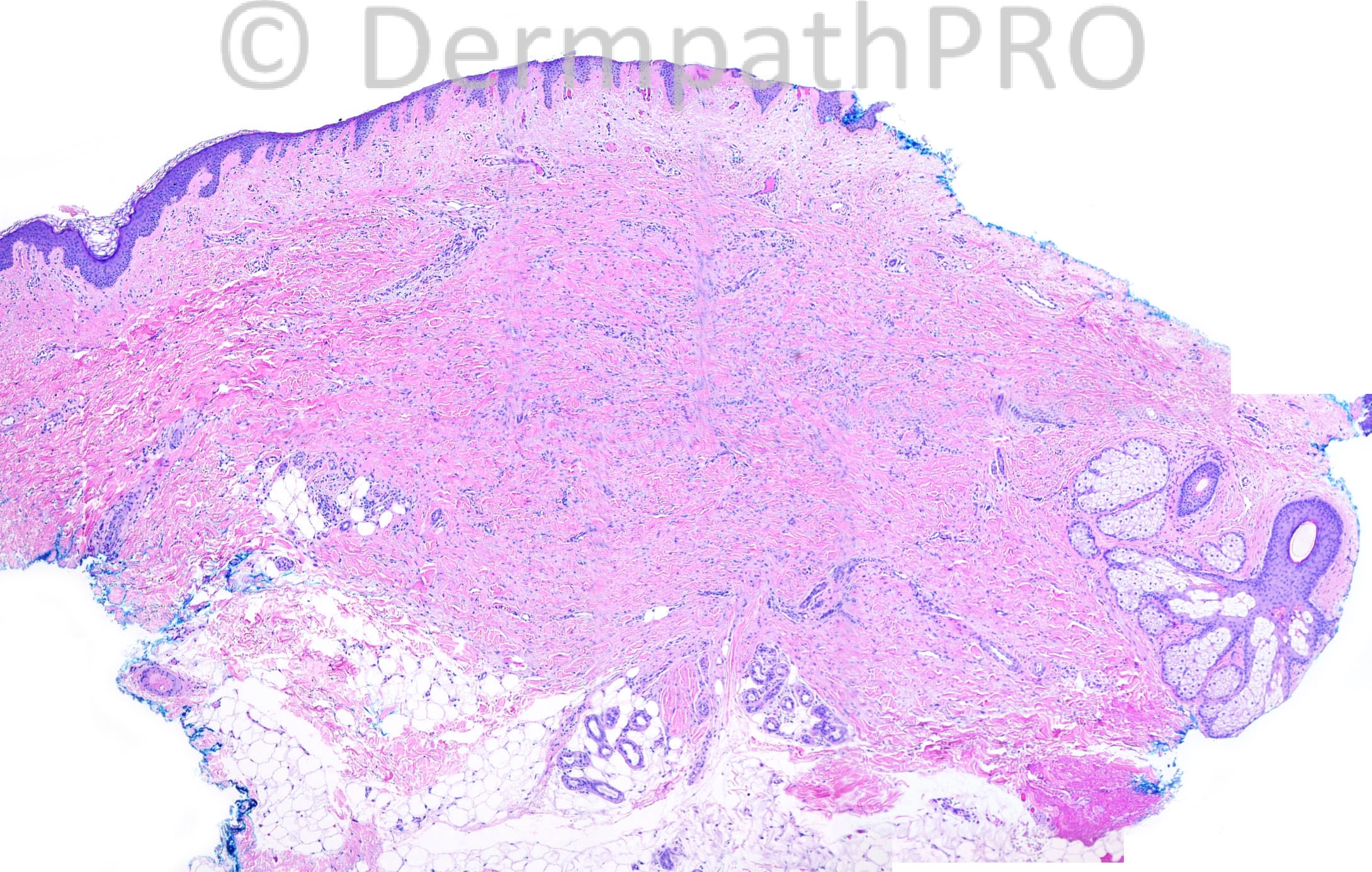
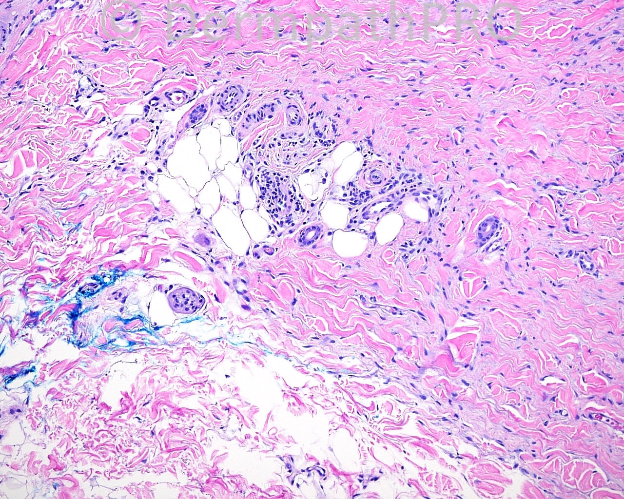
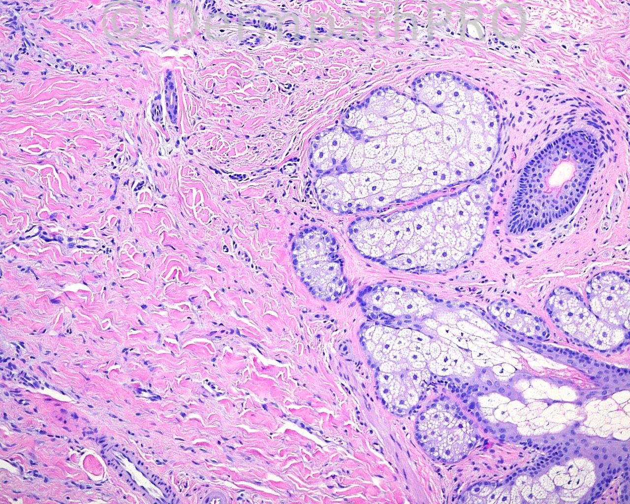
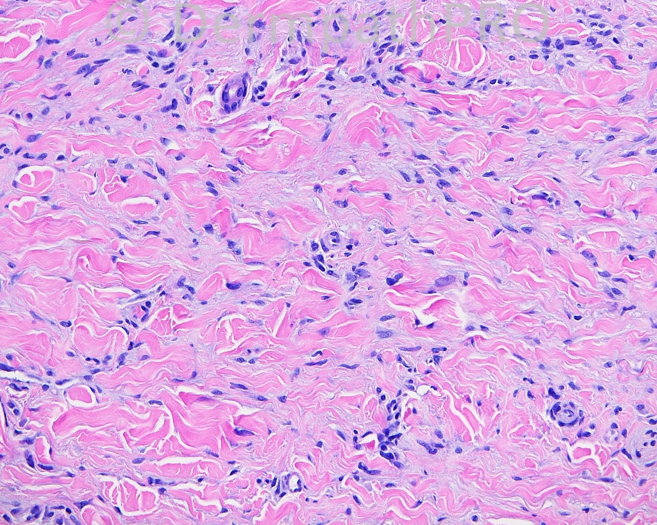
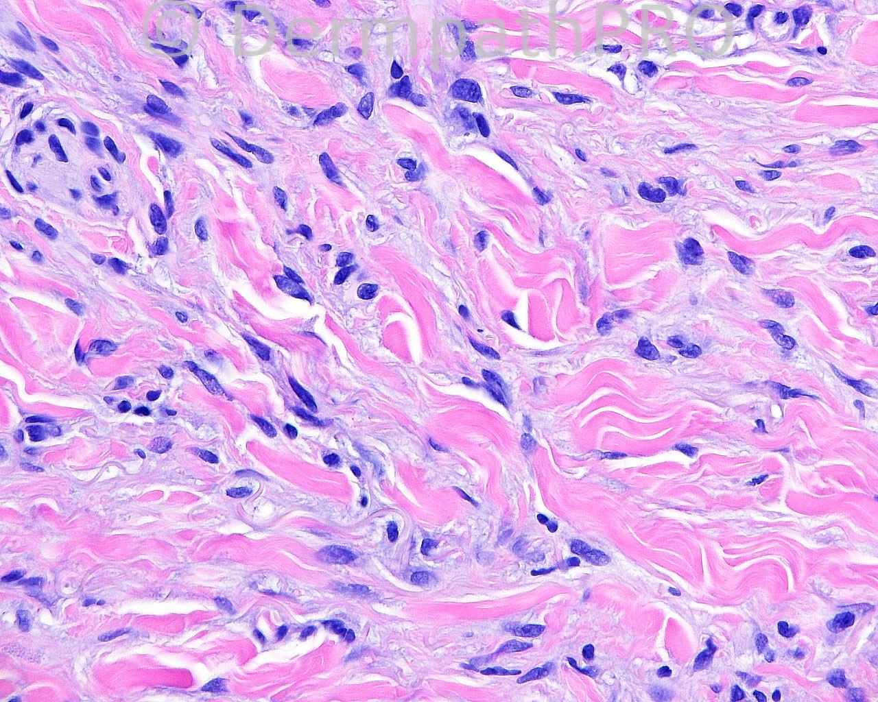
The patient is a 61 year old woman with an excision with margin exam if malignant of a lesion on the midline lower abdomen.

Case posted by Dr. Mark Hurt.


  Report Record

User Feedback


Robledo F. Rocha

Posted

Interstitial granulomatous dermatitis, if an underlying systemic illness exists, or interstitial granulomatous drug reaction, if related to pharmacological therapy.

Share this comment


Link to comment
share_externally

Guest Romualdo

Posted

These stains point to a diagnosis of dermatofibroma.

Share this comment


Link to comment
share_externally

Dr. Richard Carr

Posted

Assuming S100 is also negative would go for a dermatofibroma.

Share this comment


Link to comment
share_externally

Dr. Mona Abdel-Halim

Posted

Dermatofibroma..

Share this comment


Link to comment
share_externally

Eman El-Nabarawy

Posted

Myofibroblastic dermatofibroma.

Share this comment


Link to comment
share_externally

Dr. Mona Abdel-Halim

Posted

Dr Mark, I think dermatomyofibroma has a different morphology with a plaque like dermal growth pattern parallel to the epidermis composed of fascicles of spindle cells with abundant esinophilic cytoplasm. I think the lesion here has a typical morphology of a dermatofibroma with keloidal like collagen entrapped by tumor cells. I believe two components are found in this lesion,a spindle cell one and a histiocytic one. The SMA positivity is reported in some dermatofibromas and I do not think it is against the diagnosis. What do you think?

Share this comment


Link to comment
share_externally


Join the conversation

You can post now and register later. If you have an account, sign in now to post with your account.

Guest
Add a comment...

×   Pasted as rich text.   Paste as plain text instead

  Only 75 emoji are allowed.

×   Your link has been automatically embedded.   Display as a link instead

×   Your previous content has been restored.   Clear editor

×   You cannot paste images directly. Upload or insert images from URL.

Loading...

×
×
  • Create New...