Jump to content
In this section we have spot diagnoses posted on a daily basis since June 2010, now over 4000! You can review the archived cases and read the suggested diagnoses by users and the final comment by the contributors.
Case are uploaded each week day by 10 am UK time with the correct diagnosis will generally be posted at 8 pm UK time. Why not view the most recent spot diagnosis and proffer a diagnosis?

Case Number : Case 1012 - 9th May Posted By: Guest

Please read the clinical history and view the images by clicking on them before you proffer your diagnosis.
Submitted Date :
   (0 reviews)

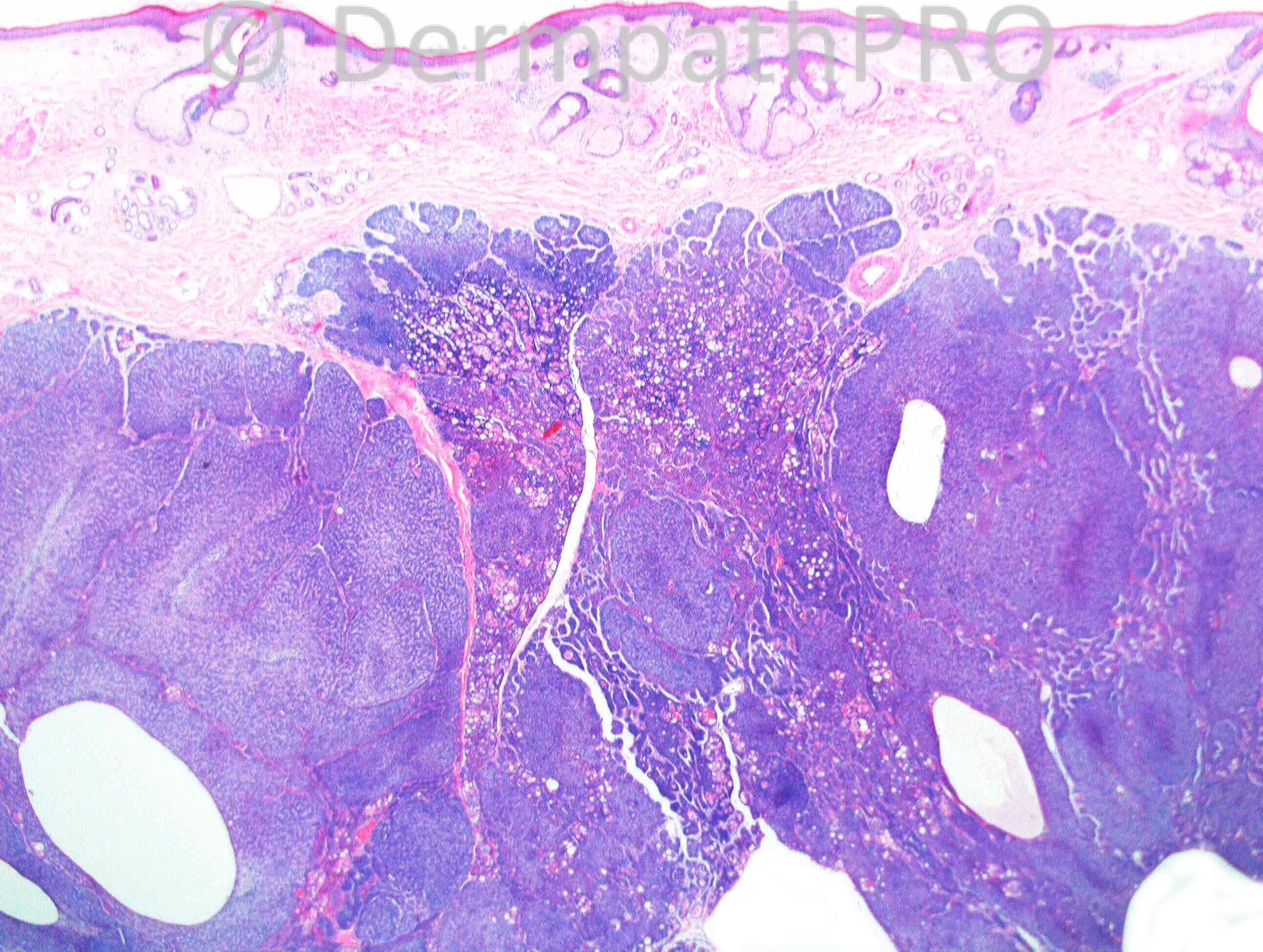
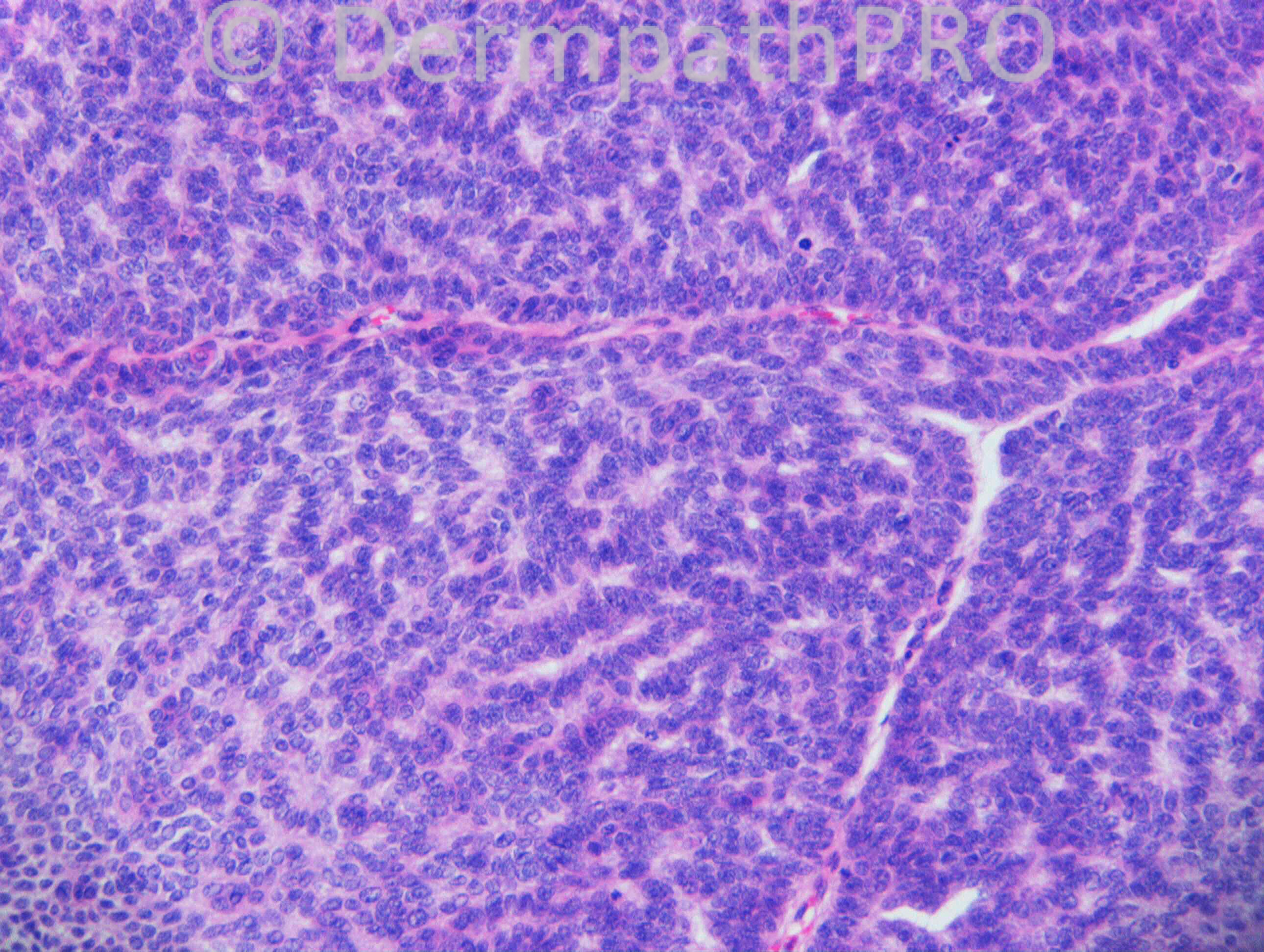
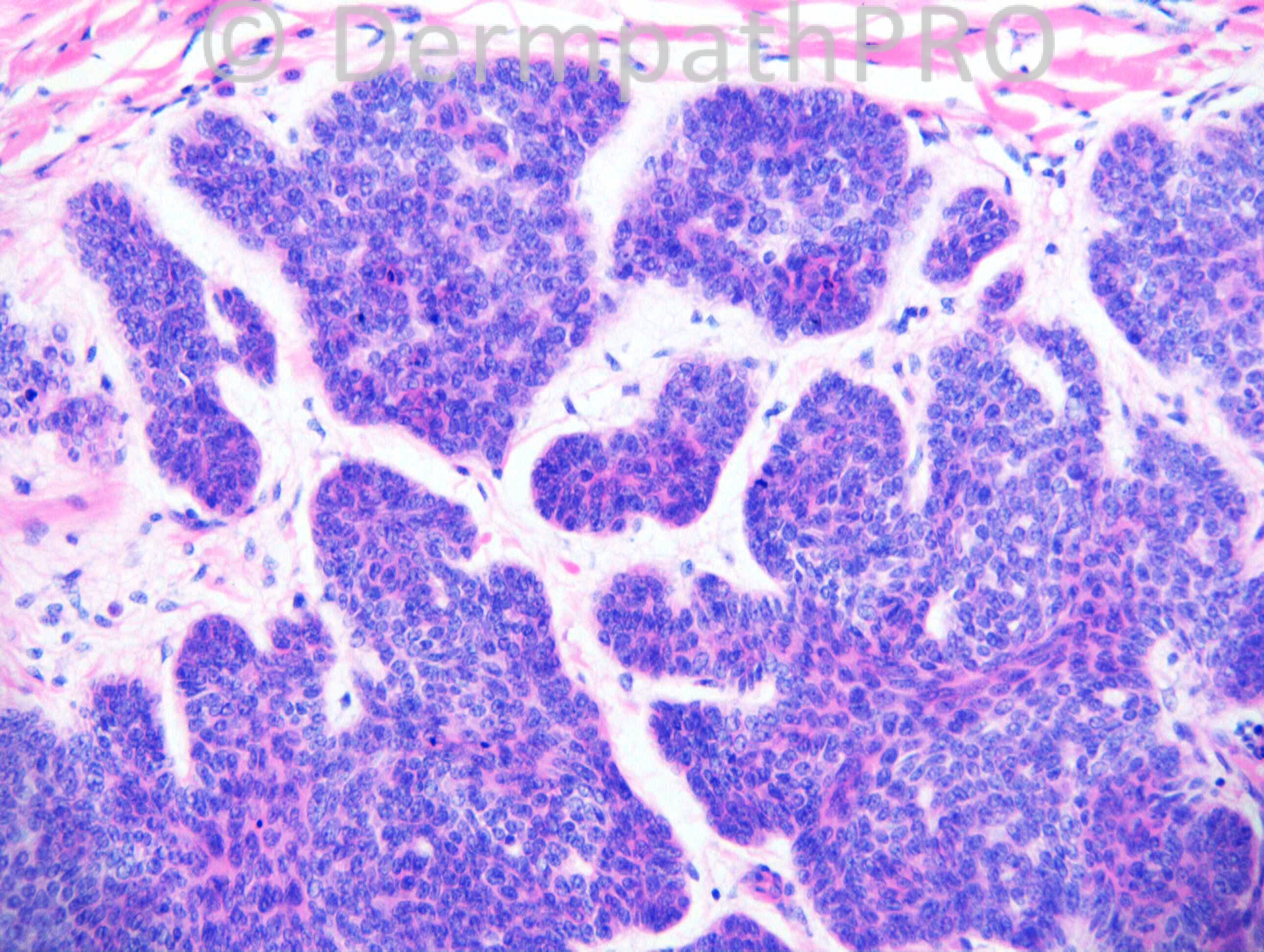
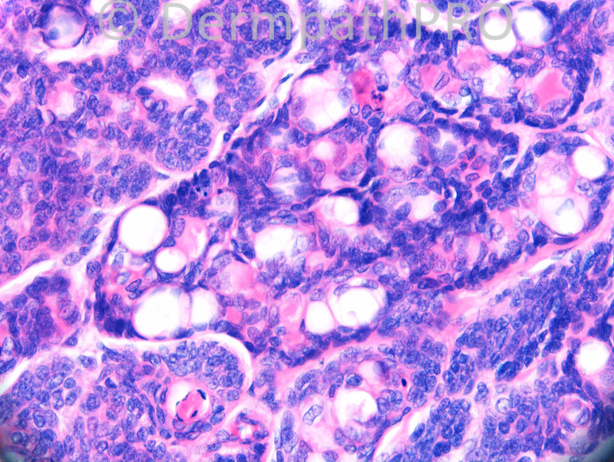
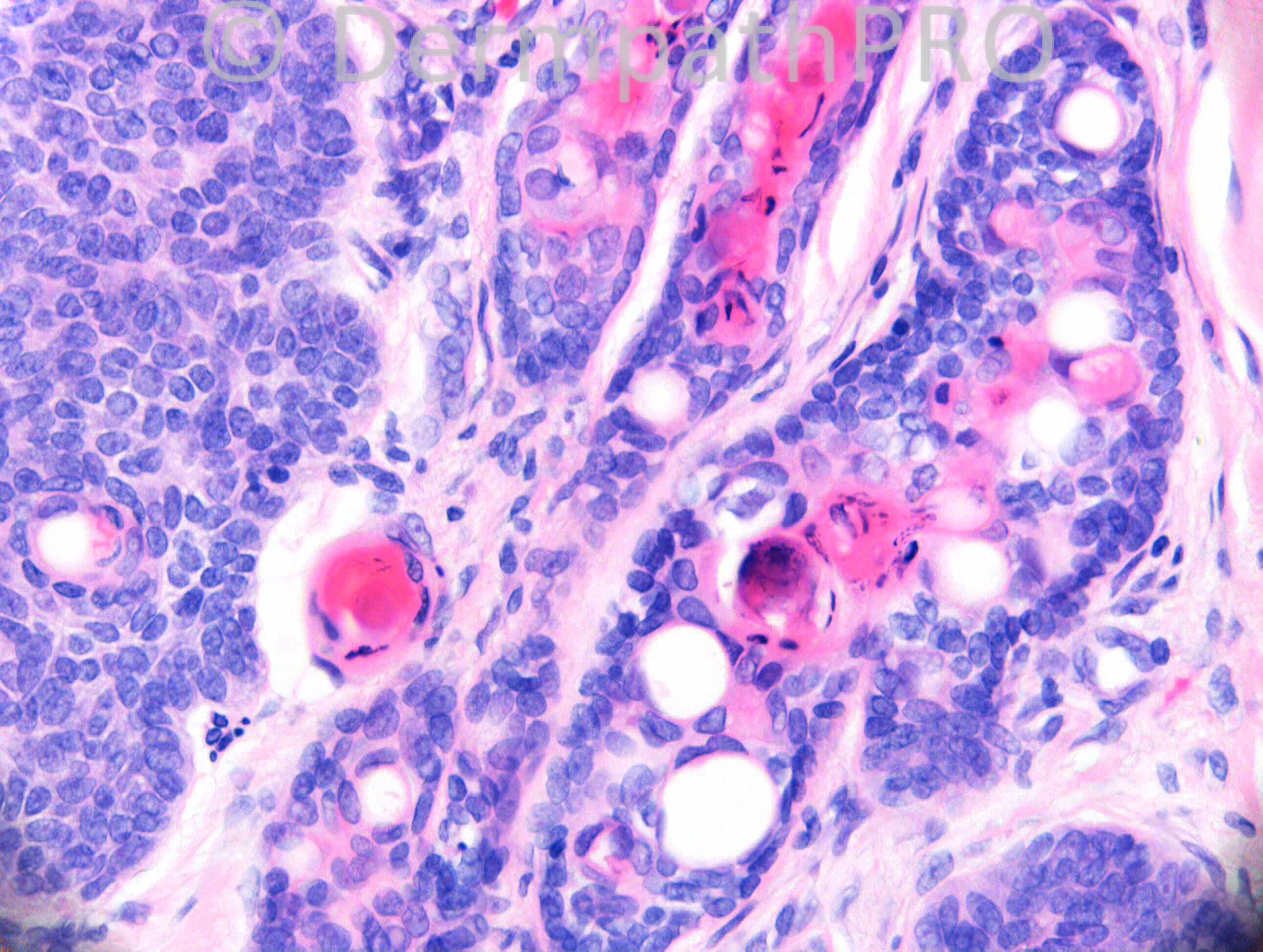
M79. Referred case with no clinical information provided.

Case posted by Dr. Richard Carr.

  Report Record

User Feedback


nick turnbull

Posted

Large basaloid tumour with rippled pattern. Ductal differentiation. I think i see sebocytes also with indented nuclei. Very pink?, ?matrical differenttation. I also wondered if there was background nevus sebaceous but not sure about that. I would think about requesting a BerEP4 and EMA to start with and my working diagnosis is this is a sebaceoma, rippled pattern with ductal differentiation. DDX rippled pattern BCC with duactal and sebaceous differentiation (which would be very rare)

Share this comment


Link to comment
share_externally

I did think there was a carcinoid-like (rippled) architecture in the upper right image, but I didn't think there was sebaceous differentiation. My first thought was basaloid hidradenoma. Would EMA differentiate between hidradenoma and sebaceoma?

Share this comment


Link to comment
share_externally

Dr. Mona Abdel-Halim

Posted

To me, the first impression with the large basaloid tumor, deep in the dermis, with ductal differentiation and esinophilic hyaline deposits (fig 4 and 5) and trabecular pattern, was spiradenoma. However the lack of dual cell population in some areas, lack of lymphocytes within the tumor and the presence of some nuclear pleomorphism, prominent nucleoli and ? mitoses (fig 2 and 3), made me think of the possibility of low grade spiradenocarcinoma developing within a pre-existing spiradenoma. An overlapping spiradenoma/cylindroma as an origin is also possible. Interesting case. Anxious to know the correct diagnosis!!!!

Share this comment


Link to comment
share_externally

Eman El-Nabarawy

Posted

Spiradenocarcinoma vs BCC with adnexal differentiation.

Share this comment


Link to comment
share_externally

Dr. Richard Carr

Posted

IHC will be posted at 4pm British Summer Time

Share this comment


Link to comment
share_externally

Dr. Richard Carr

Posted

[img]https://dermpathpro.com/uploads/spot_diagnosis_comment_img/CASE1012_%20RAC6814x10_EMA_Label.jpg[/img]

[img]https://dermpathpro.com/uploads/spot_diagnosis_comment_img/CASE1012_%20RAC6814x10b_CEA_Label.jpg[/img]

[img]https://dermpathpro.com/uploads/spot_diagnosis_comment_img/CASE1012_%20RAC6814x20_PASD_Label.jpg[/img]

[img]https://dermpathpro.com/uploads/spot_diagnosis_comment_img/CASE1012_%20RAC6814x40_EMA_Label.jpg[/img]

[img]https://dermpathpro.com/uploads/spot_diagnosis_comment_img/CASE1012_%20RAC6814x5_BerEP4_Label.jpg[/img]

Share this comment


Link to comment
share_externally

BCC with ductal differentiation, as Dr El-Nabarawy suggested.

Share this comment


Link to comment
share_externally

Dr. Mona Abdel-Halim

Posted

So it is BCC with ductal differentiation... Lovely one as usual Dr Carr...

Share this comment


Link to comment
share_externally

Robledo F. Rocha

Posted

[font=Arial, sans-serif][size=4]Basal cell carcinoma with areas showing organoid pattern and focal ductal differentiation with squamoid lining.[/size][/font]

Share this comment


Link to comment
share_externally

Guest Jim Davie MD

Posted

Agree with BCC with eccrine differentiation.
The ductal areas show dual positivity with EMA and CEA with some showing hylaline eosinophilic linings (sebaceous would be CEA negative). The basaloid cells are EMA negative and BER-EP4 positive, and have scattered mitotic activity.

Wouldn't rule out a coexistent progenitor trichoblastoma if this was a nevus sebaceus background (as Nick suggested).

Share this comment


Link to comment
share_externally

Dr. Richard Carr

Posted

Well a great case (yes BCC with striking ductal differentiation) even if I say so myself and great responses today. I think you have all made my points for me. Remember ductal differentiation is not that uncommon in BCC. This one was a referred slide with rather pale haematoxylin so the mucin in the stroma of the third image cannot be easily made out and palisading is rather pathetic. I have not yet come across rippled hidradenoma or rippled spiradenoma (remember I used the rippled pattern recently to favour sebaceoma v hidradenoma). The EMA is very helpful as the lumens should be punched out in hidradenoma / ductal differentiation (as in this BCC) whereas we like to see multiple intra-cytoplasmic vacuoles indenting the nucleus on EMA in true sebaceous differentation (exceedingly rare in BCC although I do have 3 cases collected). CEA can be nice for ductal differentiation but is often not sensitive and it does stain squamous cells rather non-specifically. I am not too sure what the dull, lightly PASD positive globular material is - but maybe a keratin derived cuticular type material? Finally we do not have papillary mesenchymal cells for rippled pattern trichoblastoma. There was a low grade mitotic rate and apoptotic cells (not shown well in these images) so I was happy after seeing the BerEP4 to confirm my favoured H&E diagnosis of BCC with ductal differentiation. In my experience rippled pattern BCC is very rare (only collected two or three). Regards to all and enjoy your weekends.

Share this comment


Link to comment
share_externally


Join the conversation

You can post now and register later. If you have an account, sign in now to post with your account.

Guest
Add a comment...

×   Pasted as rich text.   Paste as plain text instead

  Only 75 emoji are allowed.

×   Your link has been automatically embedded.   Display as a link instead

×   Your previous content has been restored.   Clear editor

×   You cannot paste images directly. Upload or insert images from URL.

Loading...

×
×
  • Create New...