Jump to content
In this section we have spot diagnoses posted on a daily basis since June 2010, now over 4000! You can review the archived cases and read the suggested diagnoses by users and the final comment by the contributors.
Case are uploaded each week day by 10 am UK time with the correct diagnosis will generally be posted at 8 pm UK time. Why not view the most recent spot diagnosis and proffer a diagnosis?

Case Number : Case 1013 - 12th May Posted By: Guest

Please read the clinical history and view the images by clicking on them before you proffer your diagnosis.
Submitted Date :
   (0 reviews)

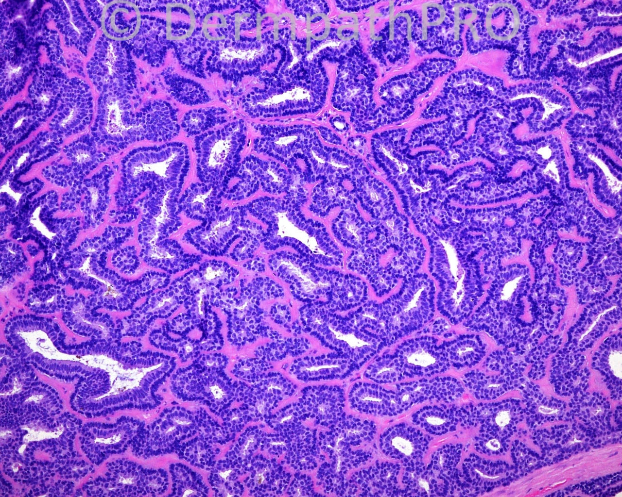
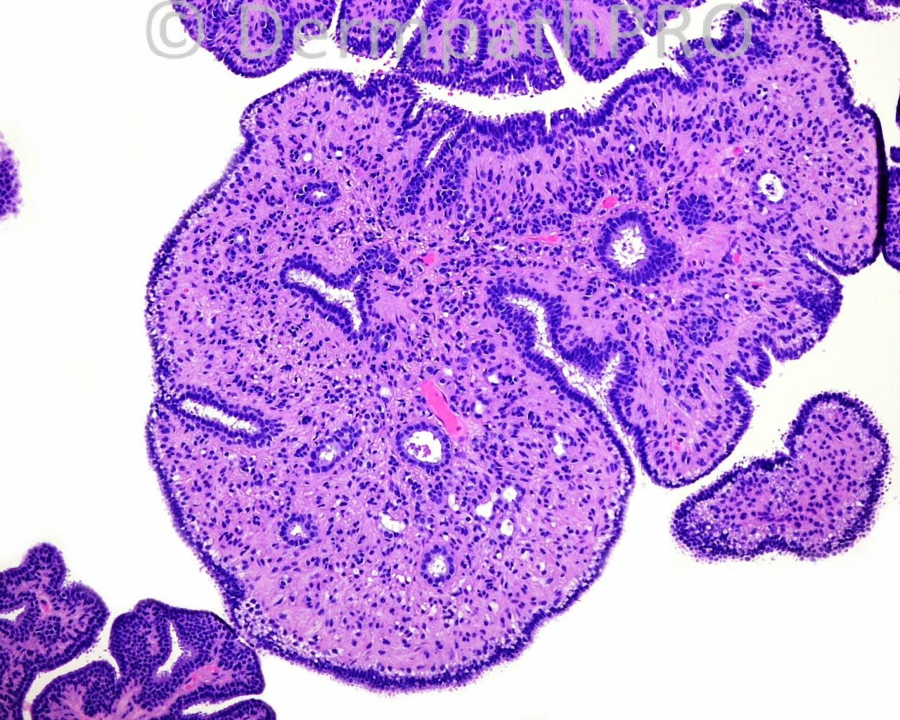
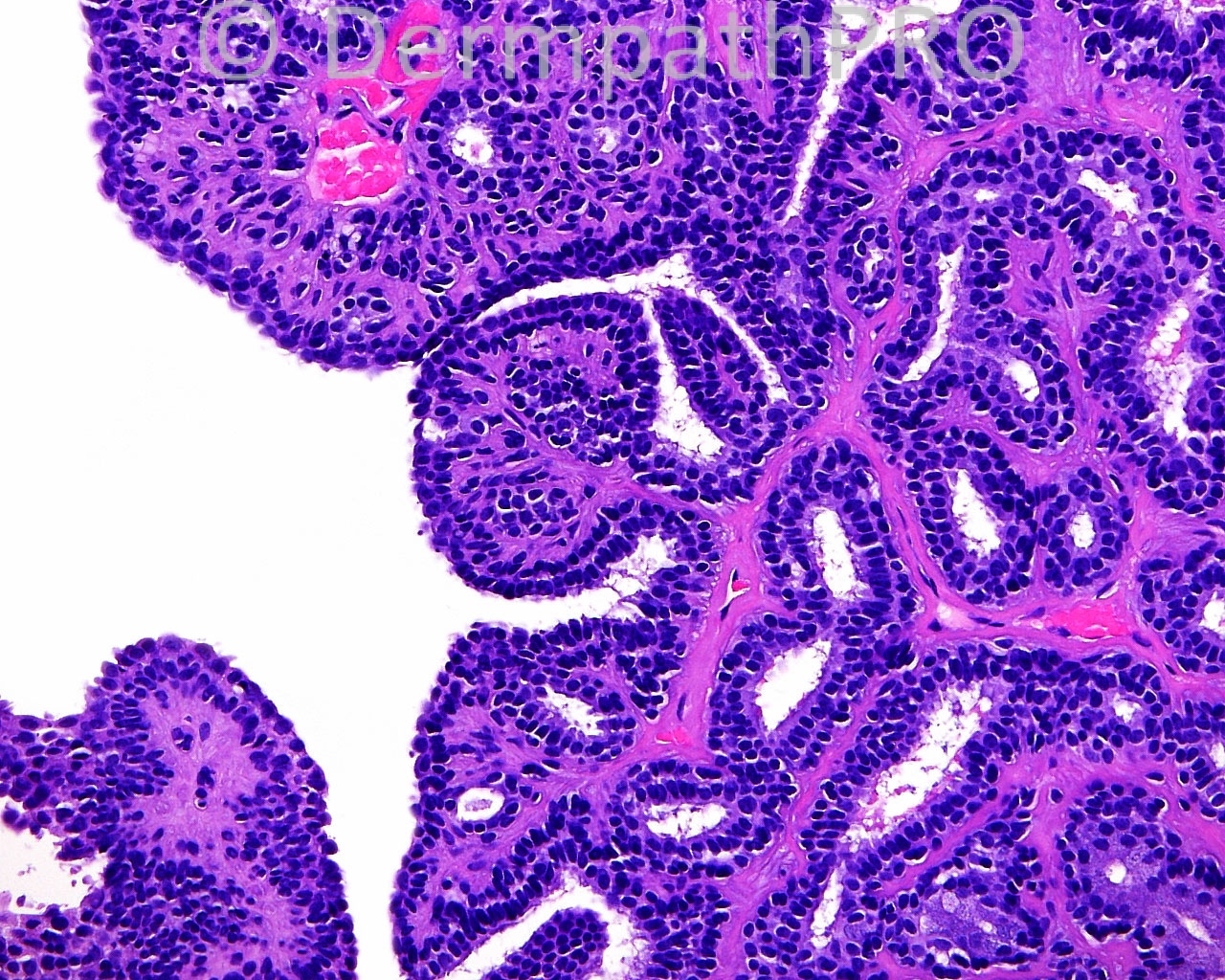
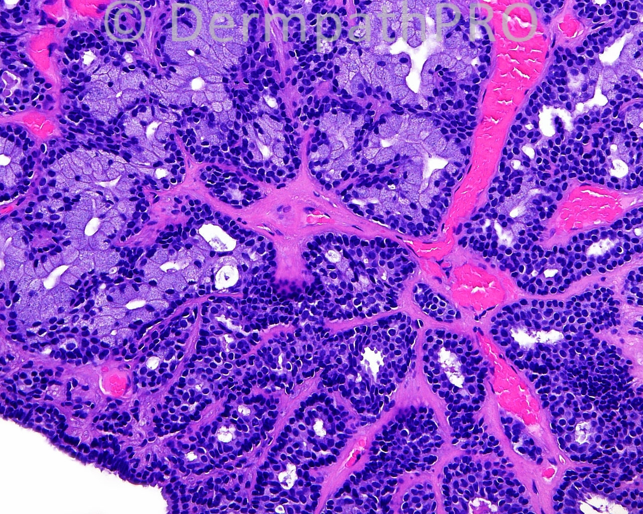
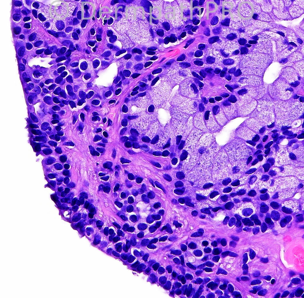
The patient is a 57 year old white woman with a punch biopsy taken from the right posterior medial scalp.

Case posted by Dr. Mark Hurt.

  Report Record

User Feedback


Apocrine papillary cystadenoma with focal mucinous metaplasia.

Share this comment


Link to comment
share_externally

Robledo F. Rocha

Posted

[size=4][color=#000000][font=Arial, sans-serif]Solid-cystic apocrine hidrocystoma showing papillae with focal mucinous change that project into cystic lumen and tubular solid areas.[/font][/color][/size]

Share this comment


Link to comment
share_externally

Dr. Mona Abdel-Halim

Posted

Apocrine papillary cystadenoma with focal mucinous metaplasia

Share this comment


Link to comment
share_externally

Apocrine papillary cystadenoma with focal mucinous metaplasia. the lesion not totaly excised see fig a

Share this comment


Link to comment
share_externally

Dr. Richard Carr

Posted

Apocrine tubulopapillary cystadenoma with mucinous metaplasia.

Share this comment


Link to comment
share_externally

Guest Jim Davie MD

Posted

Agree. Tubulopapillary hidradenoma. (Too many synonyms for this exist....tubular apocrine adenoma, tubulopapillary apocrine adenoma, papillary tubular adenoma, apocrine cystadenoma, etc. in addition to others, above.) Location on scalp is common.

Well circumscribed nodule with hyaline collagenous, non-inflammatory stroma lacking inflammation, and numerous telangiectatic vessels. Variable glandular, cribriform, cystic, and arborizing papillary architecture. Dual-layer sweat-gland architecture with myoepithelial layer, and cuboidal vs. columnar lumenal cells, some with apocrine snouting. Foamy lumenal cells with goblet-cell features occasionally present, consistent with incidental mucinous metaplasia. No significant cytologic atypia or proliferative activity.

Share this comment


Link to comment
share_externally


Join the conversation

You can post now and register later. If you have an account, sign in now to post with your account.

Guest
Add a comment...

×   Pasted as rich text.   Paste as plain text instead

  Only 75 emoji are allowed.

×   Your link has been automatically embedded.   Display as a link instead

×   Your previous content has been restored.   Clear editor

×   You cannot paste images directly. Upload or insert images from URL.

Loading...

×
×
  • Create New...