Jump to content
In this section we have spot diagnoses posted on a daily basis since June 2010, now over 4000! You can review the archived cases and read the suggested diagnoses by users and the final comment by the contributors.
Case are uploaded each week day by 10 am UK time with the correct diagnosis will generally be posted at 8 pm UK time. Why not view the most recent spot diagnosis and proffer a diagnosis?

Case Number : Case 1017 - 16th May Posted By: Guest

Please read the clinical history and view the images by clicking on them before you proffer your diagnosis.
Submitted Date :
   (0 reviews)

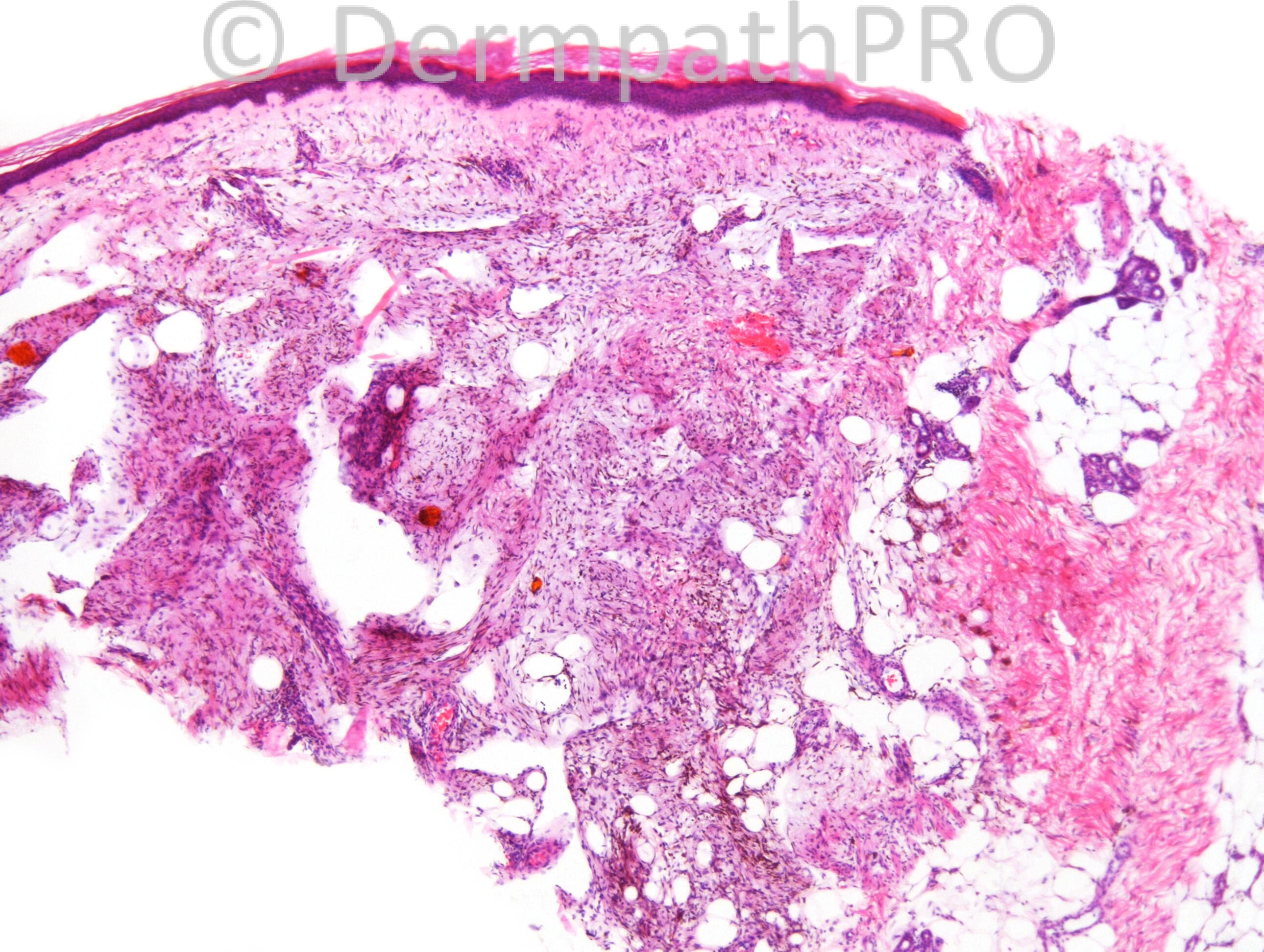
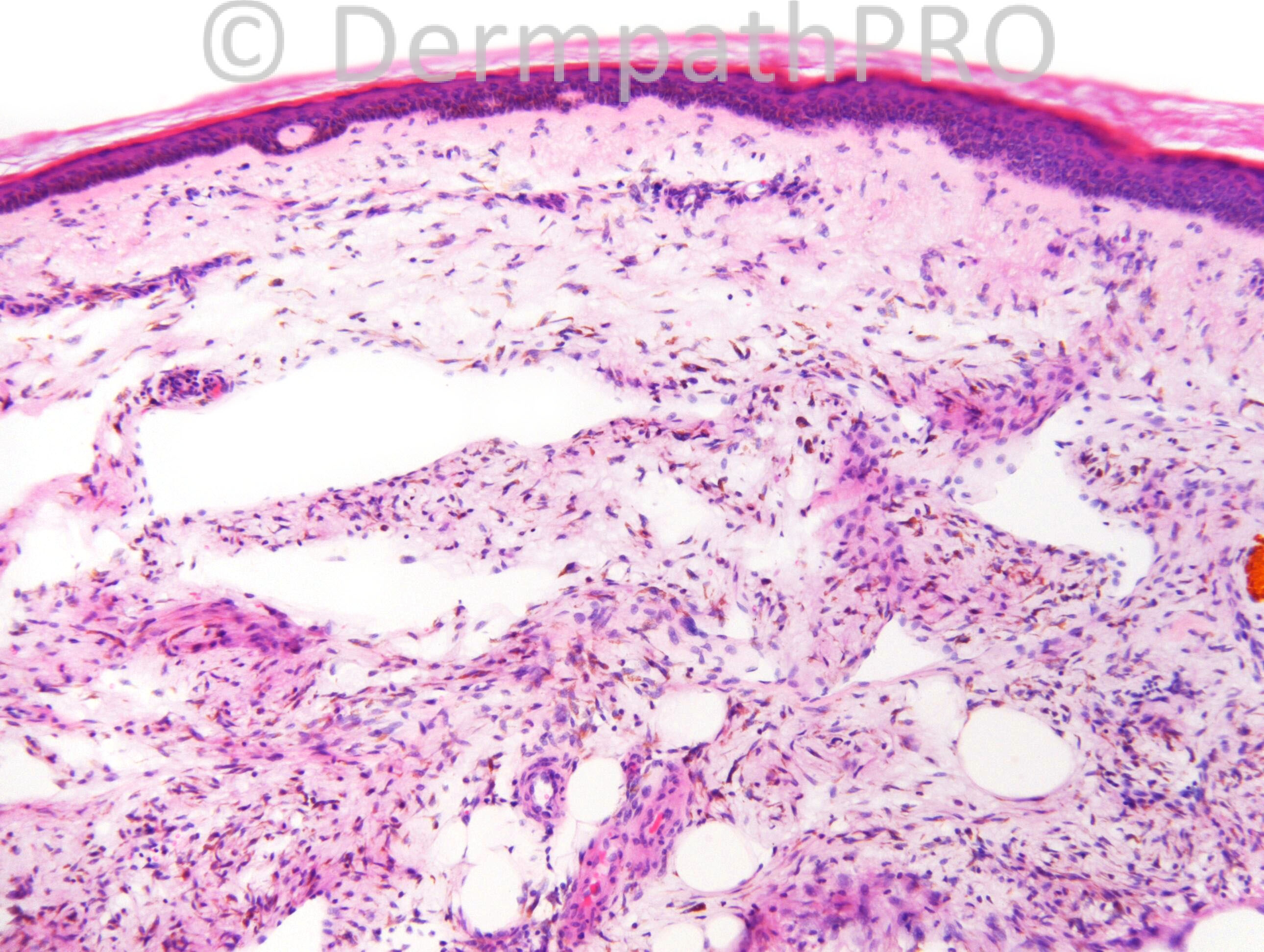
90 years old female.

Case posted by Dr. Richard Carr.


  Report Record

User Feedback


Eman El-Nabarawy

Posted

Malignant peripheral nerve sheath tumor with probable melanocytic differentiation .

Share this comment


Link to comment
share_externally

Haemosiderotic fibrohistiocytic lipomatous lesion.

Share this comment


Link to comment
share_externally

Dr. Mona Abdel-Halim

Posted

I am thinking of hemosiderotic fibrohistiocytic lipomatous tumor, but I am unaware of its possible dermal extension. Can not think in something else. It has to be it!!!! Careful examination of the specimen to exclude possible overlap with myxoinflammatory fibroblastic sarcoma is important since both tumors are interrelated as I have learned from the last Annual Updates in Dermatopathology last April...

Share this comment


Link to comment
share_externally

Haemosiderotic fibrohistiocytic lipomatous tumor

Share this comment


Link to comment
share_externally

Guest Romualdo

Posted

I think the pigment could be melanin and the lesion a melanotic (pigmented) diffuse neurofibroma.

Share this comment


Link to comment
share_externally

Guest Jim Davie MD

Posted

Agree with hemosiderotic fibrohistiocytic lipomatous tumor.
Unusual presentation: patient is much older than the common age range (40-70 yrs).
The pigment seems refractile, favoring hemosiderin. Cells show no high grade atypia, conspicuous nucleoli, or proliferative activity. Would expect CD34(+), S100(-) staining pattern.

Share this comment


Link to comment
share_externally

dermpath1

Posted

Agree with above but Just want to add melanoma and DFSP as a differential.
Snce melaoma is a great mimik, s100 should be done.

Share this comment


Link to comment
share_externally

Robledo F. Rocha

Posted

[font=arial,helvetica,sans-serif][size=4][color=#000000]My first impression is myxoinflammatory fibroblastic sarcoma. Looking for my colleagues' opinion favoring hemosiderotic fibrohistiocytic lipomatous tumor, I discovered both can be related lesions.[/color][/size][/font]

Share this comment


Link to comment
share_externally

Dr. Richard Carr

Posted

I reported this as a haemosiderotic fibrohistiocytic lipomatous lesion / tumour.

Share this comment


Link to comment
share_externally


Join the conversation

You can post now and register later. If you have an account, sign in now to post with your account.

Guest
Add a comment...

×   Pasted as rich text.   Paste as plain text instead

  Only 75 emoji are allowed.

×   Your link has been automatically embedded.   Display as a link instead

×   Your previous content has been restored.   Clear editor

×   You cannot paste images directly. Upload or insert images from URL.

Loading...

×
×
  • Create New...