Jump to content
In this section we have spot diagnoses posted on a daily basis since June 2010, now over 4000! You can review the archived cases and read the suggested diagnoses by users and the final comment by the contributors.
Case are uploaded each week day by 10 am UK time with the correct diagnosis will generally be posted at 8 pm UK time. Why not view the most recent spot diagnosis and proffer a diagnosis?

Case Number : Case 1019 - 20th May Posted By: Guest

Please read the clinical history and view the images by clicking on them before you proffer your diagnosis.
Submitted Date :
   (0 reviews)

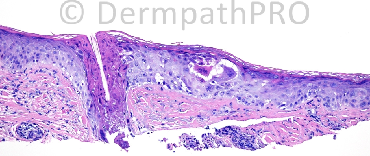
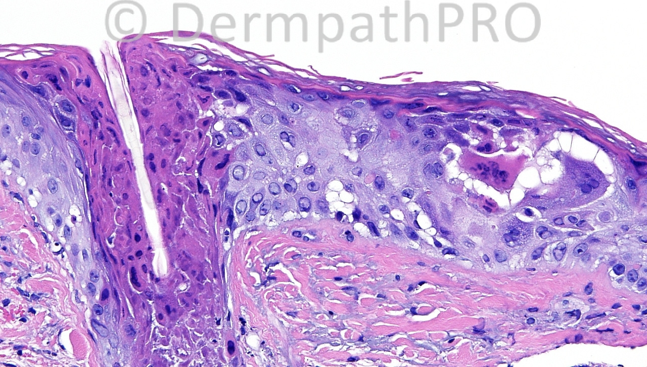
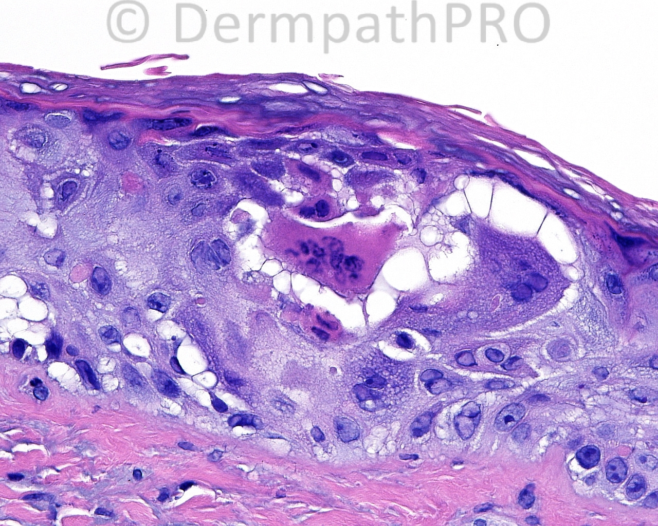
The patient is a 59 year old white woman with a shave biopsy of a large, translucent plaque on the right side of the forehead.

Case posted by Dr. Mark Hurt.


  Report Record

User Feedback


IgorSC

Posted

Agree with HSV, but i think this clinical aspect of the lesion is somewhat odd.

Share this comment


Link to comment
share_externally

Dr. Mona Abdel-Halim

Posted

Yes looks like herpetic folliculitis, very weird clinical impression...

Share this comment


Link to comment
share_externally

Robledo F. Rocha

Posted

[font=arial,helvetica,sans-serif][size=4][color=#000000]Undoubtedly, there are signs of herpes virus infection. But the peculiar clinical presentation makes me think of development of herpes virus infection in a previous inflammatory skin disease, like a spongiotic dermatitis or a burn, hence it would be eczema herpeticum or Kaposi's varicelliform eruption.[/color][/size][/font]

Share this comment


Link to comment
share_externally

dermpath1

Posted

I would include chemotherapy effect, sun burn, porokeatsi., GVHD, and most preferably, actinic keratosis in my long differential.

Share this comment


Link to comment
share_externally

Herpetic folliculitis with strange clinical feature

Share this comment


Link to comment
share_externally

Dr. Mona Abdel-Halim

Posted

Herpes zoster, missed by the clinicians !!!

Share this comment


Link to comment
share_externally

Herpes zoster. Affection of hair follicle is a clue for diagnosis of HZ by H&E as in this case that confirmed by histochemistry

Share this comment


Link to comment
share_externally

I wonder if this is an incidental finding given its focality, at the edge of a broad, superficial shave. There is also an inflammatory infiltrate across the entire biopsy and a focus of calcification on the opposite side of the biopsy. Perhaps deeper levels into this block or a deeper biopsy would identify a cause for the clinical-pathological discrepancy.

Share this comment


Link to comment
share_externally

Great! Thanks for the follow-up. Certainly an odd clinical presentation.!

Share this comment


Link to comment
share_externally

Guest Jim Davie MD

Posted

[font=arial,helvetica,sans-serif][size=4]Agree with Noonshin that there might be more at work than isolated VZV, given the 2-year history of a translucent isolated forehead plaque, and clinical absence of classic zoster features. In first image, dermis does show some sclerosis and possible patchy depletion of basophilic elastotic fibers in the center, reminiscent of a superficially represented actinic granuloma (annular elastolytic giant-cell granuloma).

Given the unusual clinical context of long-term plaque, I'd wonder if this case represented one of the following scenarios:
1) incidental zoster developing in a previous isolated inflammatory dermatosis ([b][i]inverse isotopic response[/i][/b]), or
2) a inflammatory dermatosis that may be a direct result of previous zoster ([i][b]isotopic response[/b]).[/i][/size][/font]

[size=3] [url="http://archderm.jamanetwork.com/article.aspx?articleid=539271"]Granuloma Annulare at Sites of Healing Herpes Zoster[/url]. [/size]
[size=3]Marshall A. Guill, MC; Detlef K. Goette, MC. Arch Dermatol. 1978;114(9):1383.

[url="http://www.actasdermo.org/en/pdf/90167644/S300/?pubmed=true"]Wolf's Isotopic Response: A Series of 9 Cases[/url] [i][PDF Full article][/i]
A. Jaka-Morenoa, A. López-Pestañaa, M. López-Núñeza, N. Ormaechea-Péreza, S. Vildosola-Esturoa, A. Tuneu-Vallsa, C. Lobo-Morán. Actas Dermosifiliogr. 2012;103:798-805. - Vol. 103 Num.09 [/size]

Share this comment


Link to comment
share_externally


Join the conversation

You can post now and register later. If you have an account, sign in now to post with your account.

Guest
Add a comment...

×   Pasted as rich text.   Paste as plain text instead

  Only 75 emoji are allowed.

×   Your link has been automatically embedded.   Display as a link instead

×   Your previous content has been restored.   Clear editor

×   You cannot paste images directly. Upload or insert images from URL.

Loading...

×
×
  • Create New...