Jump to content
In this section we have spot diagnoses posted on a daily basis since June 2010, now over 4000! You can review the archived cases and read the suggested diagnoses by users and the final comment by the contributors.
Case are uploaded each week day by 10 am UK time with the correct diagnosis will generally be posted at 8 pm UK time. Why not view the most recent spot diagnosis and proffer a diagnosis?

Case Number : Case 1026 - 29th May Posted By: Guest

Please read the clinical history and view the images by clicking on them before you proffer your diagnosis.
Submitted Date :
   (0 reviews)

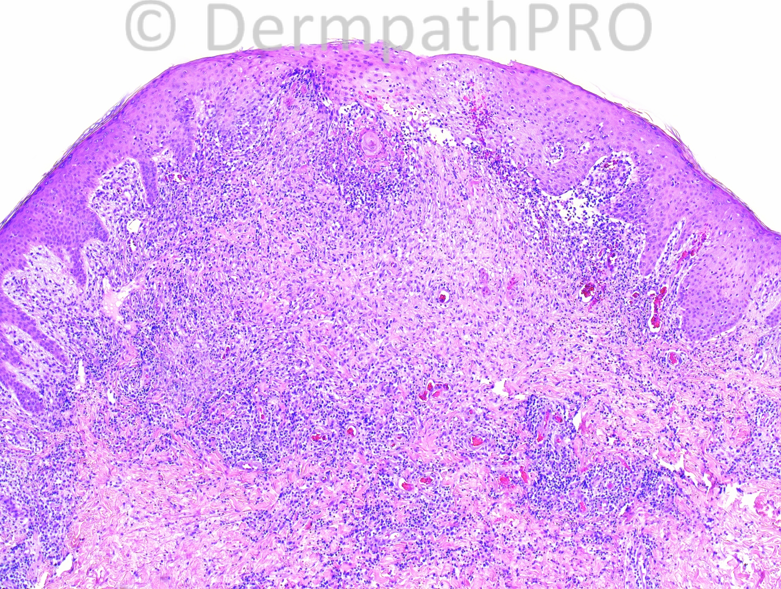
38 year-old male with multiple lesions on lower abdomen and groin.

Case posted by Dr. Hafeez Diwan.

  Report Record

User Feedback


nick turnbull

Posted

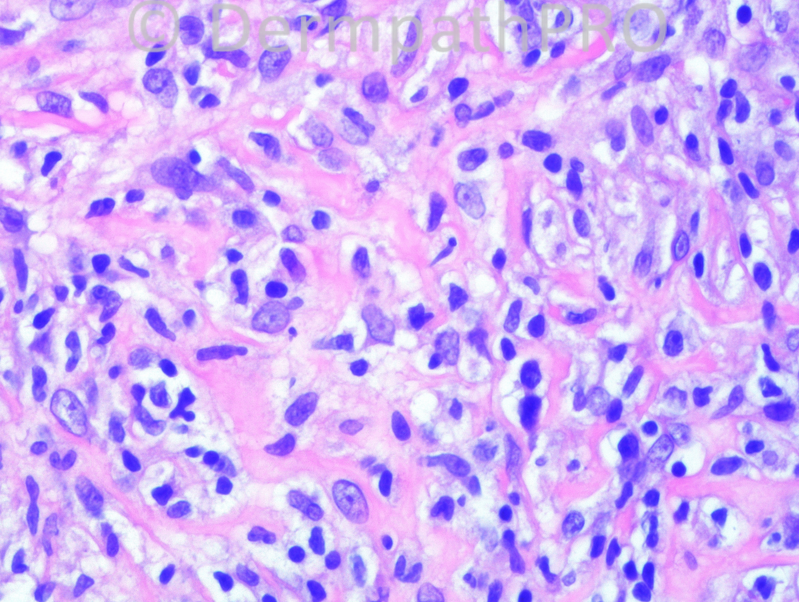
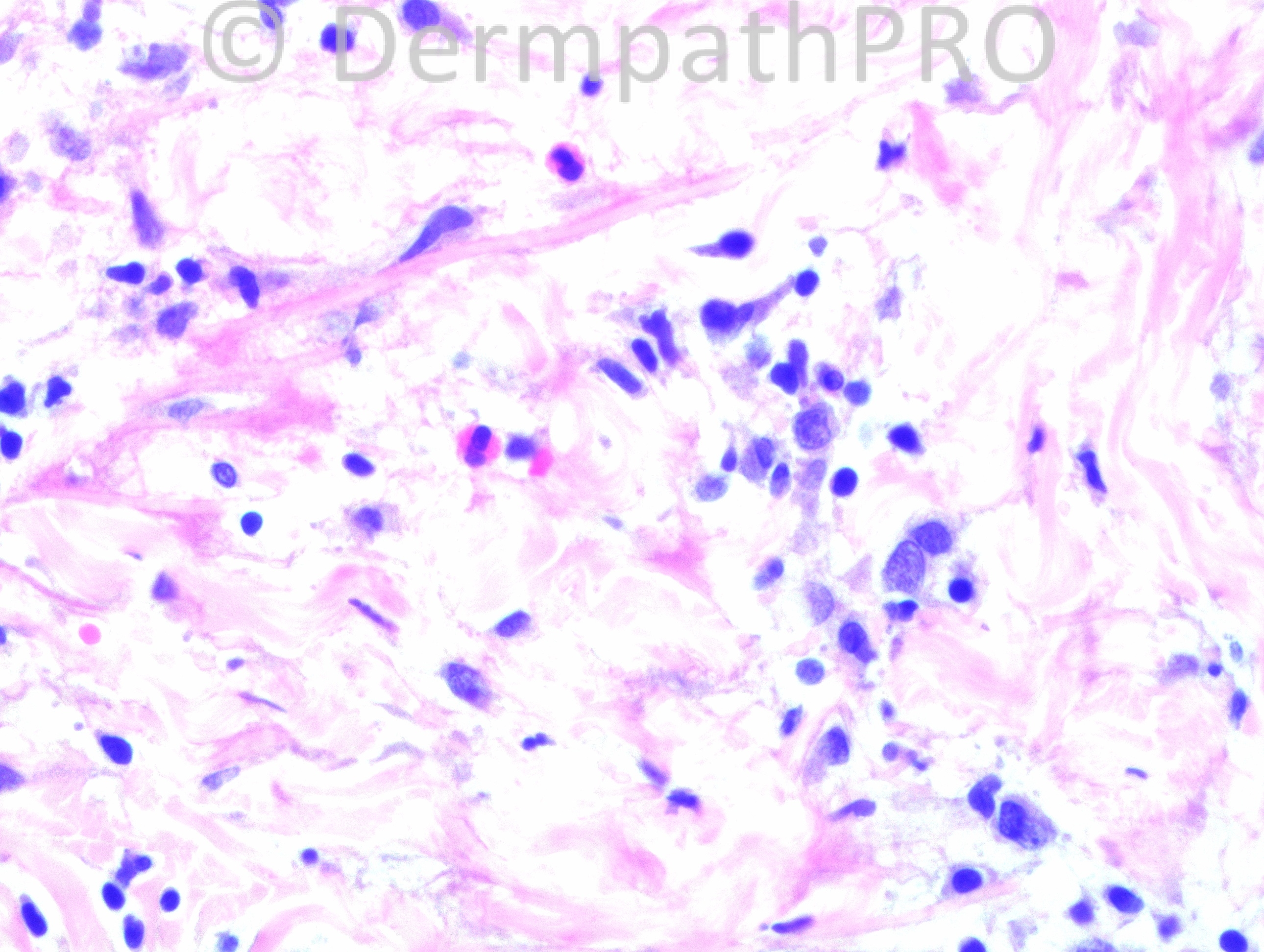
Marked interface pattern, with hydropic degeneartion of basal layer, many colloid bodies and apoptotic cells. Marked extravastion of red cells and dermal odema. Epidemotropism is present. Wedge shapped inflamtion into the dermis with small angulated lymphocytes and larger cells. Few mast cells. I was thinking about Lymphomatoid papulosis or pityriasis lichenoides maybe? but would like a bit more history or CPC before I would be making one of those diagnoses.

Share this comment


Link to comment
share_externally

Dr. Mona Abdel-Halim

Posted

Agree with the above DDx and CPC is important. Also insect bite reaction can be included

Share this comment


Link to comment
share_externally

Dr. Richard Carr

Posted

Looks reactive. First impression arthropod reaction. Looks folliculocentric that can happen with arthropods but would do some levels for folliculitis.

Share this comment


Link to comment
share_externally

Eman El-Nabarawy

Posted

Insect bite reaction.

Share this comment


Link to comment
share_externally

Guest Romualdo

Posted

Besides an insect bite reaction I would like to suggest persistent scabietic nodules as a possibility.

Share this comment


Link to comment
share_externally

Robledo F. Rocha

Posted

Wedge shape infiltrate, chiefly lymphocytes, with vacuolar alteration of the basal layer and extravasation of erythrocytes in the epidermis make me think of Mucha-Habermann disease.

Share this comment


Link to comment
share_externally

Dr. Hafeez Diwan

Posted

Lymphomatoid papulosis.

Share this comment


Link to comment
share_externally

Nice case:lymphomatiod papulosis

Share this comment


Link to comment
share_externally


Join the conversation

You can post now and register later. If you have an account, sign in now to post with your account.

Guest
Add a comment...

×   Pasted as rich text.   Paste as plain text instead

  Only 75 emoji are allowed.

×   Your link has been automatically embedded.   Display as a link instead

×   Your previous content has been restored.   Clear editor

×   You cannot paste images directly. Upload or insert images from URL.

Loading...

×
×
  • Create New...