Jump to content
In this section we have spot diagnoses posted on a daily basis since June 2010, now over 4000! You can review the archived cases and read the suggested diagnoses by users and the final comment by the contributors.
Case are uploaded each week day by 10 am UK time with the correct diagnosis will generally be posted at 8 pm UK time. Why not view the most recent spot diagnosis and proffer a diagnosis?

Case Number : Case 1027 - 30th May Posted By: Guest

Please read the clinical history and view the images by clicking on them before you proffer your diagnosis.
Submitted Date :
   (0 reviews)

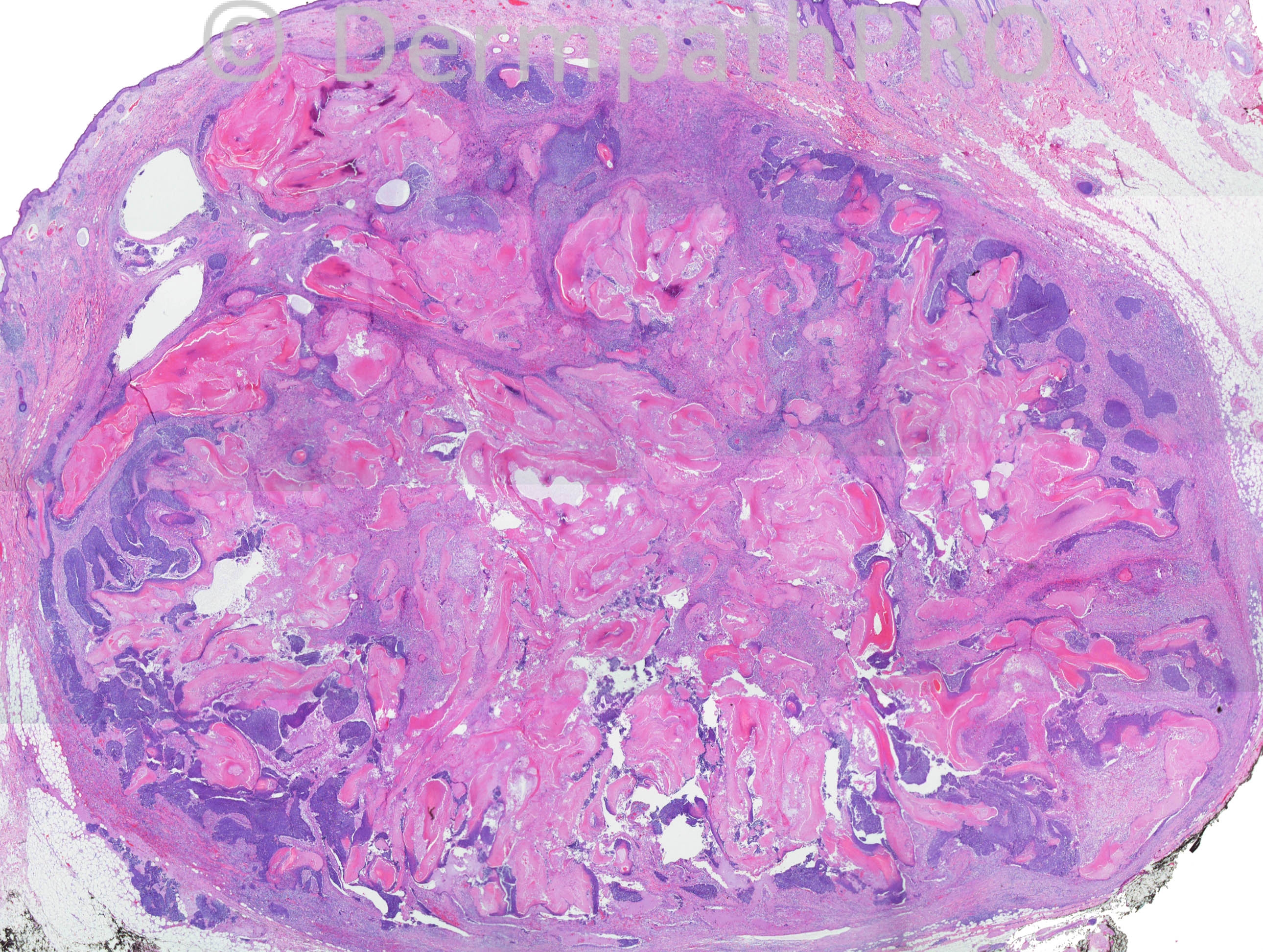
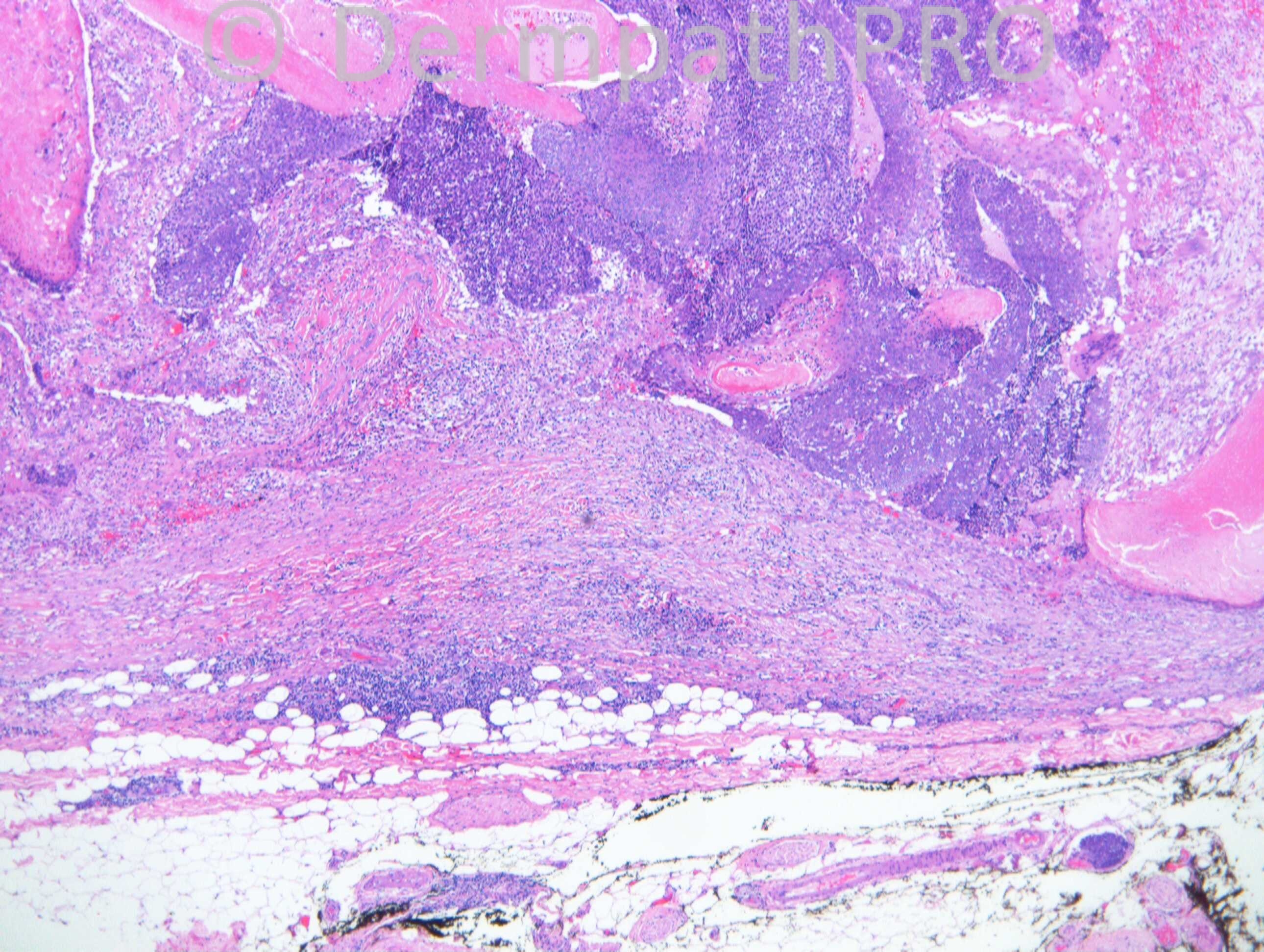
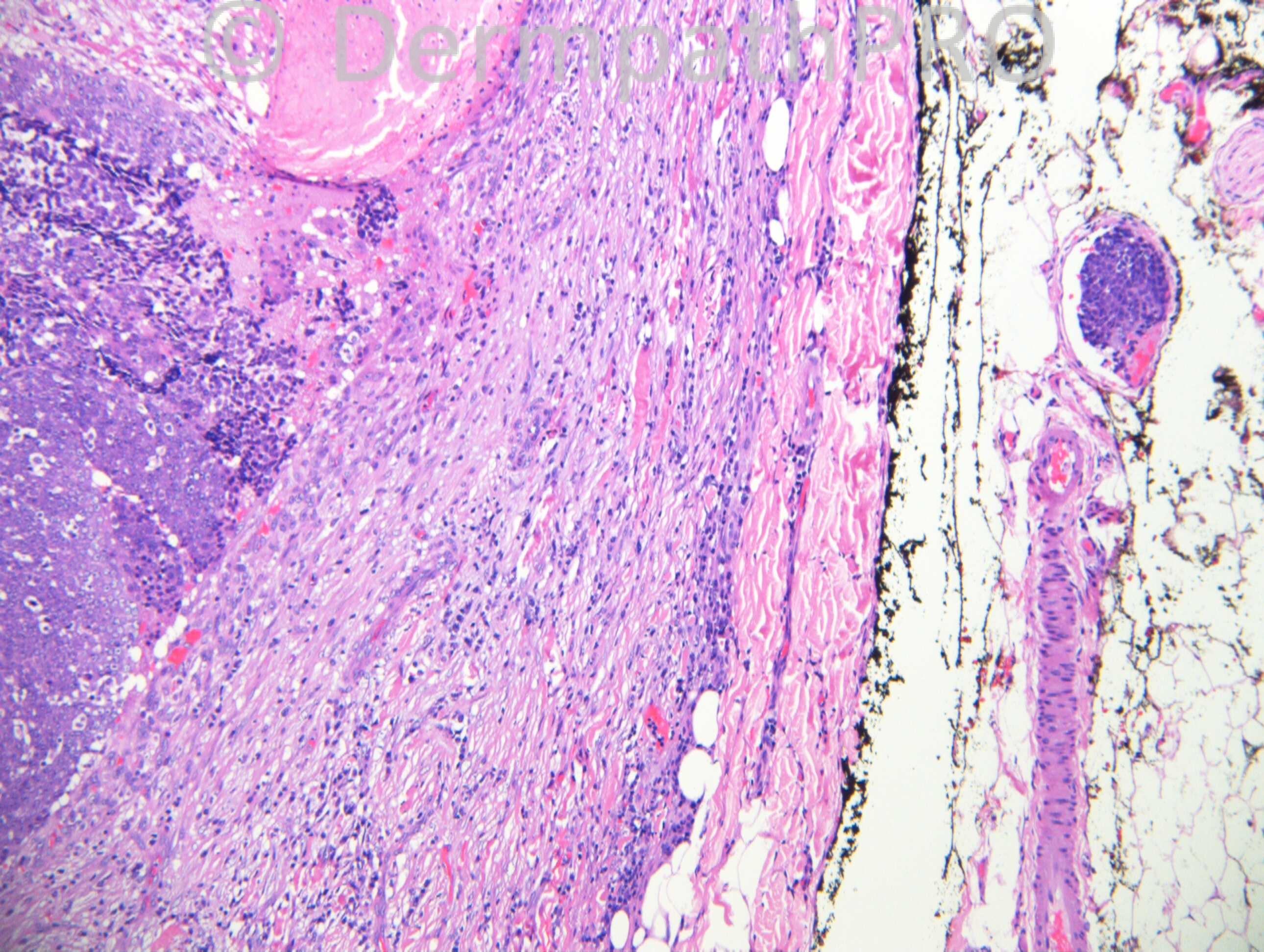
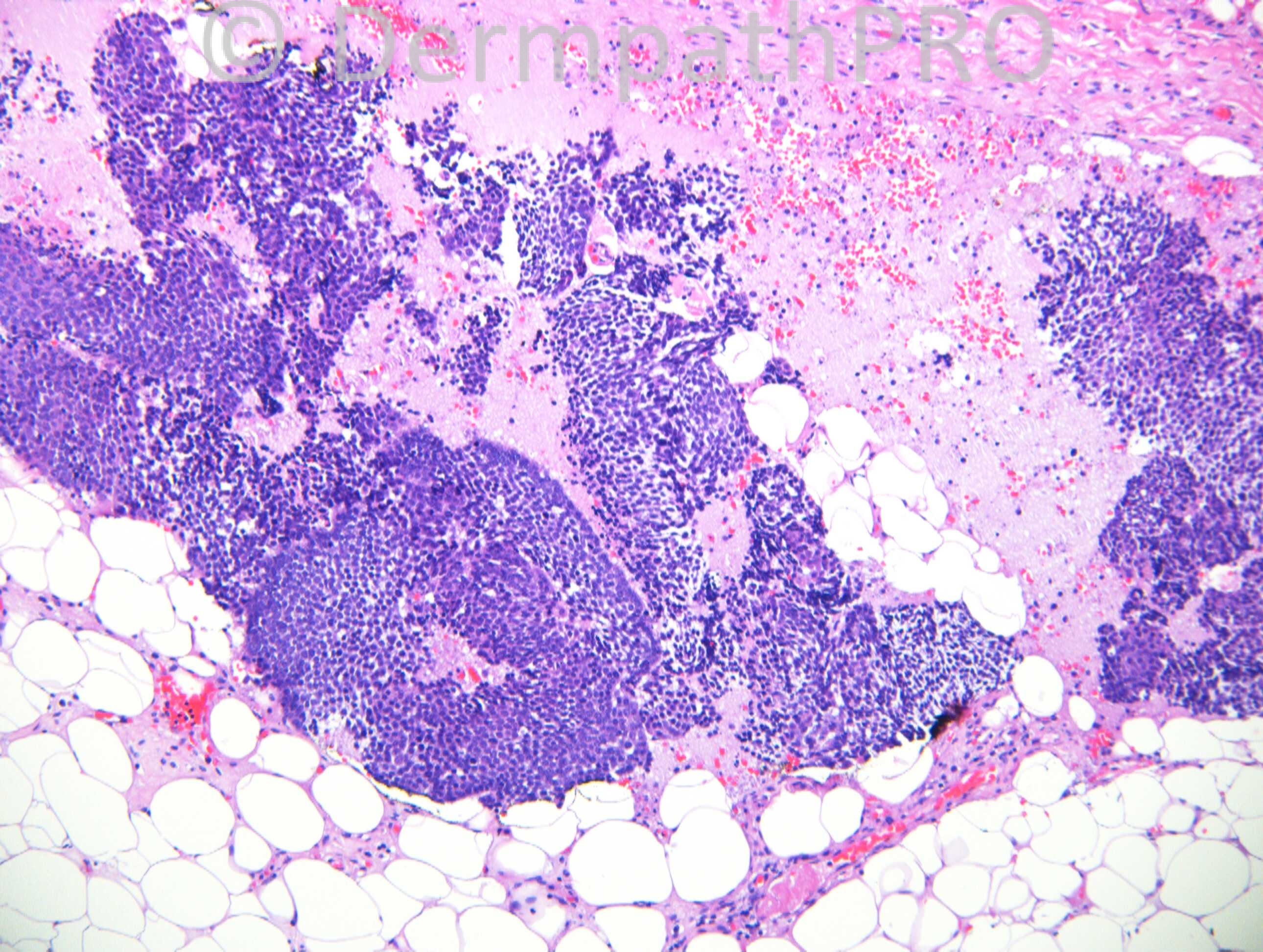
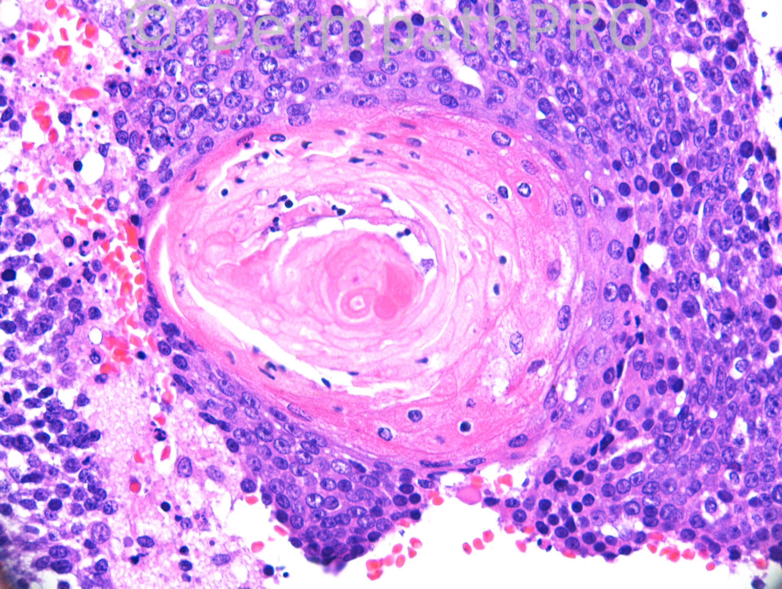
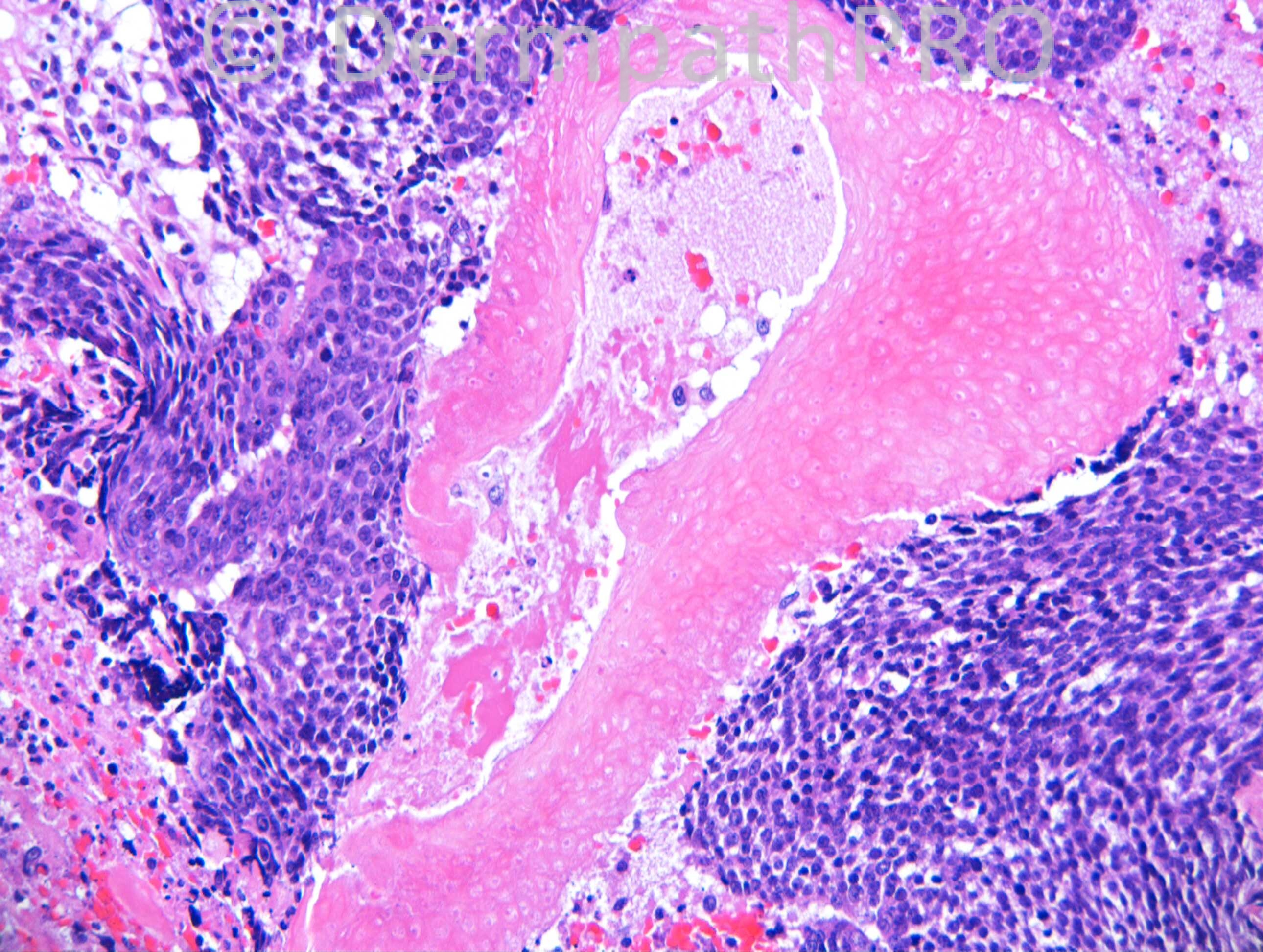
M65. Lump in neck. Malignant on FNA

Case posted by Dr. Richard Carr.

  Report Record

User Feedback


Dr. Mona Abdel-Halim

Posted

Pilomatrical carcinoma

Share this comment


Link to comment
share_externally

Agree with Mona, pilomatrical carcinoma with intravascular invasion.

Share this comment


Link to comment
share_externally

Robledo F. Rocha

Posted

Although benign pilomatricoma may have plentiful mitosis of its basaloid cells, the images above display a tumor that shows an asymmetrical silhouette due to irregular budding growth pattern into the surrounding desmoplastic stroma and into the subcutaneous fat. Geographic necrosis and vascular invasion can also be found. Moreover, basaloid cells have rather enlarged nuclei with prominent nucleoli. So, I agree with my colleagues. This is pilomatrical carcinoma.

Share this comment


Link to comment
share_externally

Guest Omobolade Obadofin

Posted

Malignant Pilomatricoma

Share this comment


Link to comment
share_externally

Guest Jim Davie MD

Posted

Agree this most likely represents a pilomatrical carcinoma, possibly arising in a pre-existing pilomatricoma. The necrosis and high-grade atypia are useful features, as well described by Robledo.

To expand the differential....
1) Pilomatrical carcinoma arising in a preexistent pilomatricoma.
2) Pilomatricoma (or proliferative pilomatricoma) with FNA artefact.
Given the history of recent FNA, one may consider procedure-related necrosis/hemorrhage, and artefactual displacement of matrical cells into vessels. There is scar-like fibrosis focally. The intravascular deposit shows small, homogeneous matrical cells lacking high-grade atypia...with trapped fibrin/red cells at the periphery.

Share this comment


Link to comment
share_externally

Arti Bakshi

Posted

The tumour appears fairly well circumscribed at low power and extension into subcutaneous fat is not unusual for pilomatricomas. As already poinetd out, mitoses can be numerous too. Not sure if the cytology is particularly high grade.
If it hadnt been for the vascular invasion, would have struggled to call this malignant!

Share this comment


Link to comment
share_externally

Dr. Richard Carr

Posted

I reported this as a tumour showing features of a proliferating pilomatricoma with intravascular foci of uncertain significance (spotted by Nick Turnbull our fellow). I could not see other criteria for malignancy and shared the case via powerpoint with Dmitry Kazakov who agreed with a non-commital appraisal. Obviously the patient needs to be followed up, I agree with Jim the intra-vascular tumour appeared banal. Typical mitoses are plentiful in the lesion as expected in the proliferative stage of a pilomatrixoma and I thought the lesion was well circumscribed and lacked tumour cell necrosis or any other criterion for malignancy. Dmitry Kazakov has reported a small number of otherwise benign mixed tumours with intravascular tumour at the peripheries with limited follow-up of no adverse outcome. I had reported another similar case to this around ten years ago and I am not aware the patient came to any harm (I only spotted the intra-vascular tumour reviewing the slides several years later). Eduardo Calonje reported a small series of otherwise typical hidradenoma with "benign" nodal metastases. In sum intravascular deposits of tumour unfortunately are not sufficient for a diagnosis of malignancy in isolation. Enjoy your weekends.

Share this comment


Link to comment
share_externally


Join the conversation

You can post now and register later. If you have an account, sign in now to post with your account.

Guest
Add a comment...

×   Pasted as rich text.   Paste as plain text instead

  Only 75 emoji are allowed.

×   Your link has been automatically embedded.   Display as a link instead

×   Your previous content has been restored.   Clear editor

×   You cannot paste images directly. Upload or insert images from URL.

Loading...

×
×
  • Create New...