Jump to content
In this section we have spot diagnoses posted on a daily basis since June 2010, now over 4000! You can review the archived cases and read the suggested diagnoses by users and the final comment by the contributors.
Case are uploaded each week day by 10 am UK time with the correct diagnosis will generally be posted at 8 pm UK time. Why not view the most recent spot diagnosis and proffer a diagnosis?

Case Number : Case 1143 - 10th November Posted By: Guest

Please read the clinical history and view the images by clicking on them before you proffer your diagnosis.
Submitted Date :
   (0 reviews)

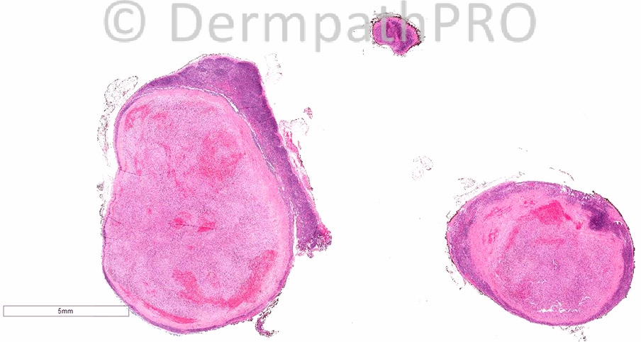
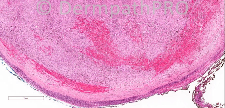
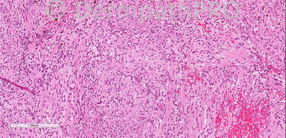
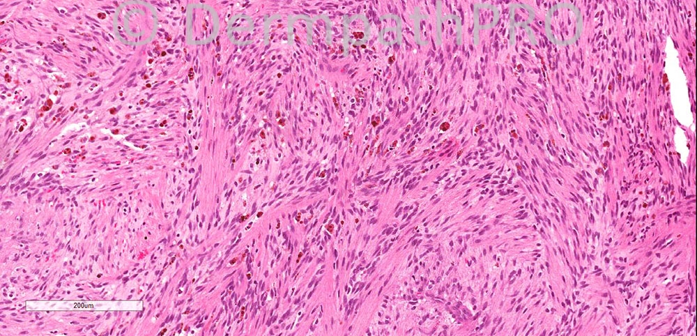
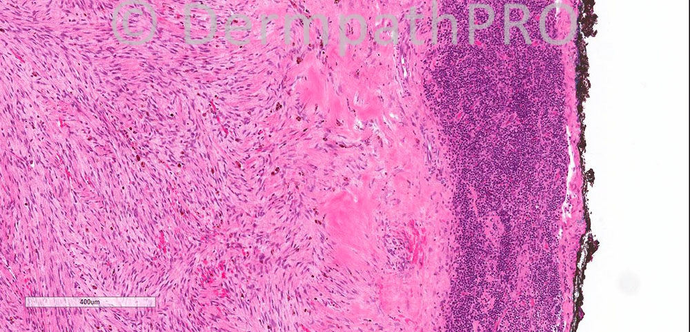
49 year old male. Enlarged nodule, left groin.

Case posted by Dr. Iskander H. Chaudhry


  Report Record

User Feedback


Dr. Mona Abdel-Halim

Posted

Agree ... Have never seen this before :-))

Share this comment


Link to comment
share_externally

Guest Romualdo

Posted

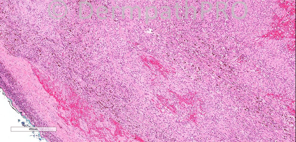
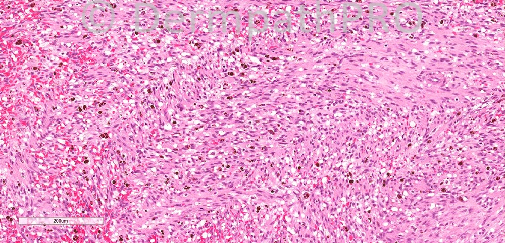
Intranodal hemorrhagic spindle cell tumor with amianthoid fibers (intranodal palisaded myofibroblastoma).

Share this comment


Link to comment
share_externally

Guest Giovanni Falconieri

Posted

Challenging case. With Romualdo input I would also favor nodal myofibroblastoma. Close resemblance to KS has been emphasized by Suster and Rosai in their AJSP seminal paper([url="http://www.ncbi.nlm.nih.gov/pubmed?term=suster%5Bauthor%5D+AND+amianthoid&TransSchema=title&cmd=detailssearch"]http://www.ncbi.nlm....d=detailssearch[/url]).

Share this comment


Link to comment
share_externally

Dr. Richard Carr

Posted

Agree with Romualdo and nice contribution of Giovanni

Share this comment


Link to comment
share_externally

Intranodal palisaded myofibroblastoma. The amianthoid fibres are seen on the last image.

Share this comment


Link to comment
share_externally

Dr. Mona Abdel-Halim

Posted

I have never seen this entity... Wounderful :-))

Share this comment


Link to comment
share_externally

Iskander H. Chaudhry

Posted

[font=arial, sans-serif][size=3]The lesion is HHV8 negative ... any other suggestions[/size][/font]

Share this comment


Link to comment
share_externally

Guest Tiberiu Tebeica

Posted

This is absolutely a new one for me :) Thank you so much for sharing and teaching!

Share this comment


Link to comment
share_externally

Dr. Richard Carr

Posted

Guess we need S100, MelanA and maybe a few more markers!

Share this comment


Link to comment
share_externally

Dr. Mona Abdel-Halim

Posted

So from Dr Iskander's comment, it seems that still the case is undiagnosed !!!!
Another idea crossed my mind just now, what if this is not a lymph node, what if this is angiomatoid fibrous histiocytoma. Thick pseudo capsule, lymphoid follicle aggregates, bland spindle cells , histiocytoid cells and blood filled spaces ...

Share this comment


Link to comment
share_externally

Iskander H. Chaudhry

Posted

[font=arial, sans-serif][size=3]The diagnosis is[/size][/font][b] Palisaded myofibroblastoma/intranodal haemorrhagic spindle cell tumour with amianthoid fibres. [/b]

[font=arial, sans-serif][size=3]Well done to all who got this right. [/size][/font]
[font=arial, sans-serif][size=3]
We also wrote a paper on that - Intranodal palisaded myofibroblastoma--an unusual entity. Thiryayi SA, Andrews B, Hall GL, Chaudhry IH. [/size][/font][font=arial, sans-serif][size=3]
[url="http://www.ncbi.nlm.nih.gov/pubmed/21217093"]http://www.ncbi.nlm.nih.gov/pubmed/21217093[/url][/size][/font]

Share this comment


Link to comment
share_externally


Join the conversation

You can post now and register later. If you have an account, sign in now to post with your account.

Guest
Add a comment...

×   Pasted as rich text.   Paste as plain text instead

  Only 75 emoji are allowed.

×   Your link has been automatically embedded.   Display as a link instead

×   Your previous content has been restored.   Clear editor

×   You cannot paste images directly. Upload or insert images from URL.

Loading...

×
×
  • Create New...