Jump to content
In this section we have spot diagnoses posted on a daily basis since June 2010, now over 4000! You can review the archived cases and read the suggested diagnoses by users and the final comment by the contributors.
Case are uploaded each week day by 10 am UK time with the correct diagnosis will generally be posted at 8 pm UK time. Why not view the most recent spot diagnosis and proffer a diagnosis?

Case Number : Case 1147 - 14th November Posted By: Guest

Please read the clinical history and view the images by clicking on them before you proffer your diagnosis.
Submitted Date :
   (0 reviews)

Spot Diagnosis

Case Posted by Dr Richard Carr.

  Report Record

User Feedback


Dr. Richard Carr

Posted

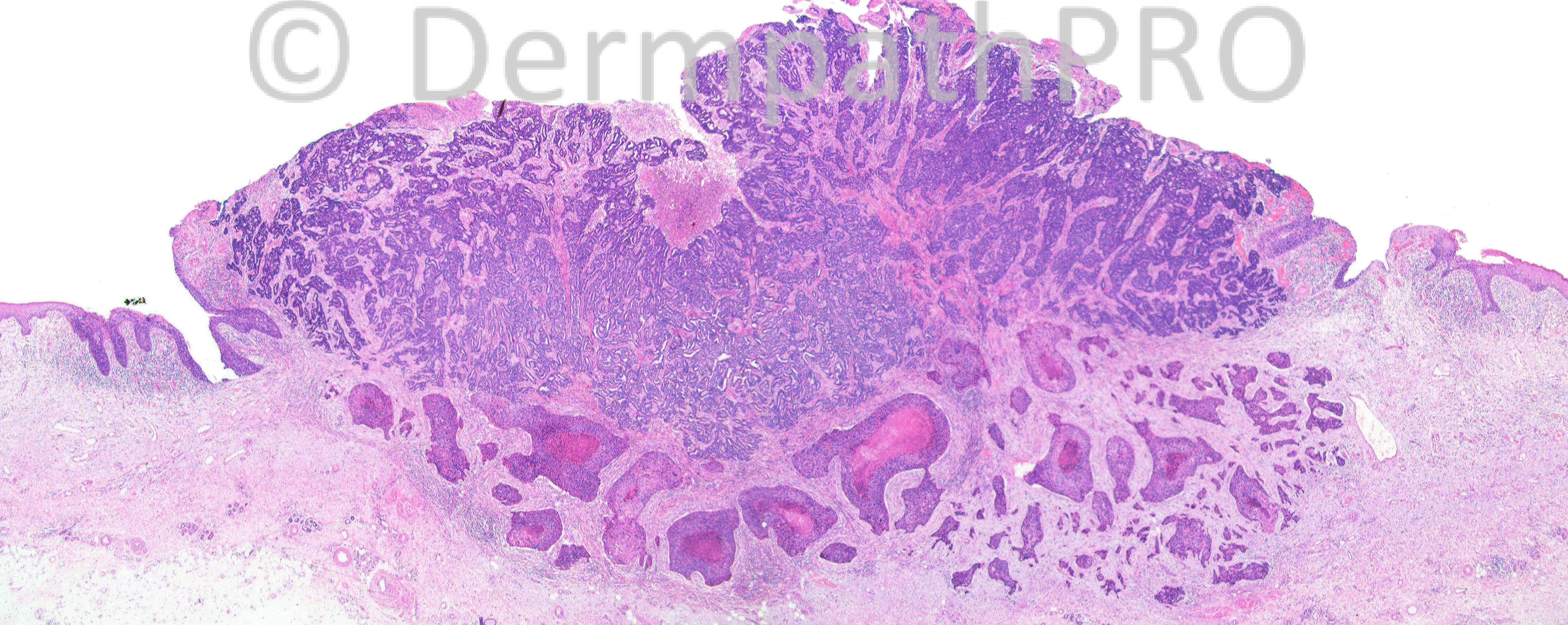
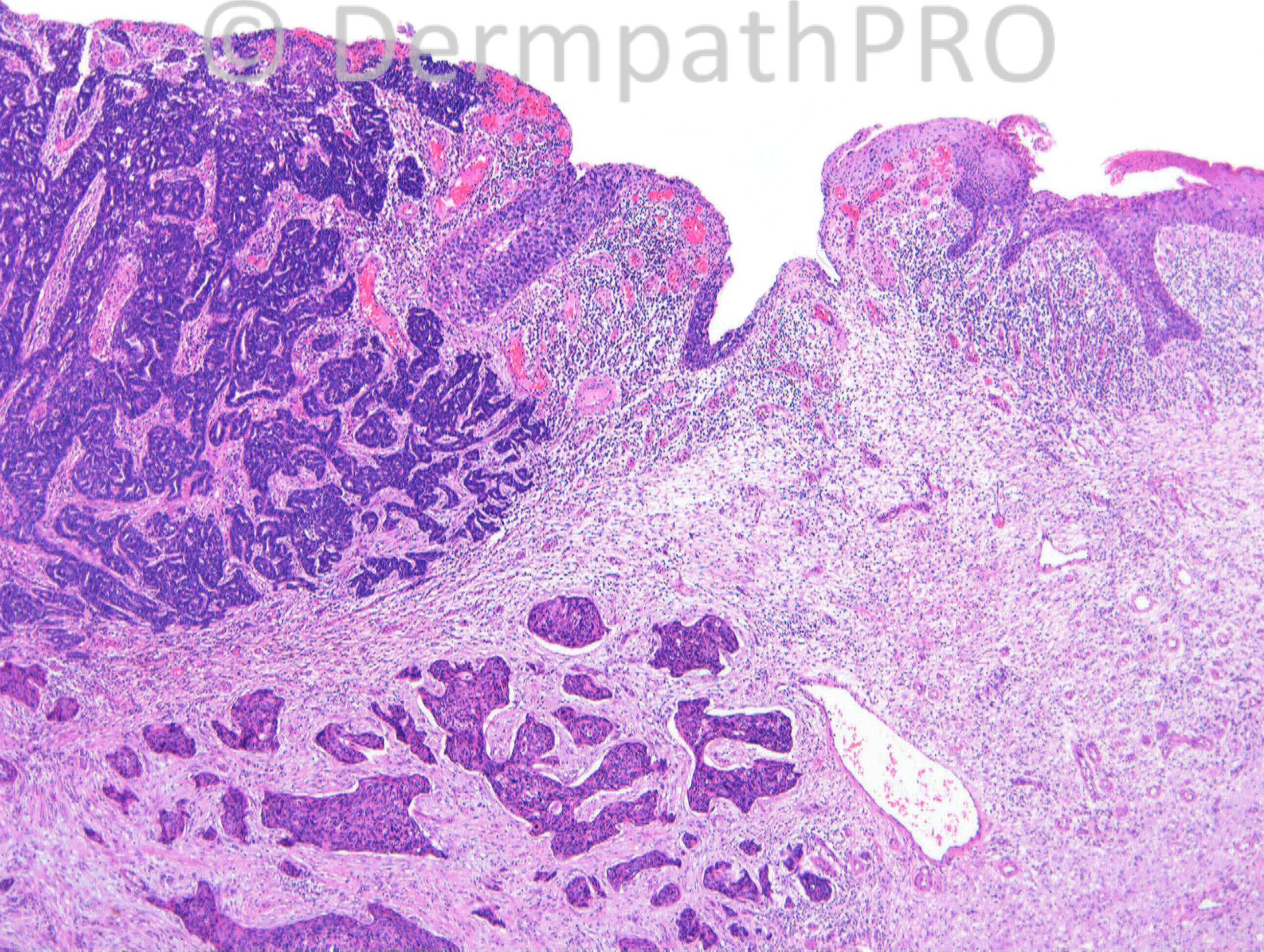
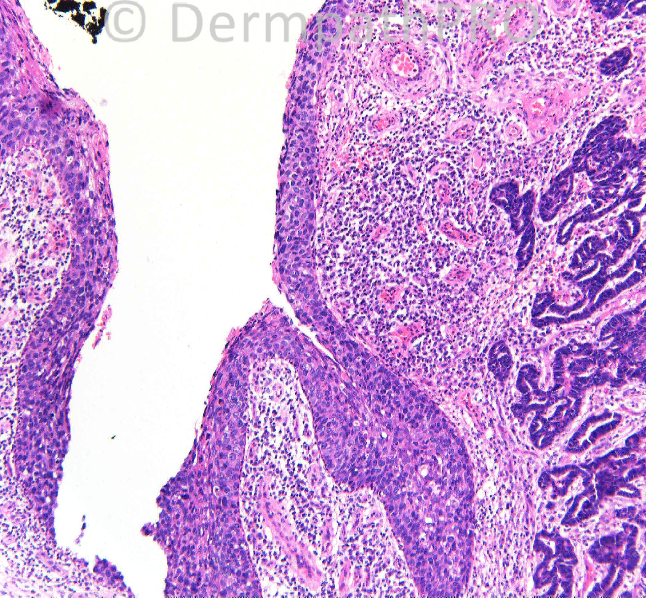
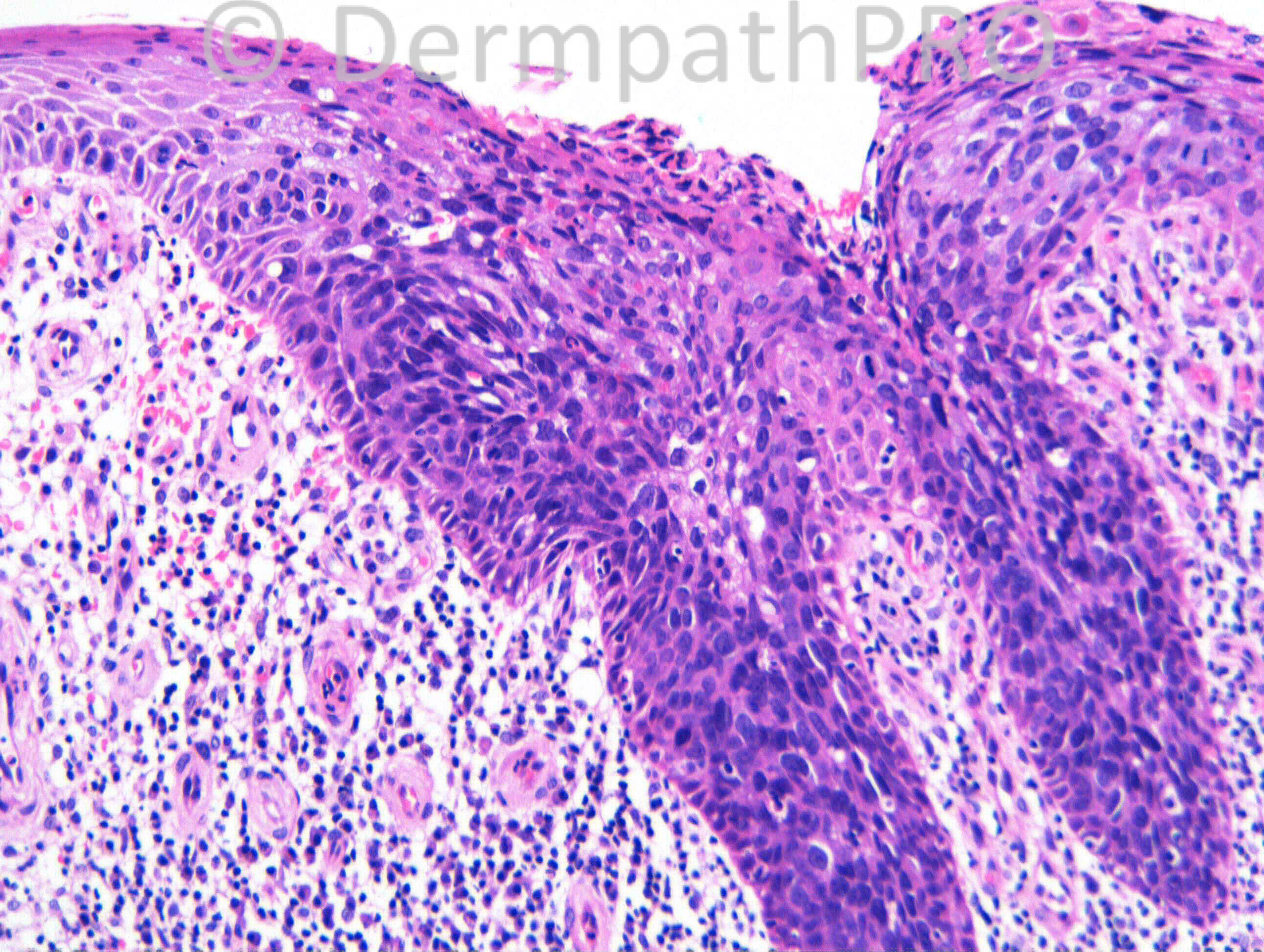
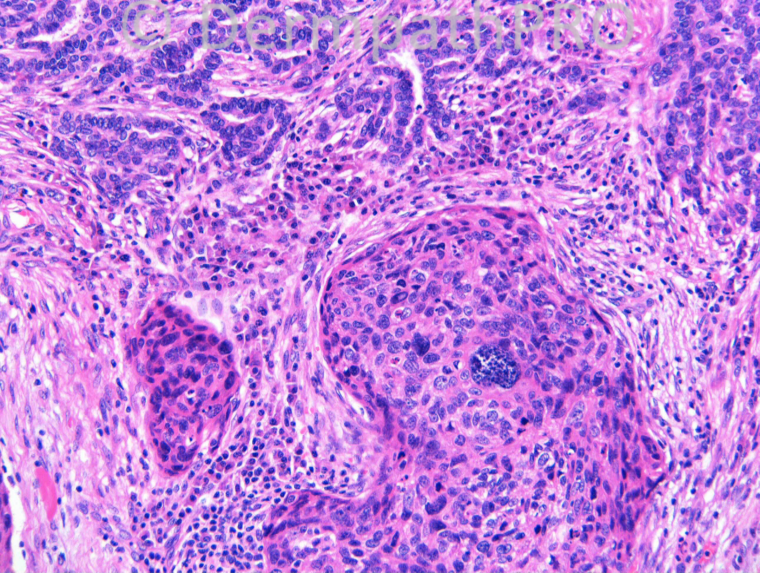
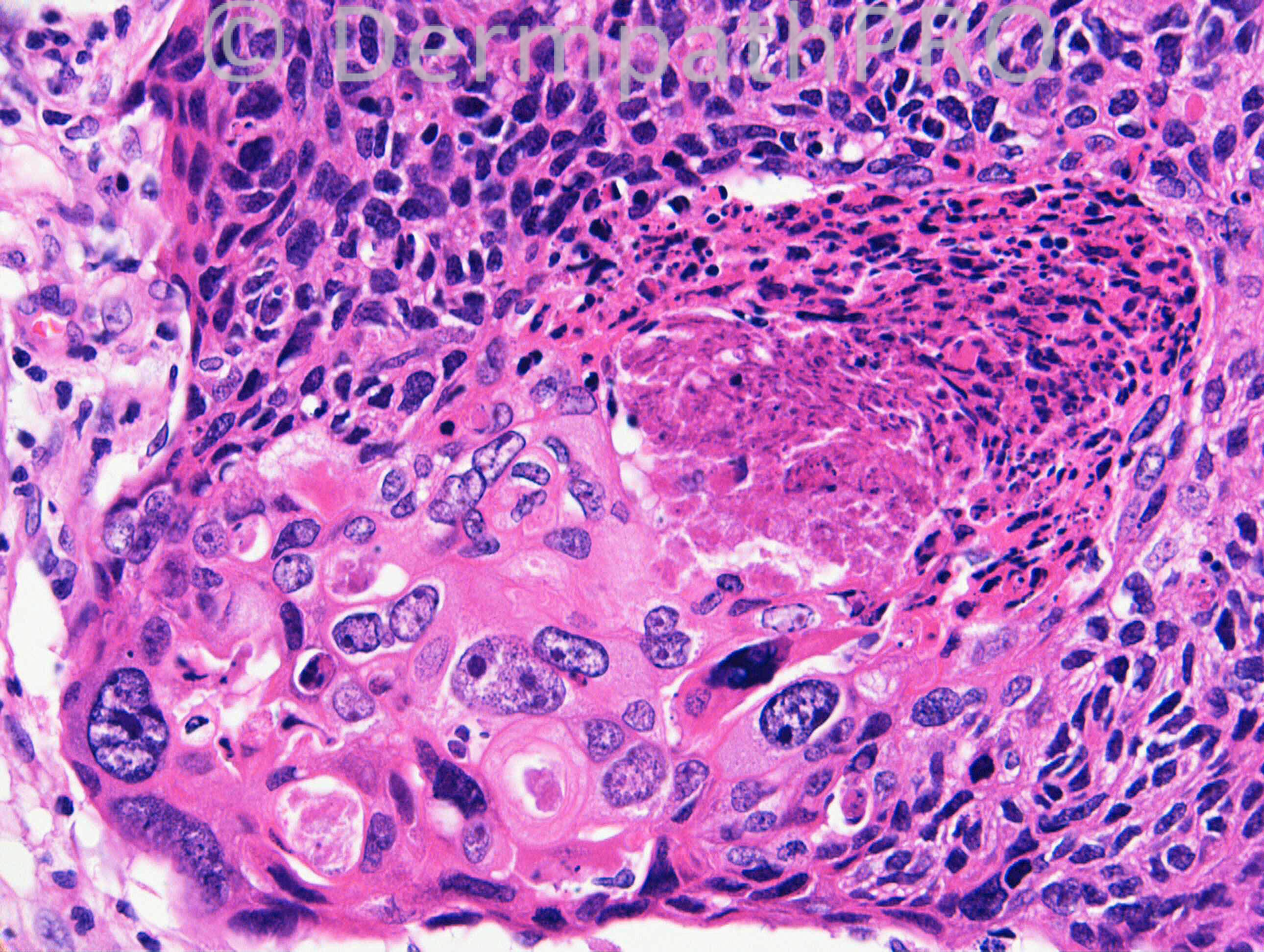
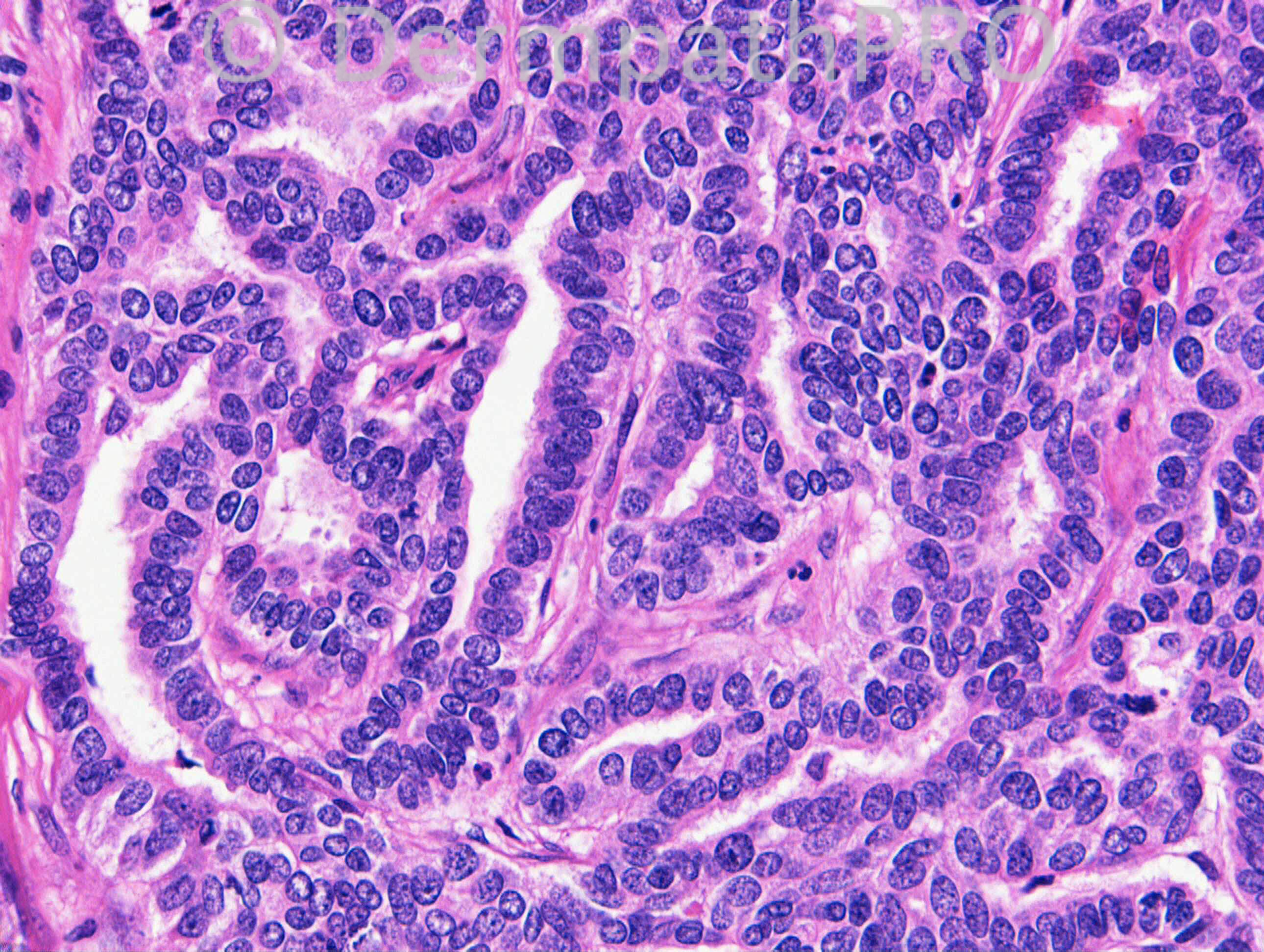
The history is of a lesion on the lower leg of an elderly female (>80years). Case kindly shared with me by Dr Florence Deroide. Some IHC at 4pm today.

Share this comment


Link to comment
share_externally

Guest Romualdo

Posted

My hypothesis: metastatic adenocarcinoma, possibly from the lung, arising within a cutaneous squamous cell carcinoma.

Share this comment


Link to comment
share_externally

Dr. Richard Carr

Posted

[img]https://dermpathpro.com/uploads/spot_diagnosis_comment_img/CASE1147_RAC6933x10_CK7_IHC_4pm.jpg[/img]

[img]https://dermpathpro.com/uploads/spot_diagnosis_comment_img/CASE1147_RAC6933x10_ER_IHC_4pm.jpg[/img]

[img]https://dermpathpro.com/uploads/spot_diagnosis_comment_img/CASE1147_RAC6933x5_p63_IHC_4pm.jpg[/img]

Share this comment


Link to comment
share_externally

Dr. Richard Carr

Posted

CK5 strong all areas, CDX2, CK20 & TTF1 negative in all areas. Welcome additional suggestions & hypotheses.

Share this comment


Link to comment
share_externally

Guest Jim Davie MD

Posted

Agree with Mark's assessment... apocrine differentiation in pre-existing in-situ and invasive SCC.

There is definitely an in-situ and invasive squamous carcinoma component. The basaloid glandular component is broad and shallow, with epidermal replacement, hinting at an origin within in-situ SCC...there is limited overlap between the two histologies.

The epicenter of the glandular component precisely matches the epicenter of the invasive SCC component, which would probabilistically favor apocrine differentiation in pre-existing in-situ/invasive SCC over an unlikely precise targeting of metastatic ACa into the center of a small well-circumscribed focus of preexisting SCC.

For immunostaining, strong CK5 in all areas would favor adnexal carcinoma (97% CK5+); metastatic ACa is positive for CK5 in 30% of cases, but staining is reportedly weaker. [size=4][ref 1][/size]

That said, negative staining for P63 would be rare for primary adnexal ACa (9% of cases); additional stains for CK15 and D2-40 (podoplanin) may help differentiate [size=4]primary adnexal ACa vs cutaneous metastasis from breast [/size][size=4](P63/CK15/D2-40: primary adnexal ACa is 91%, 40% and 44% positive, vs. mets: 0%, 6% and 0% positive) [ref 2]. [/size]

Ref:
[1] [size=4]Cytokeratin 5/6 immunostaining in cutaneous adnexal neoplasms and metastatic adenocarcinoma. [/size][i][size=4]Am J Dermatopathol. 2004 Dec;26(6):447-51.[/size][/i]
[url="http://www.ncbi.nlm.nih.gov/pubmed/15618924"]http://www.ncbi.nlm....pubmed/15618924[/url]

[2] [size=4]The diagnostic utility of immunohistochemistry in distinguishing primary skin adnexal carcinomas from metastatic adenocarcinoma to skin: an immunohistochemical reappraisal using cytokeratin 15, nestin, p63, D2-40, and calretinin. [/size][i][size=4]Modern Pathology (2010) 23, 713–719.[/size][/i]
Full article online: [url="http://www.nature.com/modpathol/journal/v23/n5/full/modpathol201046a.html"]www.nature.com/modpathol/journal/v23/n5/full/modpathol201046a.html[/url]

Share this comment


Link to comment
share_externally

Arti Bakshi

Posted

What an unusual case and what great discussion! Not sure that I can add anything to all the possibilities considered. To my eye, the 2 areas appear too distinct with completely distinct immunoprofile to reconcile with apocrine differentiation within invasive SCC. Cutaneous mets from breast Ca has to be excluded although agree that 'the precise targeting' is unusual!

Share this comment


Link to comment
share_externally

Dr. Richard Carr

Posted

Great responses. I would not argue against apocrine although the similarity to regular breast ductal carcinoma put me more in favour of calling it eccrine ductal adenocarcinoma with Bowen's and invasive bowenoid SCC. My report as follows:

We appear to have conventional Bowen's disease superficially, associated
with an invasive Bowenoid / poorly differentiated squamous cell
carcinoma. Distinct from this, but closely abutting the lesion on all
sides, is a centrally located moderately differentiated adenocarcinoma;
most in keeping with eccrine ductal type. One would have to consider
the possibility of a metastasis for the latter lesion, but the strong
staining for CK5 would support a primary adnexal origin. I think the
general broad nature of the lesion also supports that hypothesis.

I undertook additional immunostains. Interestingly, the oestrogen
receptor is moderately positive in the Bowen's disease and focally in
the adenocarcinoma, but negative in the poorly differentiated invasive
squamous cell carcinoma. CK7 was entirely limited to the adenocarcinoma
and the superficial layers of the Bowen's disease. CDX2, CK20 and TTF1
were negative in both phenotypes.

In summary, like you, I favour eccrine ductal carcinoma of primary
cutaneous origin arising in association with conventional Bowen's
disease and an invasive Bowenoid squamous cell carcinoma. It is
probably likely one tumour has developed from the other, although we
cannot rule out the possibility of two independent primaries, or the
less likely possibility of a metastasis to the skin from a visceral
primary.

Share this comment


Link to comment
share_externally


Join the conversation

You can post now and register later. If you have an account, sign in now to post with your account.

Guest
Add a comment...

×   Pasted as rich text.   Paste as plain text instead

  Only 75 emoji are allowed.

×   Your link has been automatically embedded.   Display as a link instead

×   Your previous content has been restored.   Clear editor

×   You cannot paste images directly. Upload or insert images from URL.

Loading...

×
×
  • Create New...