Jump to content
In this section we have spot diagnoses posted on a daily basis since June 2010, now over 4000! You can review the archived cases and read the suggested diagnoses by users and the final comment by the contributors.
Case are uploaded each week day by 10 am UK time with the correct diagnosis will generally be posted at 8 pm UK time. Why not view the most recent spot diagnosis and proffer a diagnosis?

Case Number : Case 1149 - 18th November Posted By: Guest

Please read the clinical history and view the images by clicking on them before you proffer your diagnosis.
Submitted Date :
   (0 reviews)

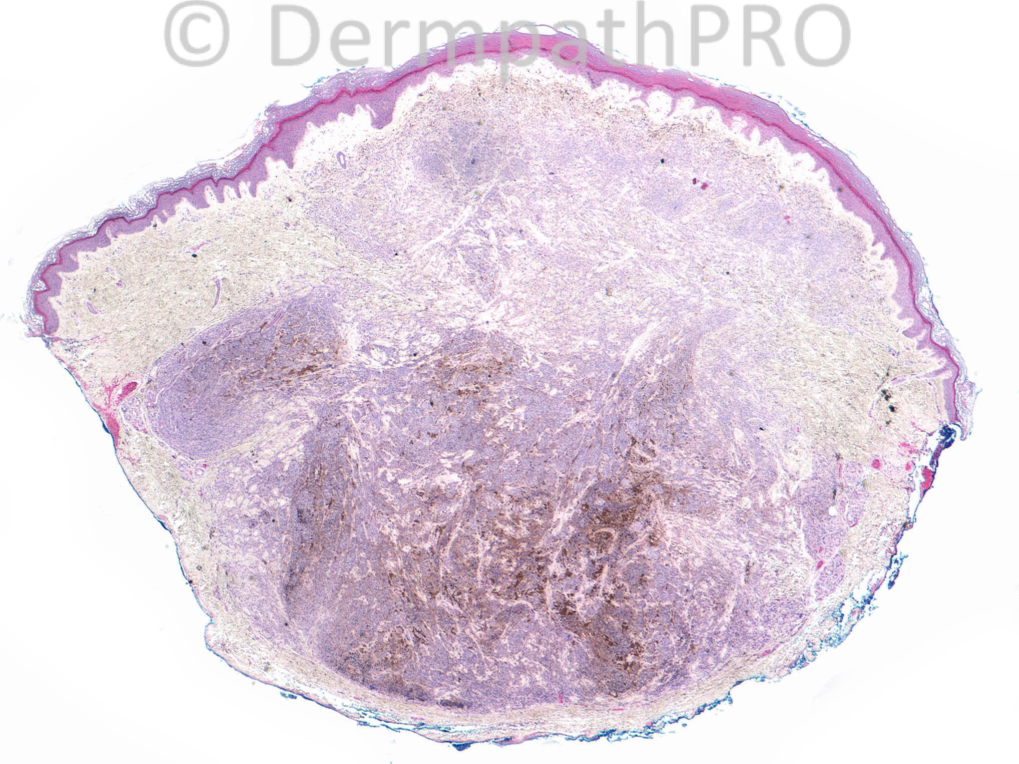
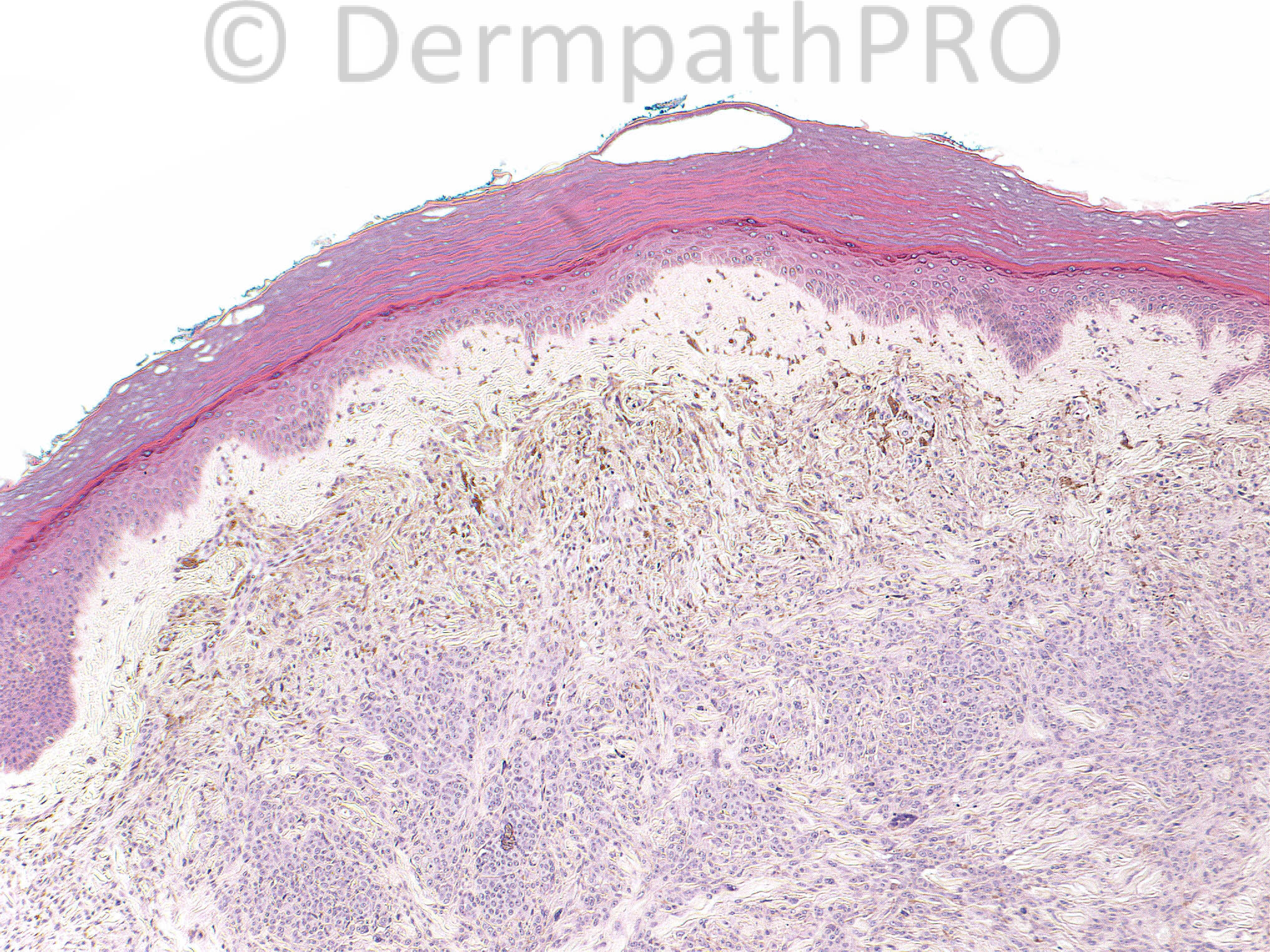
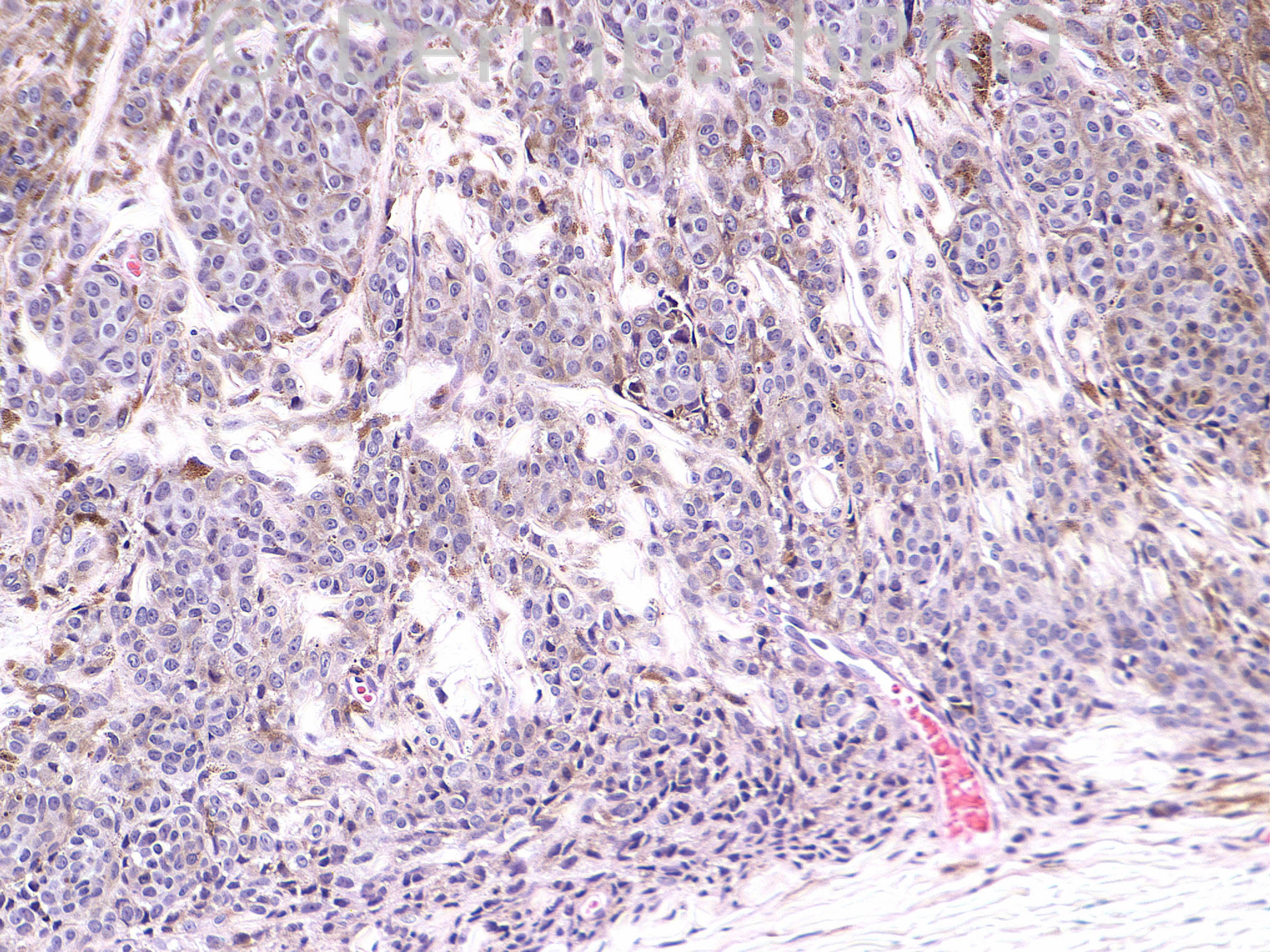
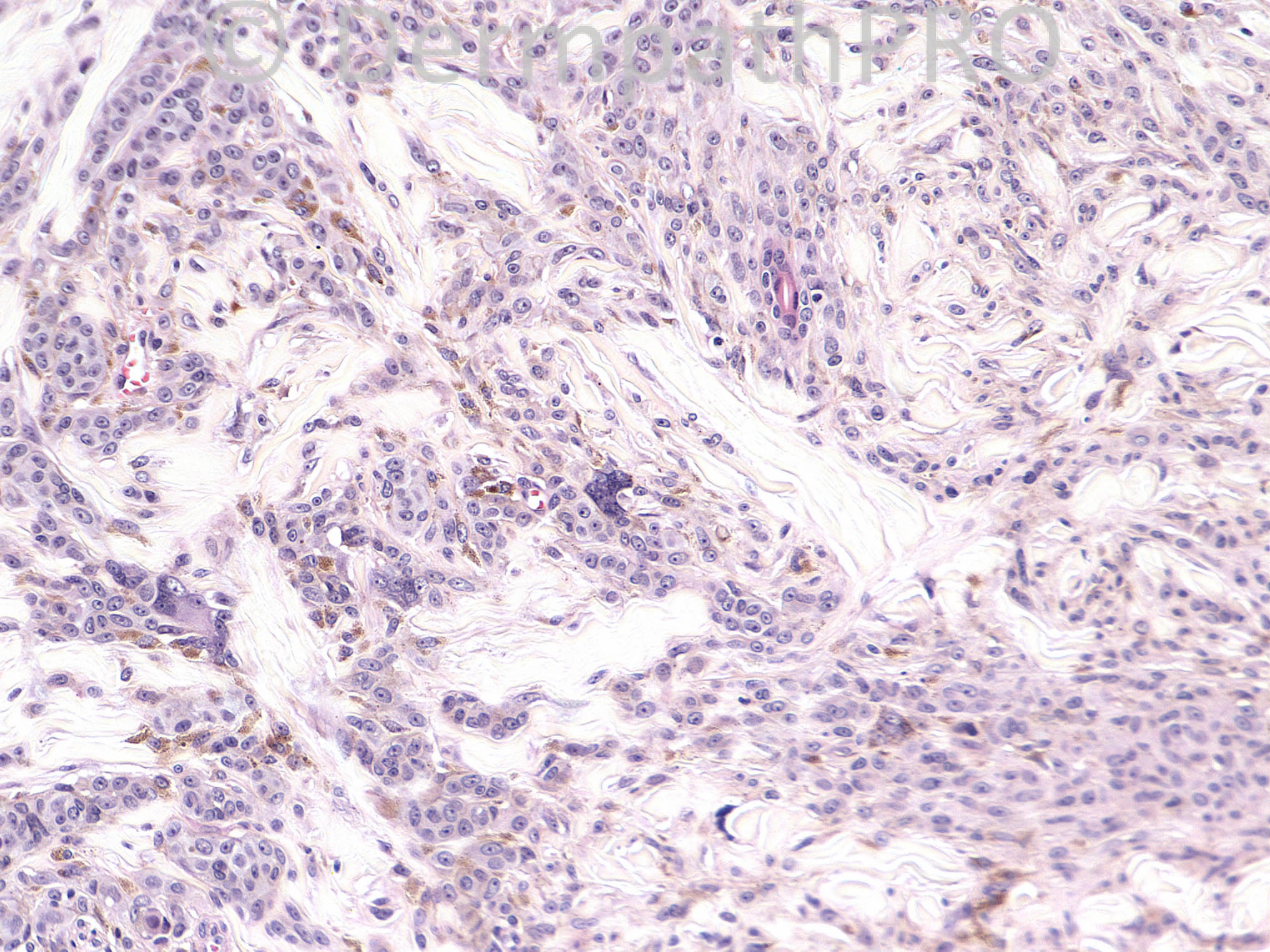
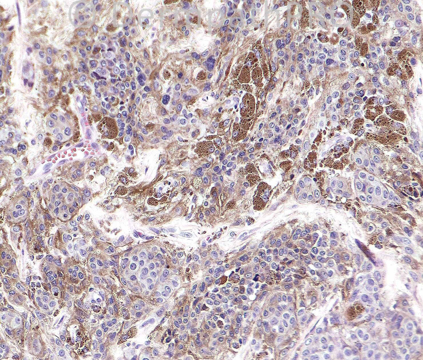
24 year old woman with rapidly enlarging left hand lesion, currently 8 mm in size, clinically suspicious for malignancy.

Case posted by Dr.Uma Sundram.


  Report Record

User Feedback


Guest Giovanni Falconieri

Posted

Epithelioid blue cell nevus/melanocytoma, to start.

Share this comment


Link to comment
share_externally

Agree. Pigmented epithelioid melanocytoma. There appears to be a common blue nevus in the upper dermis above the epithelioid component.

Share this comment


Link to comment
share_externally

Dr. Richard Carr

Posted

I find these lesions exceedingly difficult but given the biphasic character with dendritic component superficially would have to favour a cellular blue naevus. I will be interested to see if it is S100 negative as i would expect in blue naevus. Also interested to know if any molecular work-up was performed. Next question is whether I would advise a modest re-excision. I think I might not be prepared to take the risk as cellular blue naevi (buttock excepted) can recur and rarely undergo malignant transformation and i might just sleep a little easier!

Share this comment


Link to comment
share_externally

Robledo F. Rocha

Posted

It seems to have two cytologically distinct populations of melanocytes, so I go with combined nevus.

Share this comment


Link to comment
share_externally

Guest Jim Davie MD

Posted

Agree with Epithelioid blue nevus. [size=4](A bulbous architecture in the deep dermis might suggest alternate classification as deep penetrating nevus).[/size][size=4] [/size]
[size=4] There is little inflammatory reaction, small to medium nucleoli, moderate pleomorphism with some senescent-type multinucleation; the lesion shows lack of mitotic activity in the superficial and deep fields instead of the very sparse mitotic activity expected. No significant epidermal component or maturation in the mid to deep portions.[/size]

Share this comment


Link to comment
share_externally

Uma Sundram

Posted

This was an exceedingly difficult case. We did end up classifying this as a cellular blue nevus, but I must say that deep penetrating nevus and epithelioid blue nevus/pigmented epithelioid melanocytoma are both appropriate considerations. We did not favor these two latter designations because these cells seem to be spindled and nested than what we've seen in epithelioid blue nevi/pigmented epithelioid melanocytomas; the architecture is different from that of a deep penetrating nevus, which tends to have more of a columnar, wedge shaped silhouette. Molecular analysis did not favor melanoma. We did not do S100 (an excellent suggestion) but sent it to an expert who agreed with the interpretation of cellular blue nevus, although they too thought about deep penetrating nevus. We all recommended a modest re excision for the reasons suggested by Richard.

Share this comment


Link to comment
share_externally


Join the conversation

You can post now and register later. If you have an account, sign in now to post with your account.

Guest
Add a comment...

×   Pasted as rich text.   Paste as plain text instead

  Only 75 emoji are allowed.

×   Your link has been automatically embedded.   Display as a link instead

×   Your previous content has been restored.   Clear editor

×   You cannot paste images directly. Upload or insert images from URL.

Loading...

×
×
  • Create New...