Jump to content
In this section we have spot diagnoses posted on a daily basis since June 2010, now over 4000! You can review the archived cases and read the suggested diagnoses by users and the final comment by the contributors.
Case are uploaded each week day by 10 am UK time with the correct diagnosis will generally be posted at 8 pm UK time. Why not view the most recent spot diagnosis and proffer a diagnosis?

Case Number : Case 1151 - 20th November Posted By: Guest

Please read the clinical history and view the images by clicking on them before you proffer your diagnosis.
Submitted Date :
   (0 reviews)

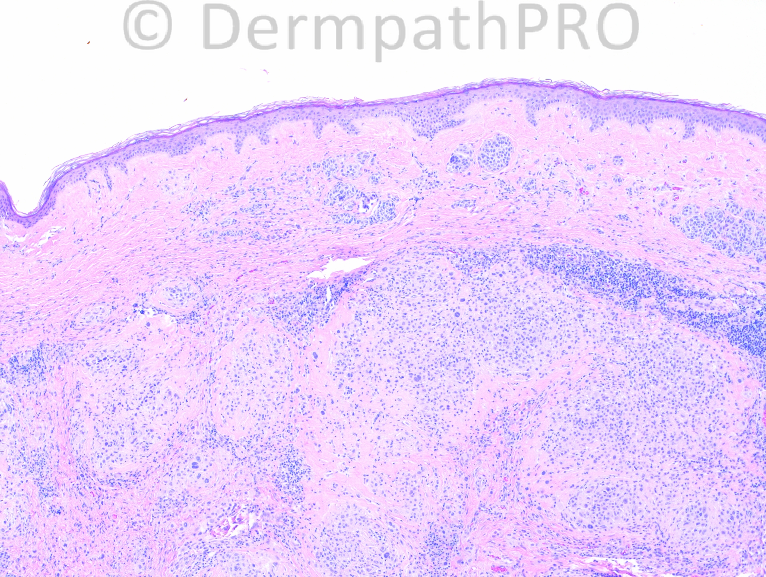
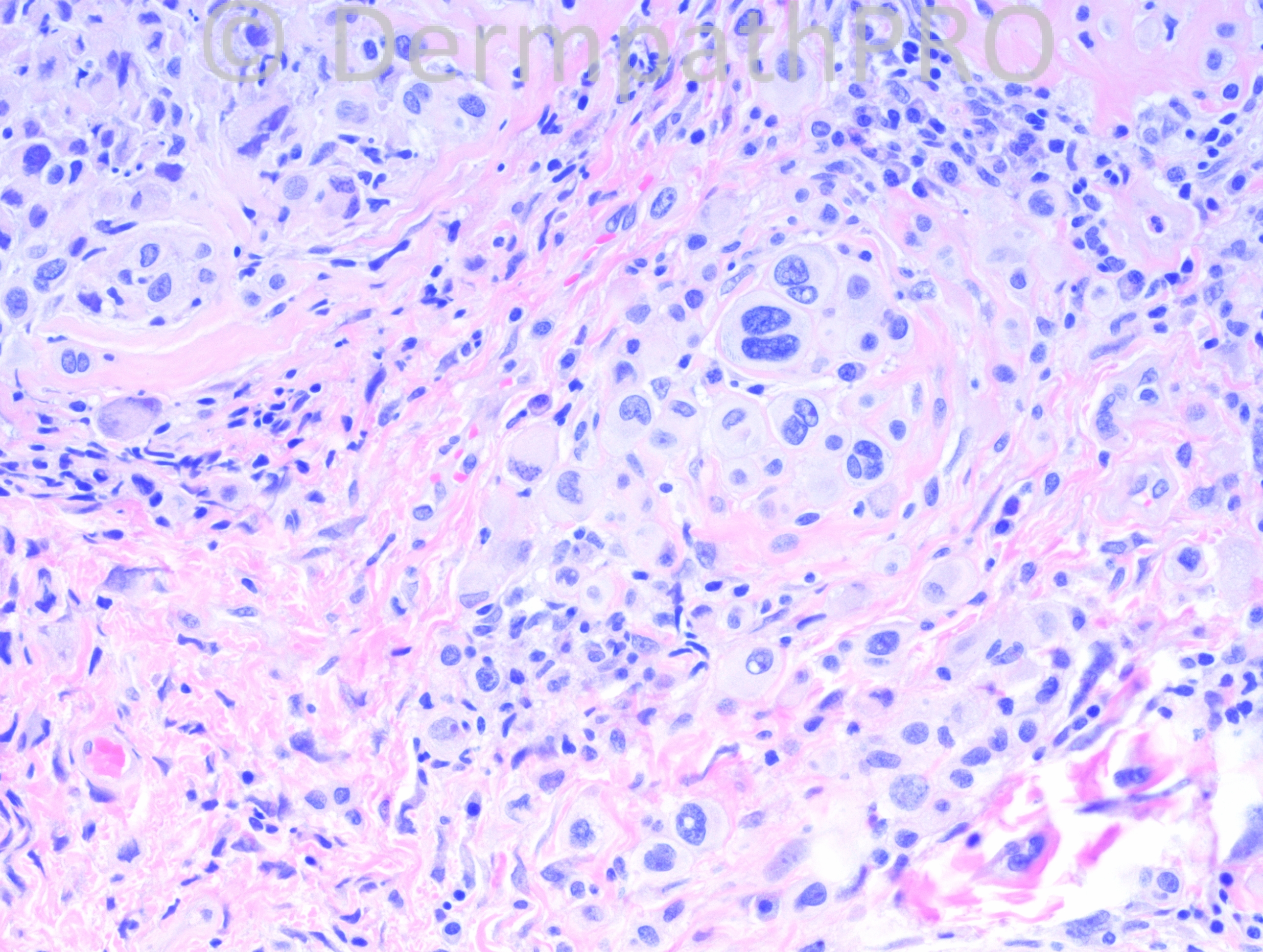
24 year-old female with right chest “mole”.

Case posted by Dr. Hafeez Diwan.

  Report Record

User Feedback


Guest Tiberiu Tebeica

Posted

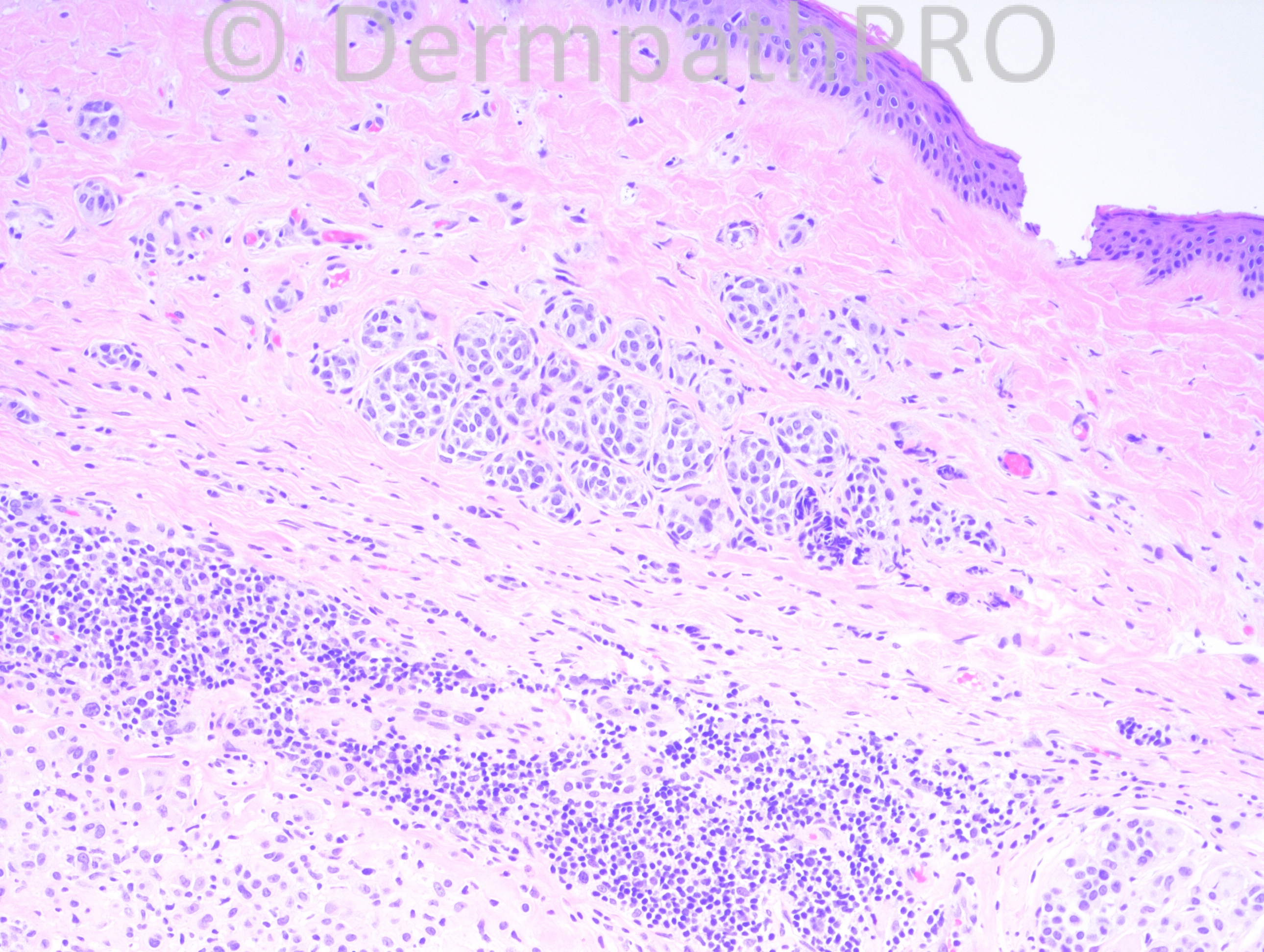
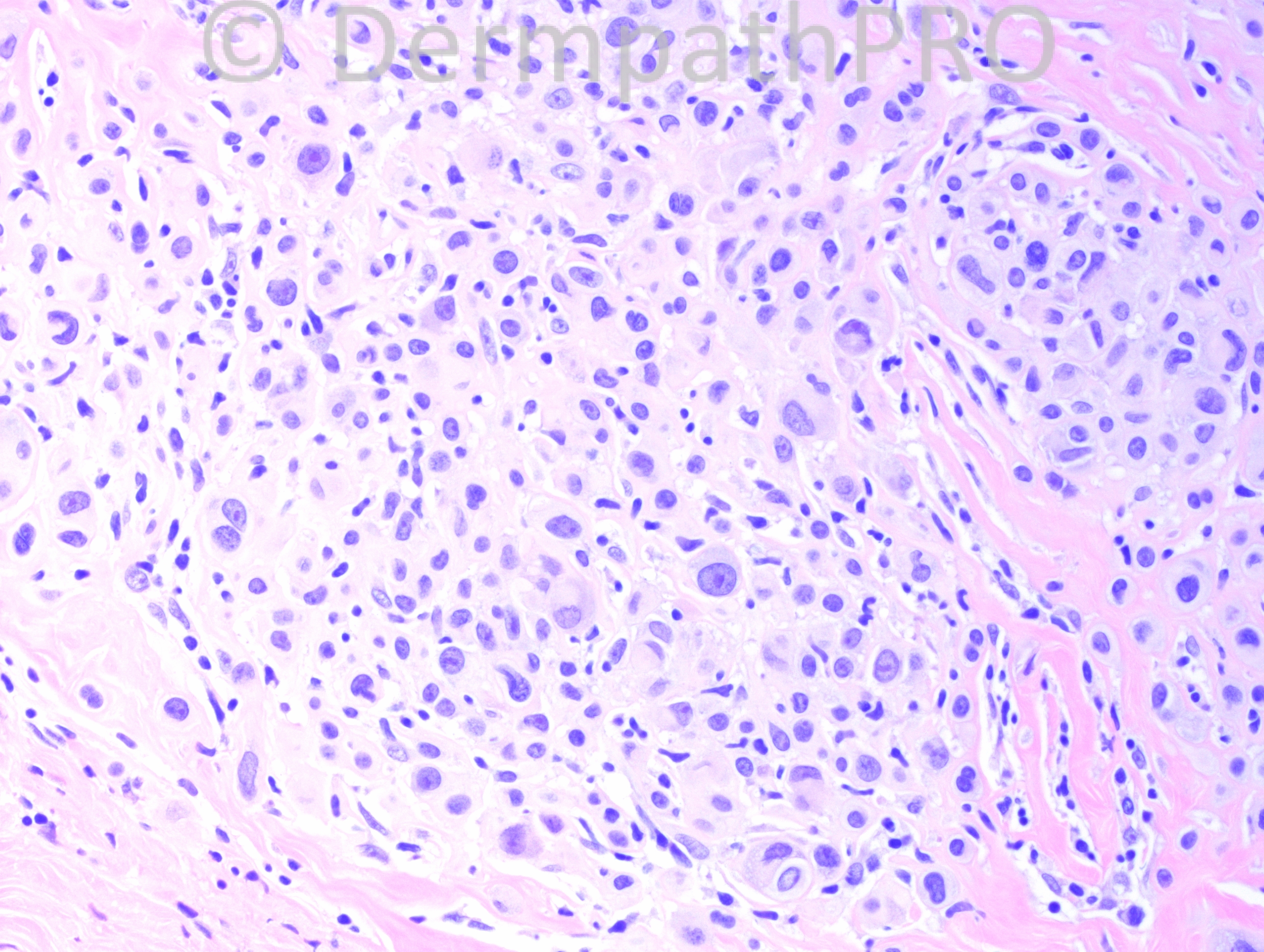
There are some histologic criteria that make me think of Wiesner nevus - another one by dr. Diwan ?!?! :) Theese criteria are: intradermal nevus with no obvious maturation, presence of epithelioid cells with nuclear pleomorphism, inflammatory infiltrate. Of course, it needs confirmatory BAP1 status. This could be also an ancient nevus, but I would not argue much with someone calling this an intradermal Spitz.

Share this comment


Link to comment
share_externally

Guest Jim Davie MD

Posted

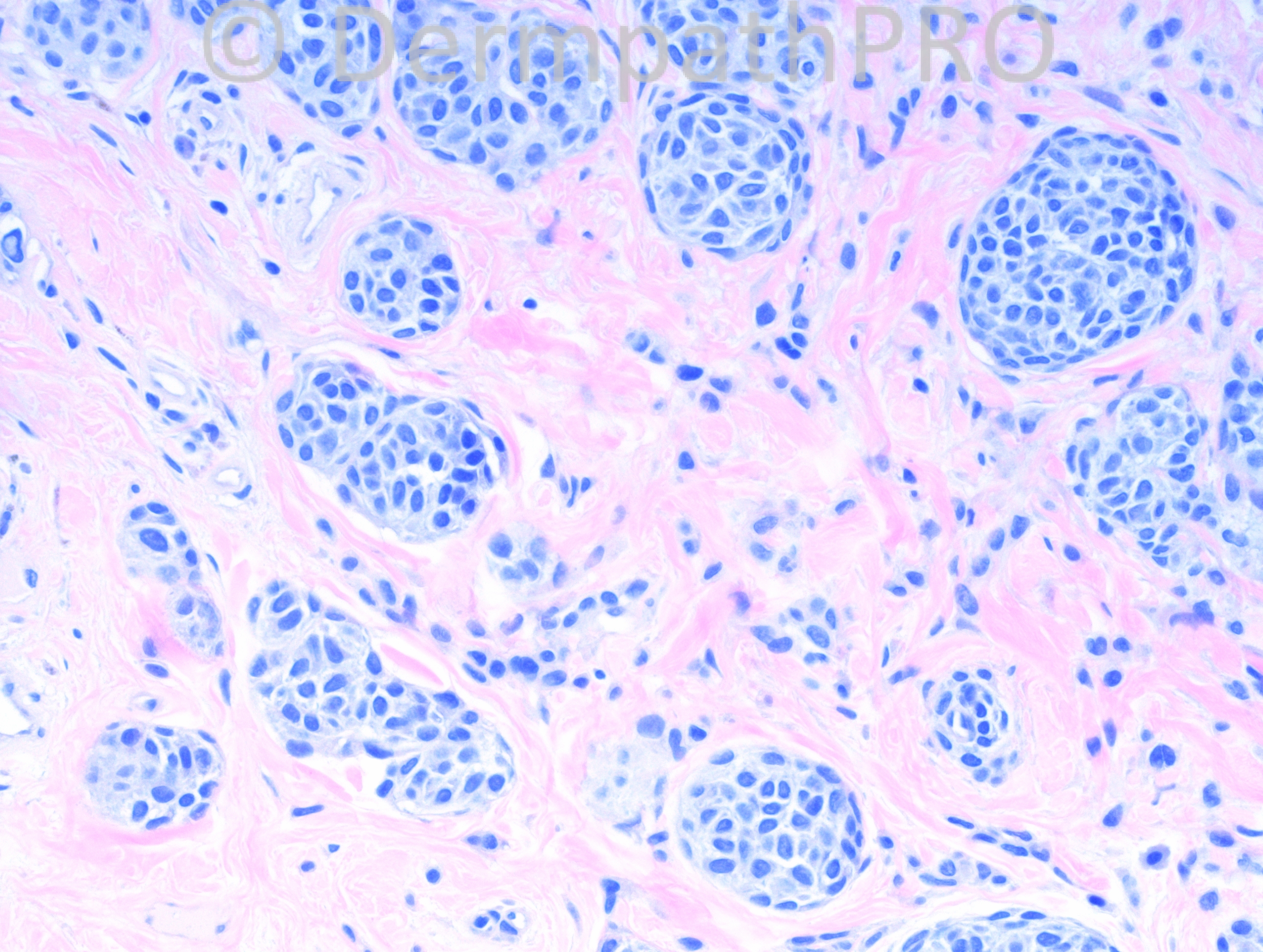
Intradermal Spitzoid melanocytic tumor of unknown malignant potential (in absence of stains/studies).

There is a hint of maturation with asymmetric melanin pigment in one asymmetric superficial dermal focus (3rd photo). The remainder of the tumor shows markedly pleomorphic epithelioid nuclei with prominent, predominantly solitary violet nucleoli, and random multinucleation, vs. smudgy senescent-type hyperchromatic round to spindled nuclei. I can't see any mitotic activity or junctional component. There is an asymmetrical chronic inflammatory response with significant plasma cell component.

I think this likely a benign Spitz nevus. However, the age range is getting high (26 yo) if a new / changing lesion. The overall asymmetry of architecture and cytology, with significant plasma cell component, are of concern and would prompt me to evaluate (1) Ki67 or other proliferative index marker, and (2) loss of P16 nuclear/cytoplasmic immunostaining (vs. FISH/CGH) to ensure this is not a wolf in sheep's clothing.

Share this comment


Link to comment
share_externally

Dr. Richard Carr

Posted

Longstanding intradermal naevus with an epithelioid clone/dermal nodule(Ball & Golitz)/phenotypic heterogeneity.

Share this comment


Link to comment
share_externally

Dr. Hafeez Diwan

Posted

Yes, this is another "Wiesner" nevus. BAP1 was negative.

Share this comment


Link to comment
share_externally


Join the conversation

You can post now and register later. If you have an account, sign in now to post with your account.

Guest
Add a comment...

×   Pasted as rich text.   Paste as plain text instead

  Only 75 emoji are allowed.

×   Your link has been automatically embedded.   Display as a link instead

×   Your previous content has been restored.   Clear editor

×   You cannot paste images directly. Upload or insert images from URL.

Loading...

×
×
  • Create New...