Jump to content
In this section we have spot diagnoses posted on a daily basis since June 2010, now over 4000! You can review the archived cases and read the suggested diagnoses by users and the final comment by the contributors.
Case are uploaded each week day by 10 am UK time with the correct diagnosis will generally be posted at 8 pm UK time. Why not view the most recent spot diagnosis and proffer a diagnosis?

Case Number : Case 1157 - 28th November Posted By: Guest

Please read the clinical history and view the images by clicking on them before you proffer your diagnosis.
Submitted Date :
   (0 reviews)

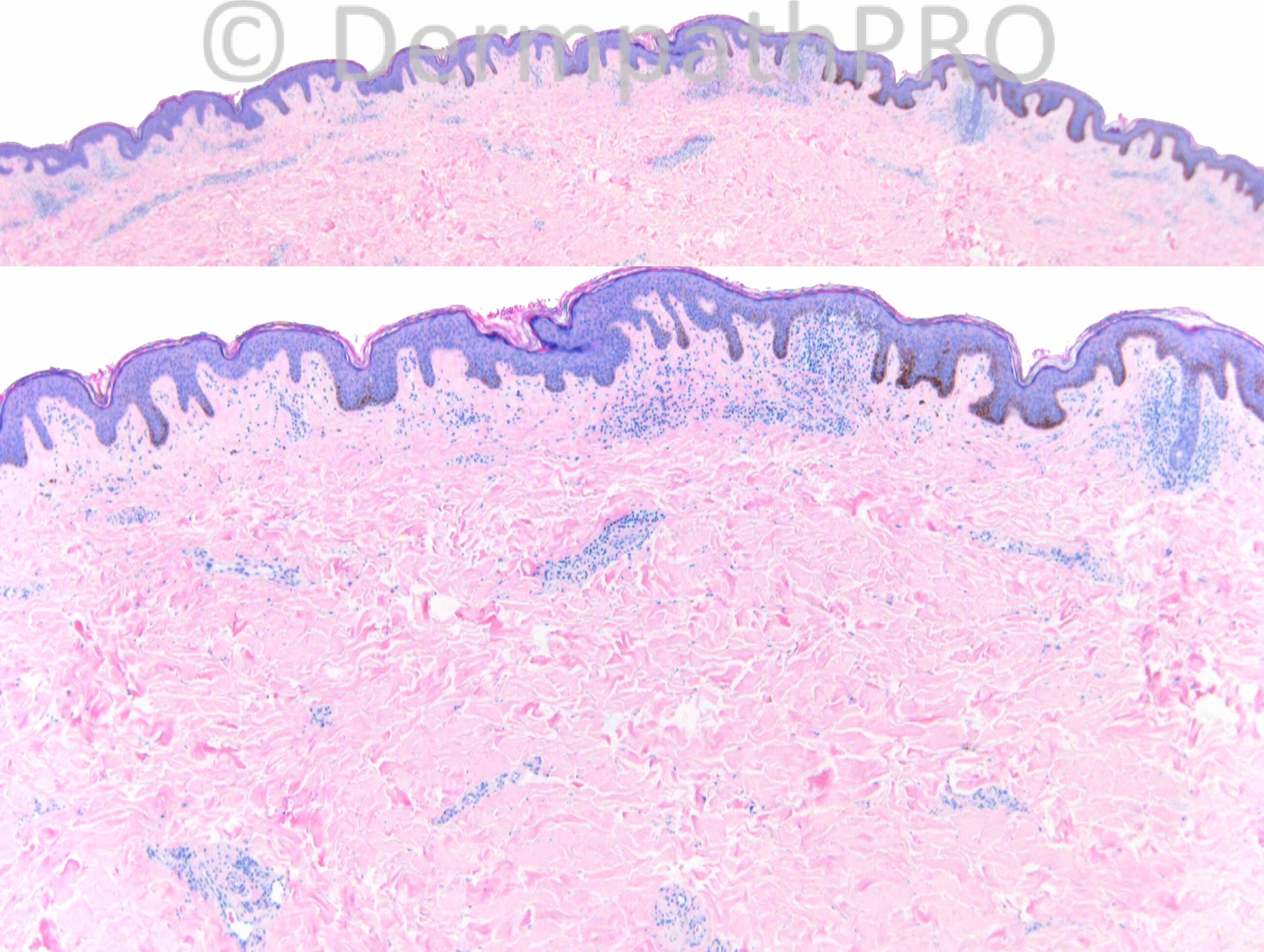
Incision biopsy Slightly atrophic, hypopigmented patch, lower abdomen. 12mths. Skin type VI ?
vitiligo,?MF

Case Posted by Dr Richard Carr


  Report Record

User Feedback


Dr. Mona Abdel-Halim

Posted

Hypopigmented MF
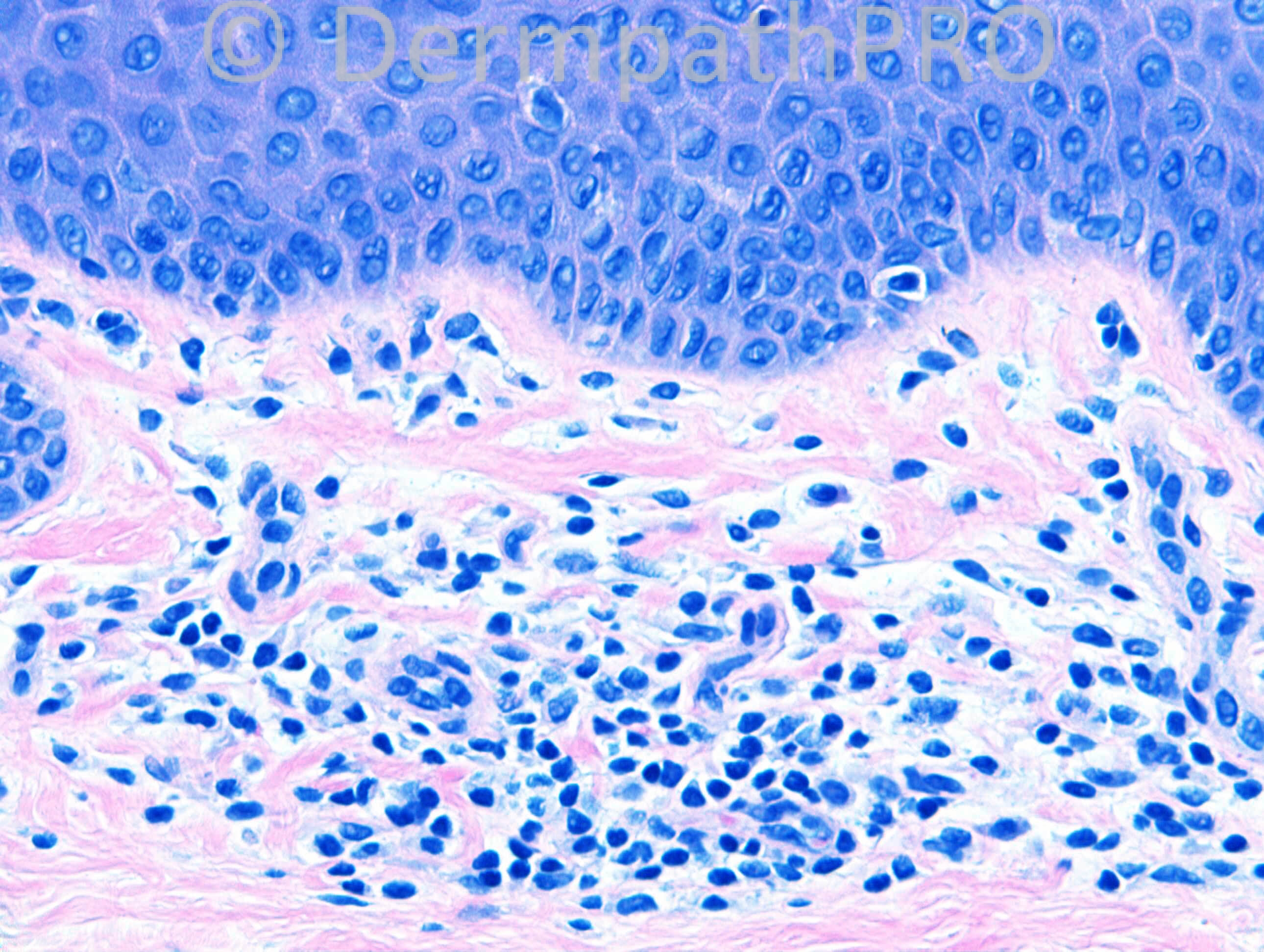
Lymphocyte atypia is perceived both in the epidermal and dermal compartments.
Wiry fibroblasia of papillary dermal collagen is an important clue that favors MF and I can perceive it here.
Clinically, the slightly atrophic nature of the lesion favors MF.

Share this comment


Link to comment
share_externally

Robledo F. Rocha

Posted

Agree with hypopigmented mycosis fungoides. Neoplastic lymphocytes are expected to express CD8+ phenotype.

Share this comment


Link to comment
share_externally

Dr. Richard Carr

Posted

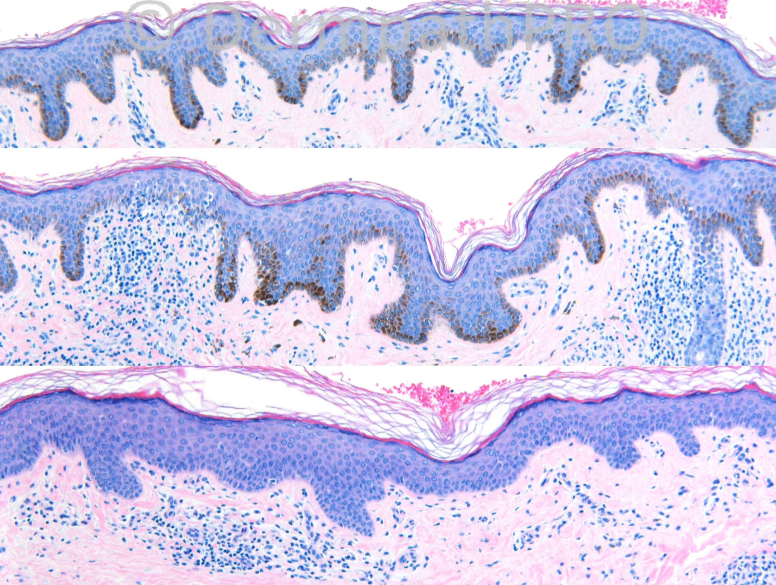
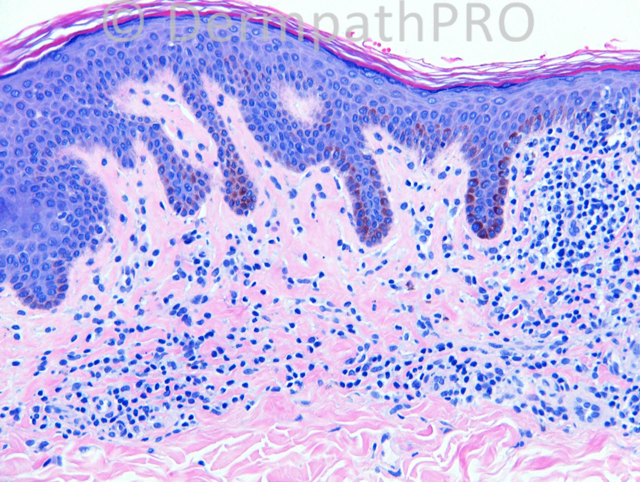
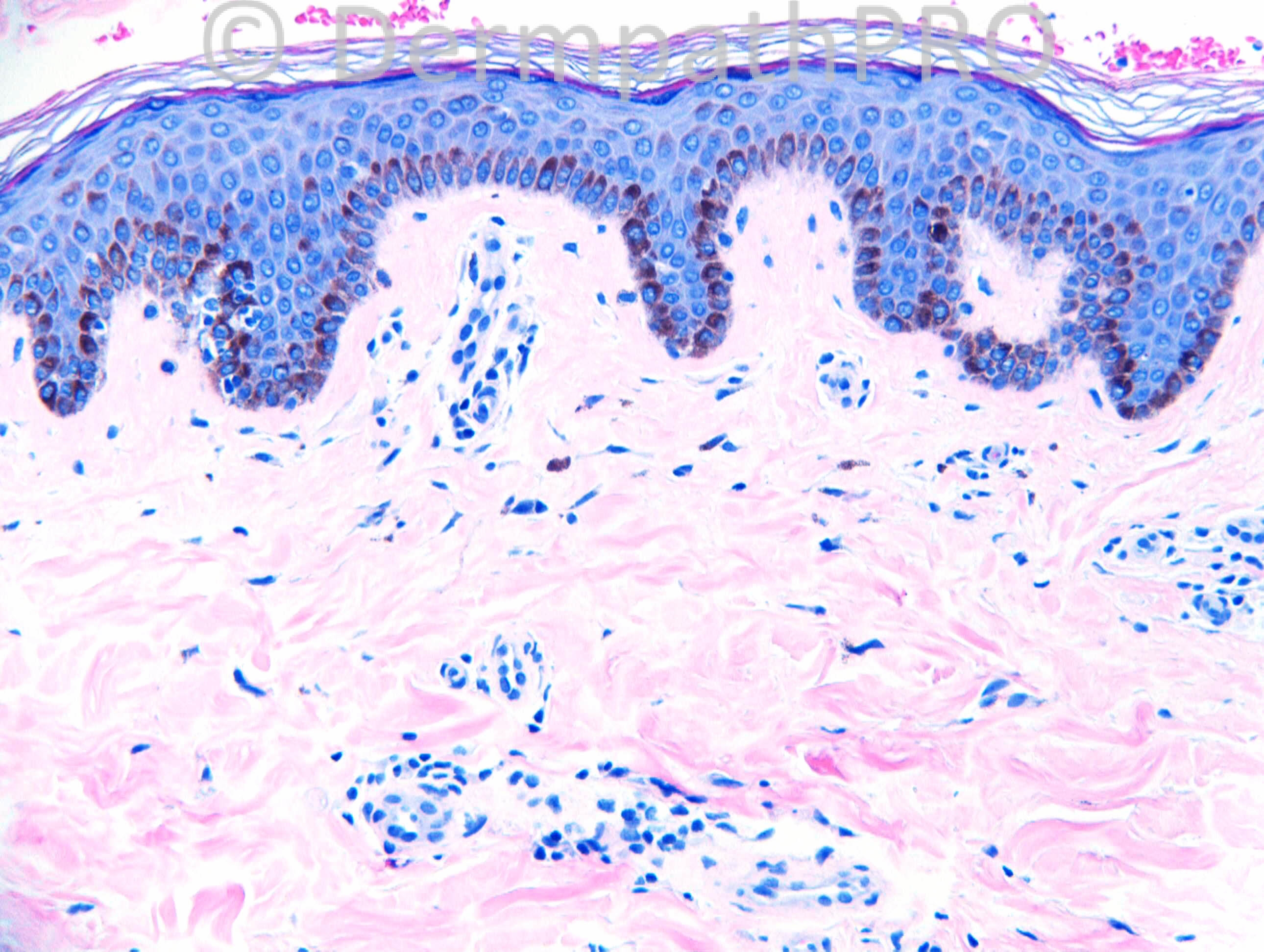
[img]https://dermpathpro.com/uploads/spot_diagnosis_comment_img/CASE1157_RAC6947_IHC_1_4pm.jpg[/img]

[img]https://dermpathpro.com/uploads/spot_diagnosis_comment_img/CASE1157_RAC6947_IHC_2_4pm.jpg[/img]

[img]https://dermpathpro.com/uploads/spot_diagnosis_comment_img/CASE1157_RAC6947_MelanA_4pm.jpg[/img]

[img]https://dermpathpro.com/uploads/spot_diagnosis_comment_img/CASE1157_RAC6947_MF_4pm.jpg[/img]

Share this comment


Link to comment
share_externally

Arti Bakshi

Posted

What about an inflammatory stage of vitiligo...this can show overlapping features with hypopigmented MF .There is striking loss of melanocytes and absence of CD8 epidermotropism, which probably favours vitiligo (although have to agree that there is some atypia in lymphoid cells)
There is loss of CD7, but not very helpful as a feature of MF as often occurs in inflammatory conditions as well.

Share this comment


Link to comment
share_externally

Dr. Mona Abdel-Halim

Posted

Inflammatory vitiligo is a vitiliginous lesion with an erythematous inflammaed advancing border. It will not show any hint of atrophic changes.
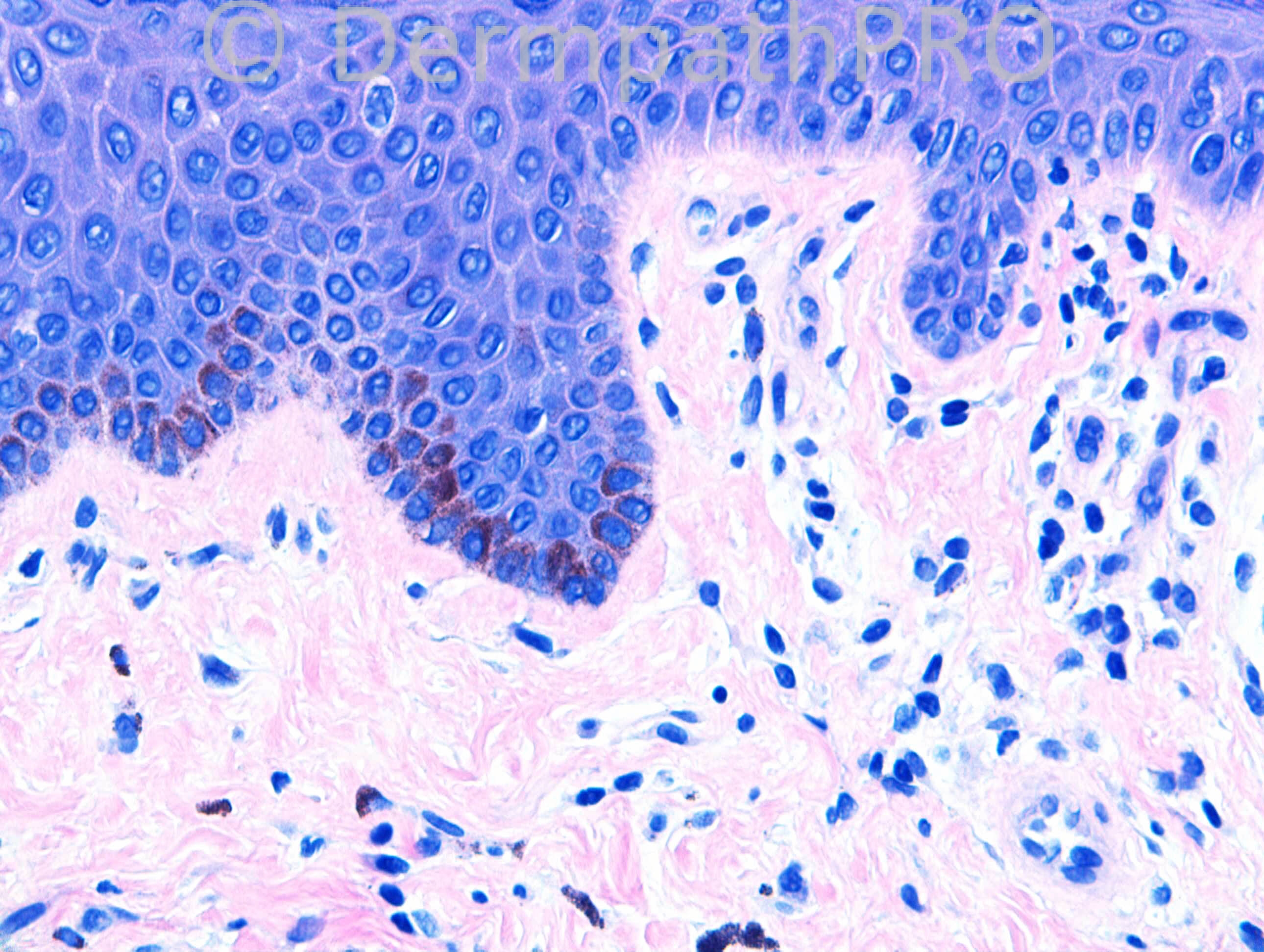
It is not expected to find melanocytes in the middle of a vitiliginous lesion, I can c Melan A positive melanocytes in the middle of the lesion and in one edge, while there is almost complete loss at the other edge.
Together with the atypia and wiry fibroplasia, I still favor hypopigmented MF.
Phenotypically, although HMF is mainly CD8 positive, cases with CD4 profile or combined profiles have been reported.

Share this comment


Link to comment
share_externally

Sasi Attili

Posted

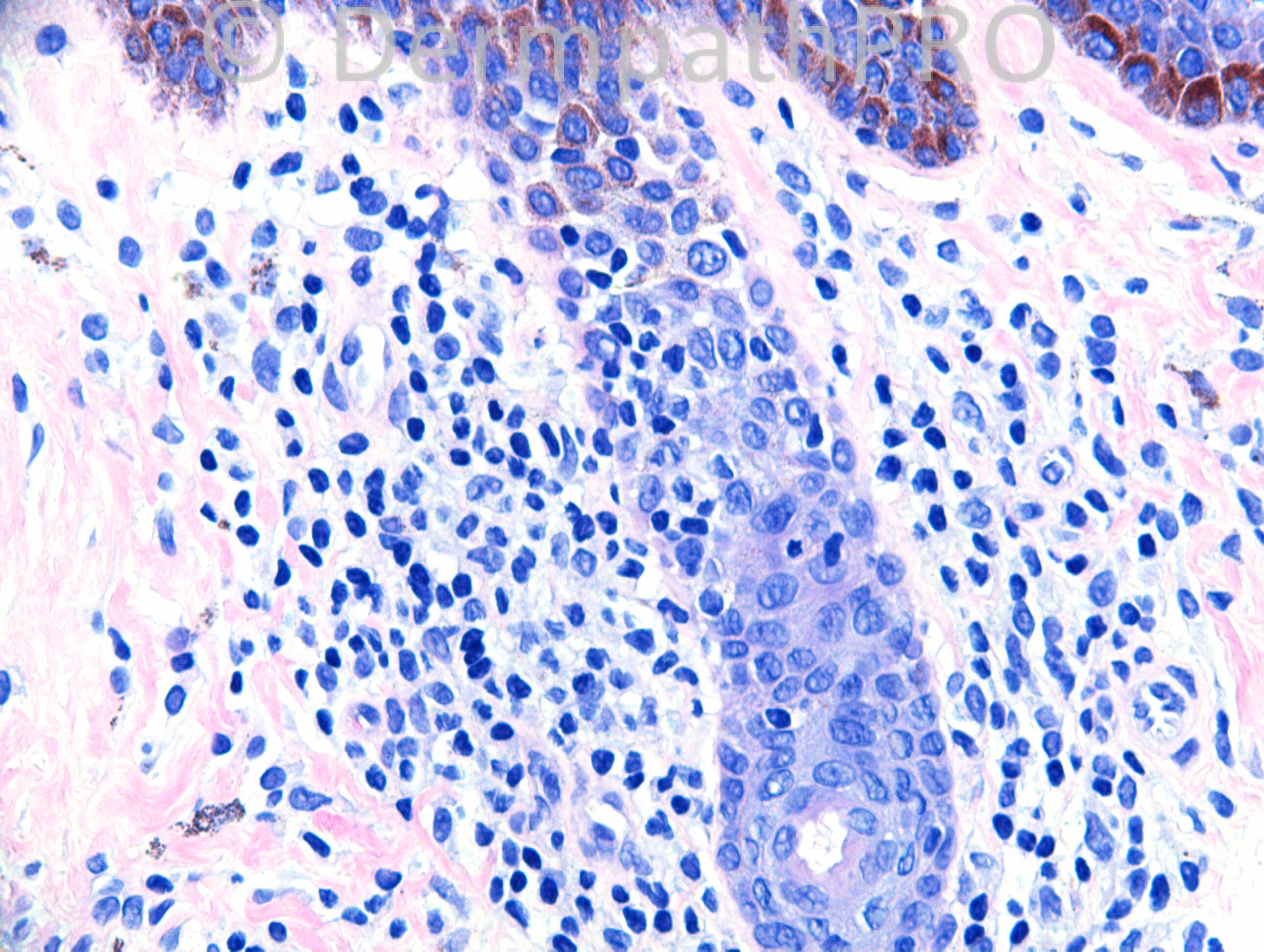
This is a tricky case. Histologically i do see focal lichenoid interface dermatitis with some epidermotropism of lymphocytes, some of which do show a perinuclear halo. However this alone cannot be taken as sine qua non for MF, especially when this is a single patch. Vitiligo and lichen Sclerosus (early stage) may show similar changes.

One can see mild papillary dermal sclerosis in cases of vitiligo and this is not an exclusion criteria. We have reported these changes in our vitiligo series published a few years ago. In fact some of the sections here can easily be taken to represent early LS. LS can show significant loss of melanocytes akin to vitiligo. http://www.ncbi.nlm.nih.gov/m/pubmed/11979069/

That infact would be my favoured diagnosis in this case, especially since the melanocytes are not completely absent. But I would raise the differential of MF and ask the clinician to follow up.

Share this comment


Link to comment
share_externally

Dr. Richard Carr

Posted

Many thanks for excellent responses. This was a recent case and I have had no contact with the clinical team unfortunately. I was suspicious for hypopigmented mycosis fungoides but would not dream of making a final diagnosis on this case. The epidermotropic cells were predominantly CD8. I am pasting my report below although you might also want to see this recent abstract from Dr Magro.

[url="http://www.ncbi.nlm.nih.gov/pubmed/?term=magro+hypopigmented+mycosis+fungoides"]http://www.ncbi.nlm.nih.gov/pubmed/?term=magro+hypopigmented+mycosis+fungoides[/url]

MY REPORT:
A challenging case showing complete loss of melanocytes (MelanA) on one side with only partial loss of melanin (Masson Fontana). There is a patchy but prominent lymphocytic infiltrate with some basal layer preference (highlighted by CD3 and CD8) and focally relatively dense lymphocytic infiltrates in the papillary dermis with CD8 > CD4. CD4 highlights weak staining of Langerhan’s cells and histiocytes but in terms of lymphocytes seems to be in the minority. Focally wiry collagen noted in the papillary dermis associated with the infiltrates. Lymphocyte atypia is only mild.
Based on the clinical comment of atrophy, focally quite dense infiltrates with wiry papillary dermal collagen and partly preserved melanin, hypopigmented mycosis fungoides is certainly a possibility in comparison to inflammatory vitiligo. The clinical history is rather short however and I suspect that the final diagnosis will await substantial follow-up and clinico-pathological correlation.
In summary: suspicious for hypopigmented mycosis fungoides rather than inflammatory vitiligo

Additional reference (in which Mona is a co-author)
Eur J Dermatol. 2006 Jan-Feb;16(1):17-22.
Vitiligo vs. hypopigmented mycosis fungoides (histopathological and immunohistochemical study, univariate analysis).
El-Darouti MA1, Marzouk SA, Azzam O, Fawzi MM, Abdel-Halim MR, Zayed AA, Leheta TM.
Author information 1Dermatology department, Faculty of Medicine, Cairo University, Egypt. [email="Mohammad_eldarouti@yahoo.com"]Mohammad_eldarouti@yahoo.com[/email]
Abstract
Vitiligo is a common skin disease characterized by the presence of well circumscribed, depigmented milky white macules devoid of identifiable melanocytes. On the other hand, hypopigmented mycosis fungoides (MF) is a rare variant of MF which presents clinically as persistent hypopigmented macules and patches. Both disorders show a predominance of CD8+ T cells in tissue samples and hence the differentiation between the two diseases on clinical, histopathological and even immunohistochemical grounds may offer great difficulty. The aim of this work is to identity certain histopathological clues which might help to differentiate between the two diseases. The study included 54 patients (26 vitiligo patients and 28 patients with Hypopigmented MF). Skin biopsies were taken and examined by hematoxylin and eosin and CD3, CD4 and CD8 markers were performed for ten vitiligo and nine MF patients. We have found that epidermotropism, hydropic degeneration of basal cells, partial loss of pigment, preservation of some melanocytes, presence of lymphocytes within the papillary dermis, increased density of the dermal infiltrate and wiry fibrosis of the papillary dermal collagen were detected with a significantly higher incidence in hypopigmented MF rather than vitiligo (P-values < 0.0001, < 0.00011, < 0.00011, = 0.001, = 0.008 and = 0.001 respectively). On the other hand, focal thickening of the basement membrane, complete loss of pigmentation, total absence of melanocytes, as well as absence or sparsness of lymphocytes in the dermal papillae were seen much more frequently in vitiligo. Statistical analysis of these differences was significant with P-values < 0.00011, < 0.00011, < 0.00011, = 0.008 respectively, regarding these pathological criteria. We conclude that differentiation of hypopigmented MF from vitiligo is possible by relying on the histopathological clues described in this study. This is particularly useful in areas of the world where cost benefit is crucial.

Share this comment


Link to comment
share_externally


Join the conversation

You can post now and register later. If you have an account, sign in now to post with your account.

Guest
Add a comment...

×   Pasted as rich text.   Paste as plain text instead

  Only 75 emoji are allowed.

×   Your link has been automatically embedded.   Display as a link instead

×   Your previous content has been restored.   Clear editor

×   You cannot paste images directly. Upload or insert images from URL.

Loading...

×
×
  • Create New...