Jump to content
In this section we have spot diagnoses posted on a daily basis since June 2010, now over 4000! You can review the archived cases and read the suggested diagnoses by users and the final comment by the contributors.
Case are uploaded each week day by 10 am UK time with the correct diagnosis will generally be posted at 8 pm UK time. Why not view the most recent spot diagnosis and proffer a diagnosis?

Case Number : Case 1117 - 3rd October Posted By: Guest

Please read the clinical history and view the images by clicking on them before you proffer your diagnosis.
Submitted Date :
   (0 reviews)

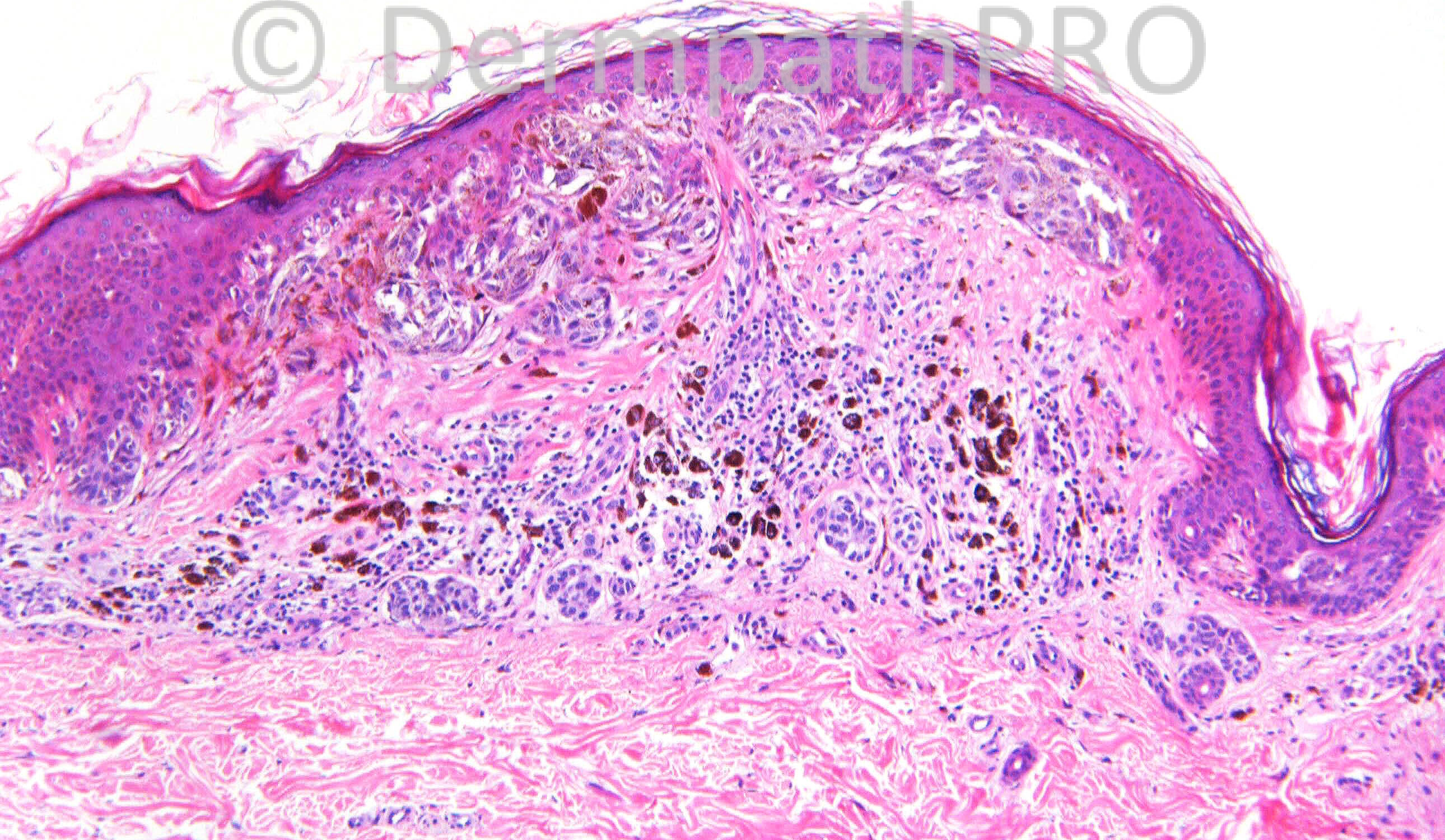
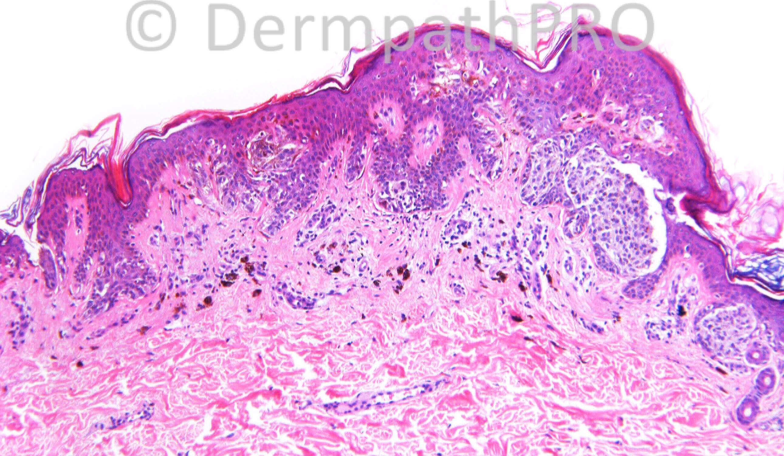
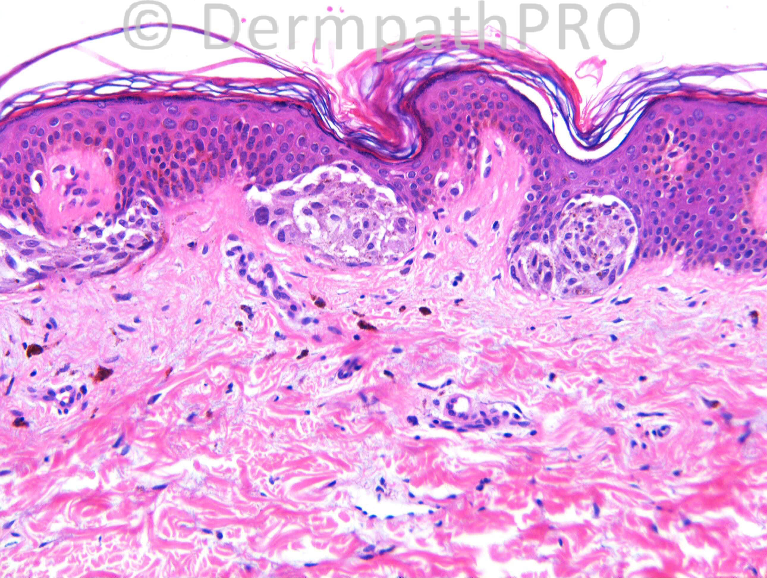
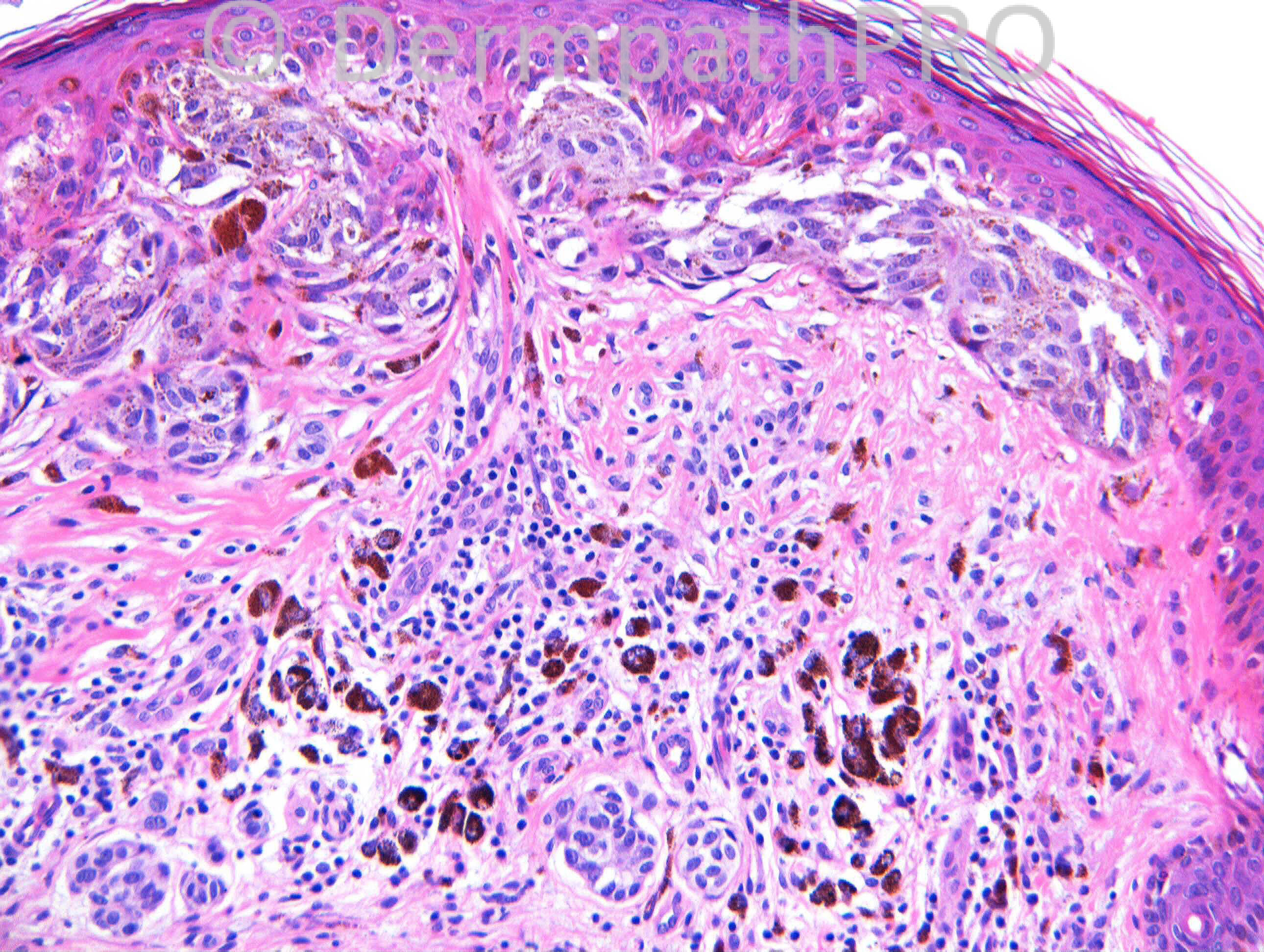
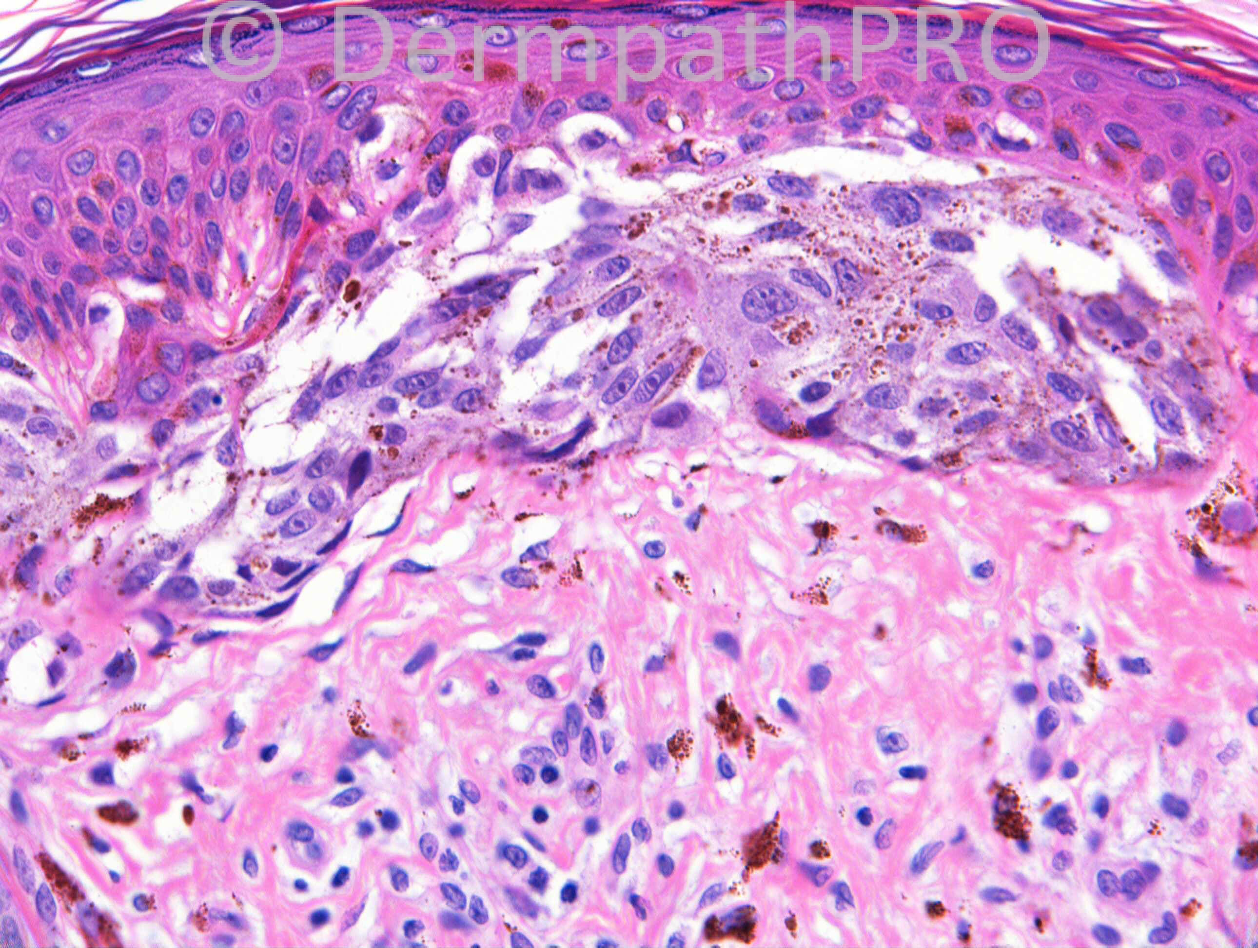
25 years old female, Pigmented lesion on breast enlarged and got darker (Case c/o Dr Monica Ahluwalia).
Case Posted by Dr Richard Carr

  Report Record

User Feedback


Looks like a special sites nevus. There's atypia, but not out of the norm for nevi of the breast/milk-line. No pagetoid spread, no dermal mitoses. If it's out there's no need for additional excision. If it goes to a margin I'd recommend conservative complete removal.

Share this comment


Link to comment
share_externally

Dr. Richard Carr

Posted

I know the dermpathpro team posted my opinion inadvertantly at the beginning of the day in the history but I would really hope to get a few more comments on the case (in any case you may not agree with my opinion). I suspect there are many of you who check on cases but never post a suggestion - now is your opportunity to contribute! I will leave it open and sincerely hope you enjoy your weekends.

Share this comment


Link to comment
share_externally

Robledo F. Rocha

Posted

I didn’t read Dr. Carr’s diagnosis that was posted early, so I write my opinion blindly.
It´s a [url="http://onlinelibrary.wiley.com/doi/10.1111/j.0303-6987.2004.00159.x/abstract"]site-specific nevus from breast[/url]. Junctional melanocytic nests that are large, horizontally-oriented and coalescent may cause concern, and melanoma or [url="https://dermpathpro.com/blog/3/entry-126-dysplastic-nevus-as-a-legitimate-phrase/"]so-called “dysplastic nevus”[/url] may therefore be signed out. However, these findings are indicative of site-specific nevus within the overall context of a symmetric lesion that begins and ends with a nest, melanocytes that are bland and monomorphic, and a case history of a pigmented lesion on a breast of a young woman.

Share this comment


Link to comment
share_externally

Guest Giovanni Falconieri

Posted

Agree with all the considerations above. Irregularly shaped and spaced nests, focal epidermal consumption, mild nuclear atypia, and dust-like pigment although generically indicating "dysplasia" are pretty much consistent with a special site nevus, given the clinical context

Share this comment


Link to comment
share_externally

Dr. Richard Carr

Posted

Yes I also reported this as a special site naevus. She had two similar lesions excised at the same time.

Share this comment


Link to comment
share_externally


Join the conversation

You can post now and register later. If you have an account, sign in now to post with your account.

Guest
Add a comment...

×   Pasted as rich text.   Paste as plain text instead

  Only 75 emoji are allowed.

×   Your link has been automatically embedded.   Display as a link instead

×   Your previous content has been restored.   Clear editor

×   You cannot paste images directly. Upload or insert images from URL.

Loading...

×
×
  • Create New...