Jump to content
In this section we have spot diagnoses posted on a daily basis since June 2010, now over 4000! You can review the archived cases and read the suggested diagnoses by users and the final comment by the contributors.
Case are uploaded each week day by 10 am UK time with the correct diagnosis will generally be posted at 8 pm UK time. Why not view the most recent spot diagnosis and proffer a diagnosis?

Case Number : Case 1122 - 10th October Posted By: Admin_Dermpath

Please read the clinical history and view the images by clicking on them before you proffer your diagnosis.
Submitted Date :
   (0 reviews)

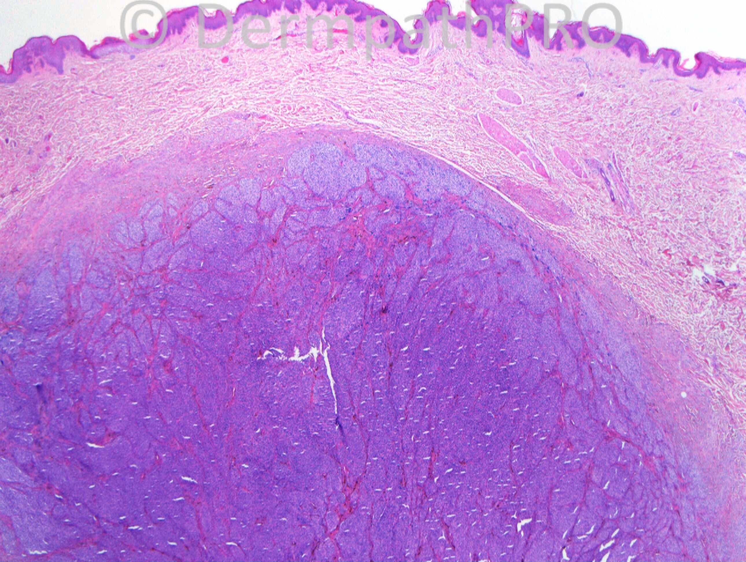
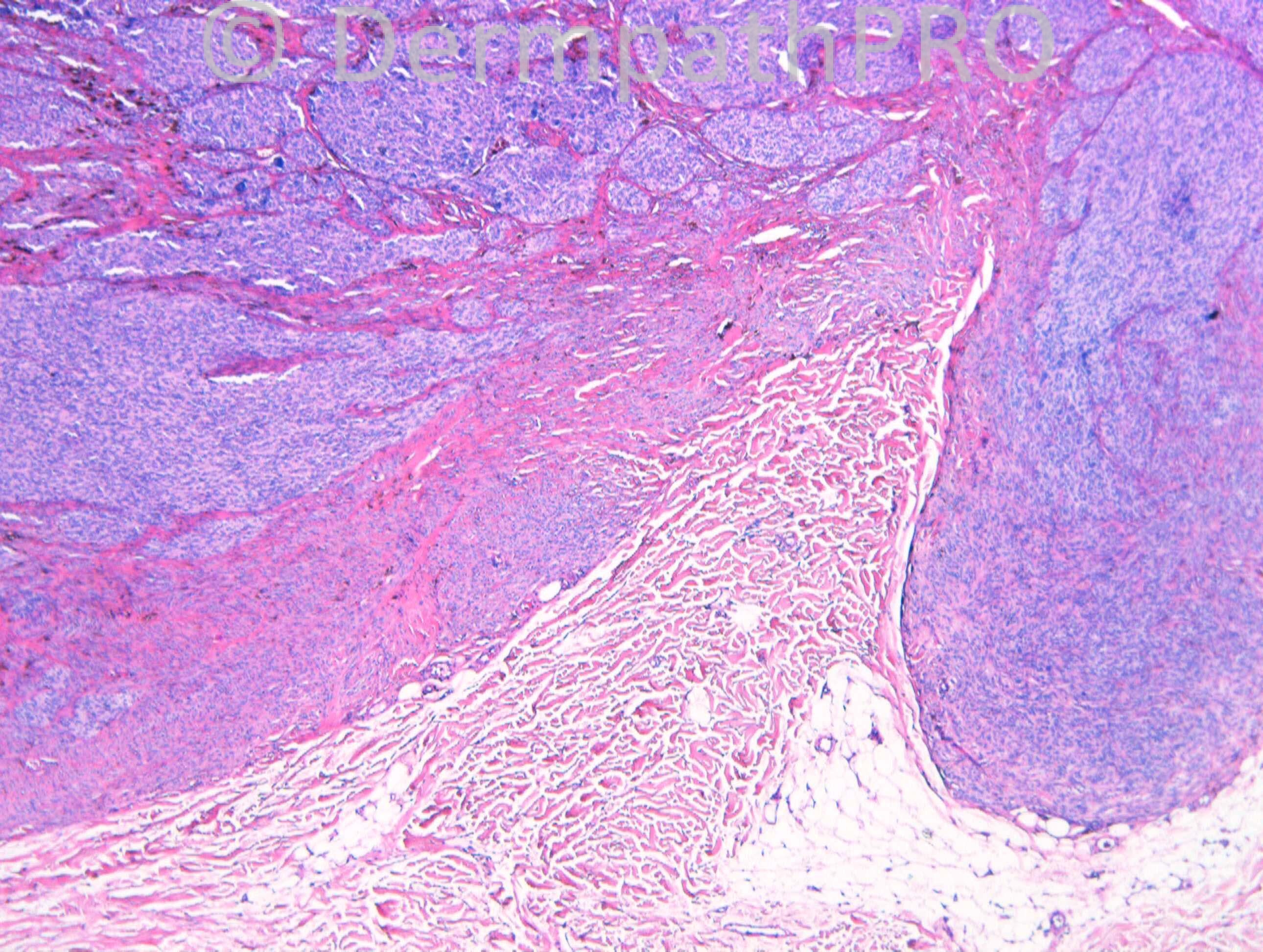
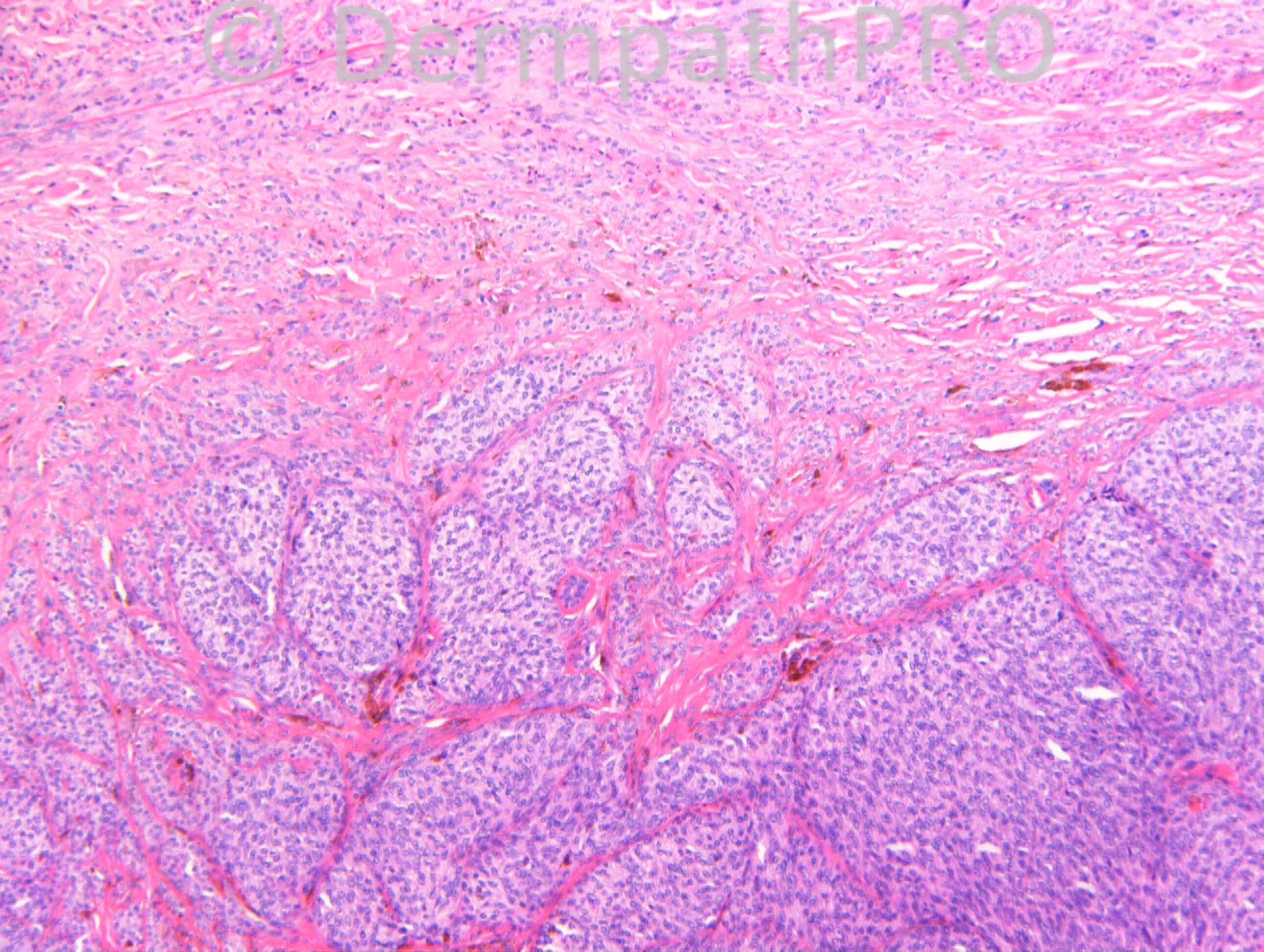
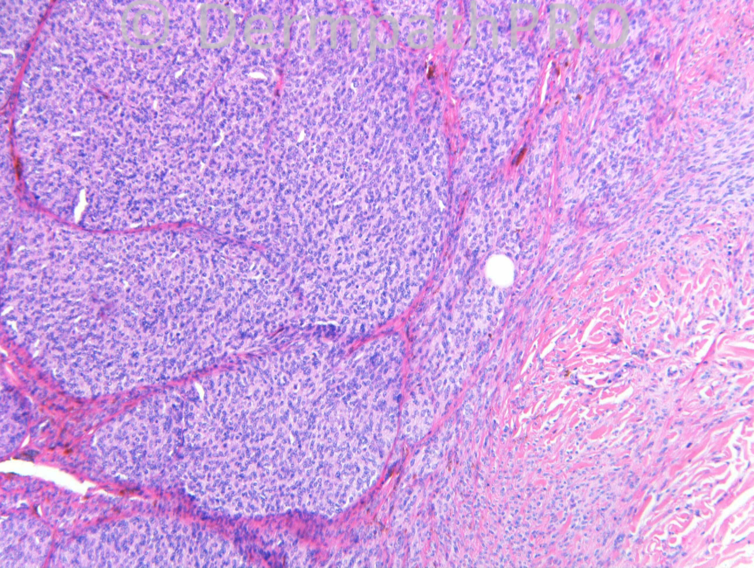
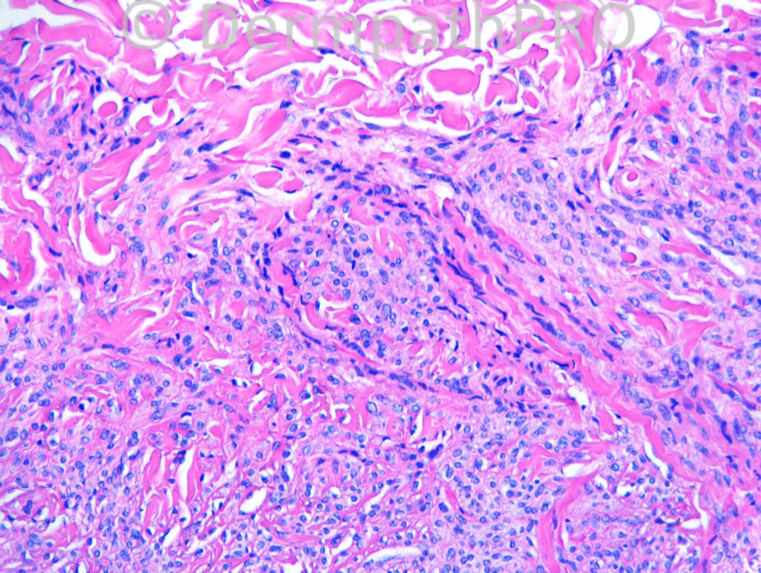
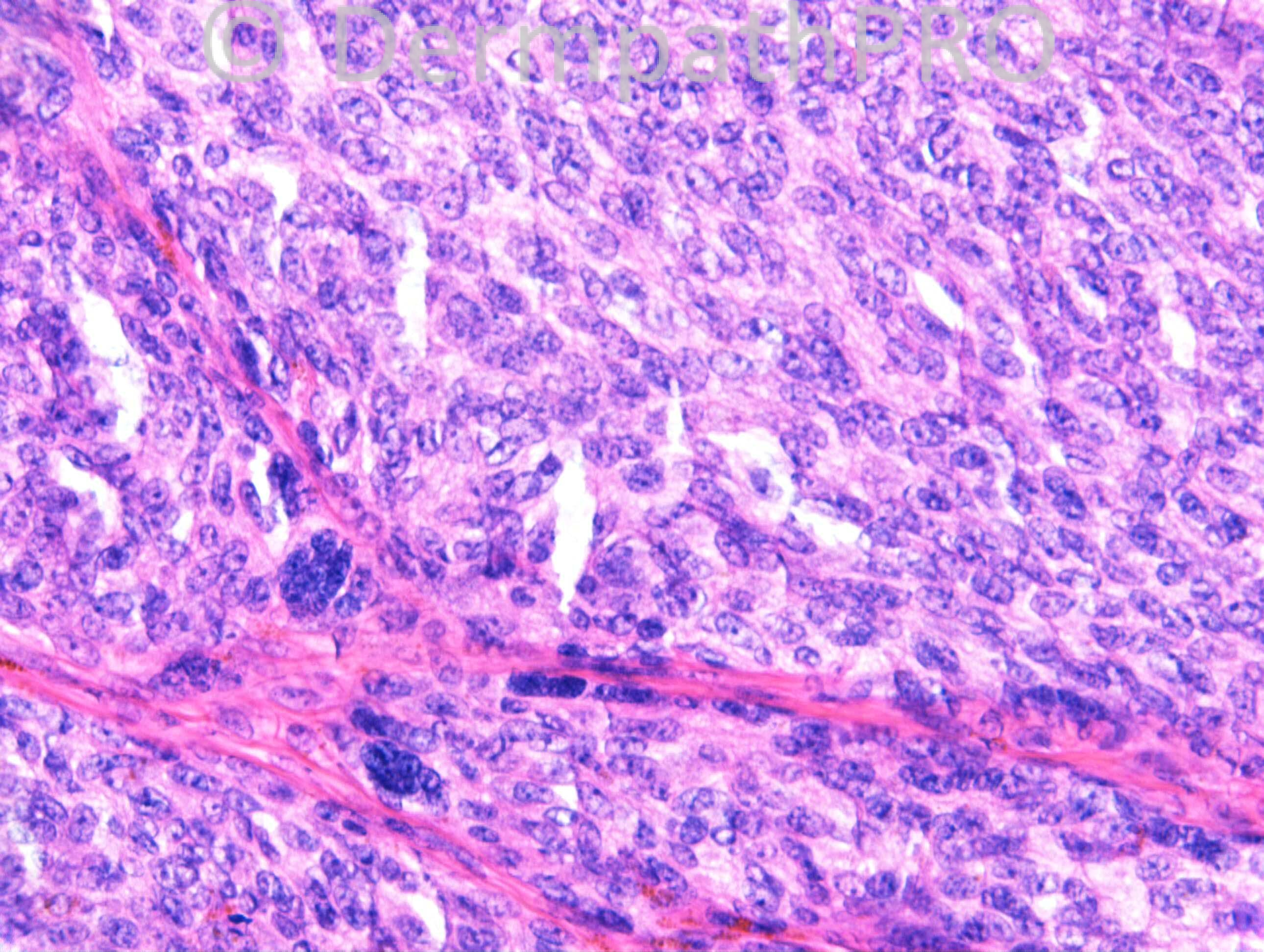
22 years old female. Cystic lump on buttock. (c/o Dr Ujjwala Mohite)

Case Posted by Dr Richard Carr


  Report Record

User Feedback


Robledo F. Rocha

Posted

Cellular neurothekeoma.

Share this comment


Link to comment
share_externally

Guest Romualdo

Posted

The following features made me think of atypical cellular blue nevus and not of conventional cellular blue nevus:
1. A strikingly nodular cellular appearance at scanning magnification
2.The presence of larger expansile melanocyte nodules
3. The marked cellularity seen in some of these nodules
4. The slight nuclear atypia present
Although I have not seen mitotic figures I hope they are present in other areas of the tumor.

Share this comment


Link to comment
share_externally

Dr. Richard Carr

Posted

[img]https://dermpathpro.com/uploads/spot_diagnosis_comment_img/CASE1122_RAC6919x20_MelanA_Label.jpg[/img]

[img]https://dermpathpro.com/uploads/spot_diagnosis_comment_img/CASE1122_RAC6919x20_S100_Label.jpg[/img]

Share this comment


Link to comment
share_externally

Dr. Mona Abdel-Halim

Posted

I think it goes with clear cell sarcoma more than cellular blue nevus

Share this comment


Link to comment
share_externally

Guest Romualdo

Posted

The vertically oriented and very well demarcated bulbous expansion of tumor, pointing to the subcutaneous tissue, seen in figure 2, is very suggestive of cellular blue nevus. The eosinophilic collagenized area at the superficial part of the tumor, seen in figure 3, is consistent with an area of common blue nevus, which is frequently seen in association with cellular blue nevus. By the other side, S100 may be negative in blue nevi, common and cellular variants, and constitute a diagnostic pitfall. S100 is positive in most, if not all cases of clear cell sarcomas, which are the most important differentials, together with melanomas, in apropriated contexts.

Share this comment


Link to comment
share_externally

Dr. Richard Carr

Posted

Yes this was a referral case that I reported as typical of cellular blue naevus. Mitoses can be moderately numerous which can add to the difficulty if you are not aware of the typical features described by Romualdo nicely. The anatomic location is also typical and preservation of adnexal structures (not illustrated here) are also helpful. The lesion was shelled out and I commented that there may be a possibility of local recurrence but a wait and watch could be adopted.

Share this comment


Link to comment
share_externally


Guest
This is now closed for further comments

×
×
  • Create New...