Jump to content
In this section we have spot diagnoses posted on a daily basis since June 2010, now over 4000! You can review the archived cases and read the suggested diagnoses by users and the final comment by the contributors.
Case are uploaded each week day by 10 am UK time with the correct diagnosis will generally be posted at 8 pm UK time. Why not view the most recent spot diagnosis and proffer a diagnosis?

Case Number : Case 1123 - 13th October Posted By: Guest

Please read the clinical history and view the images by clicking on them before you proffer your diagnosis.
Submitted Date :
   (0 reviews)

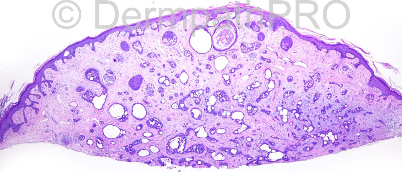
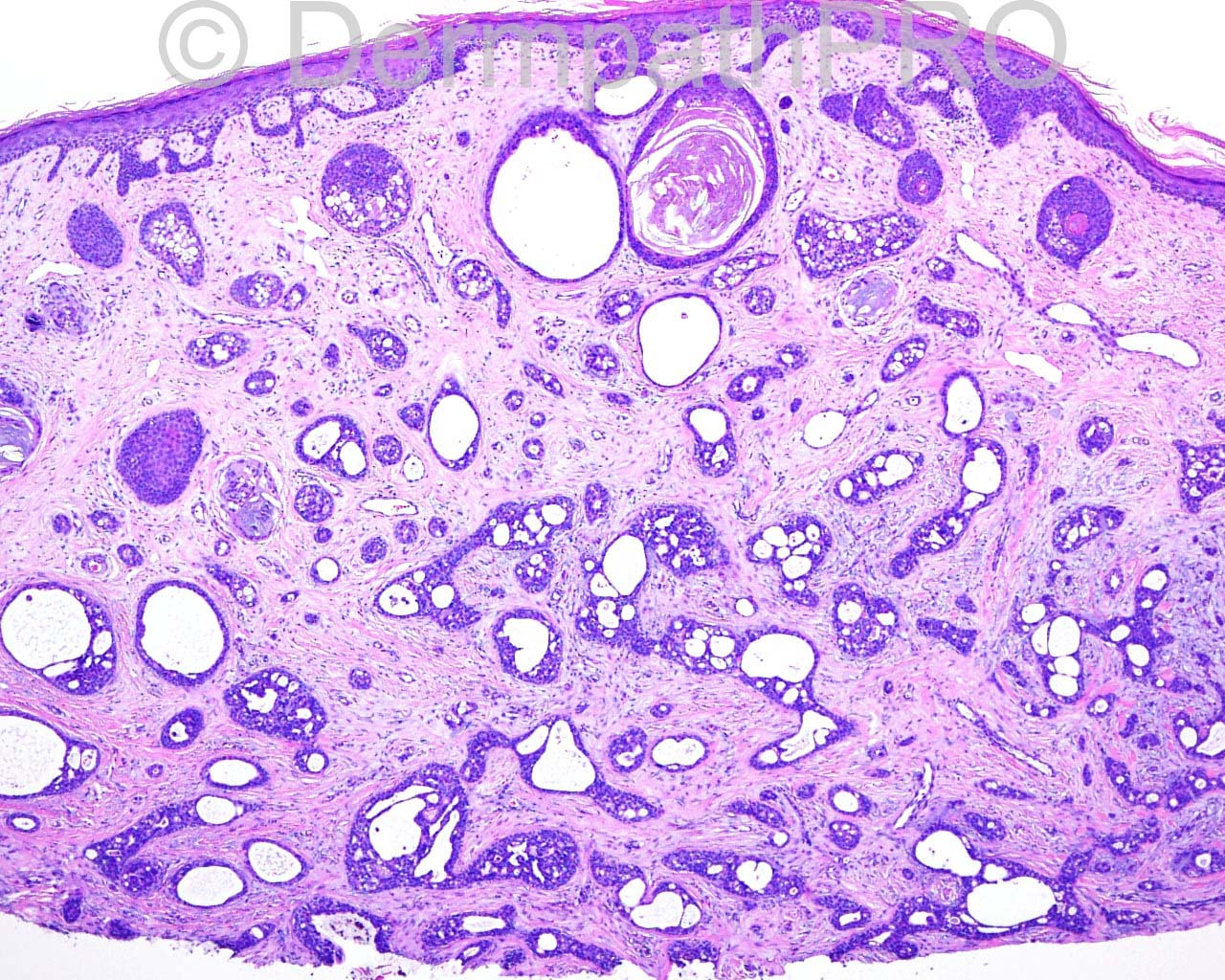
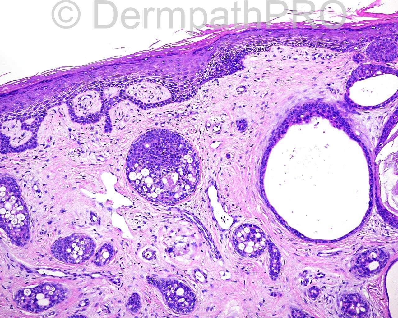
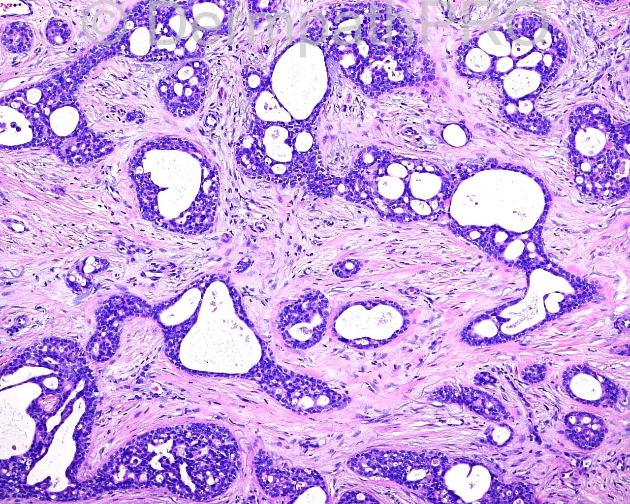
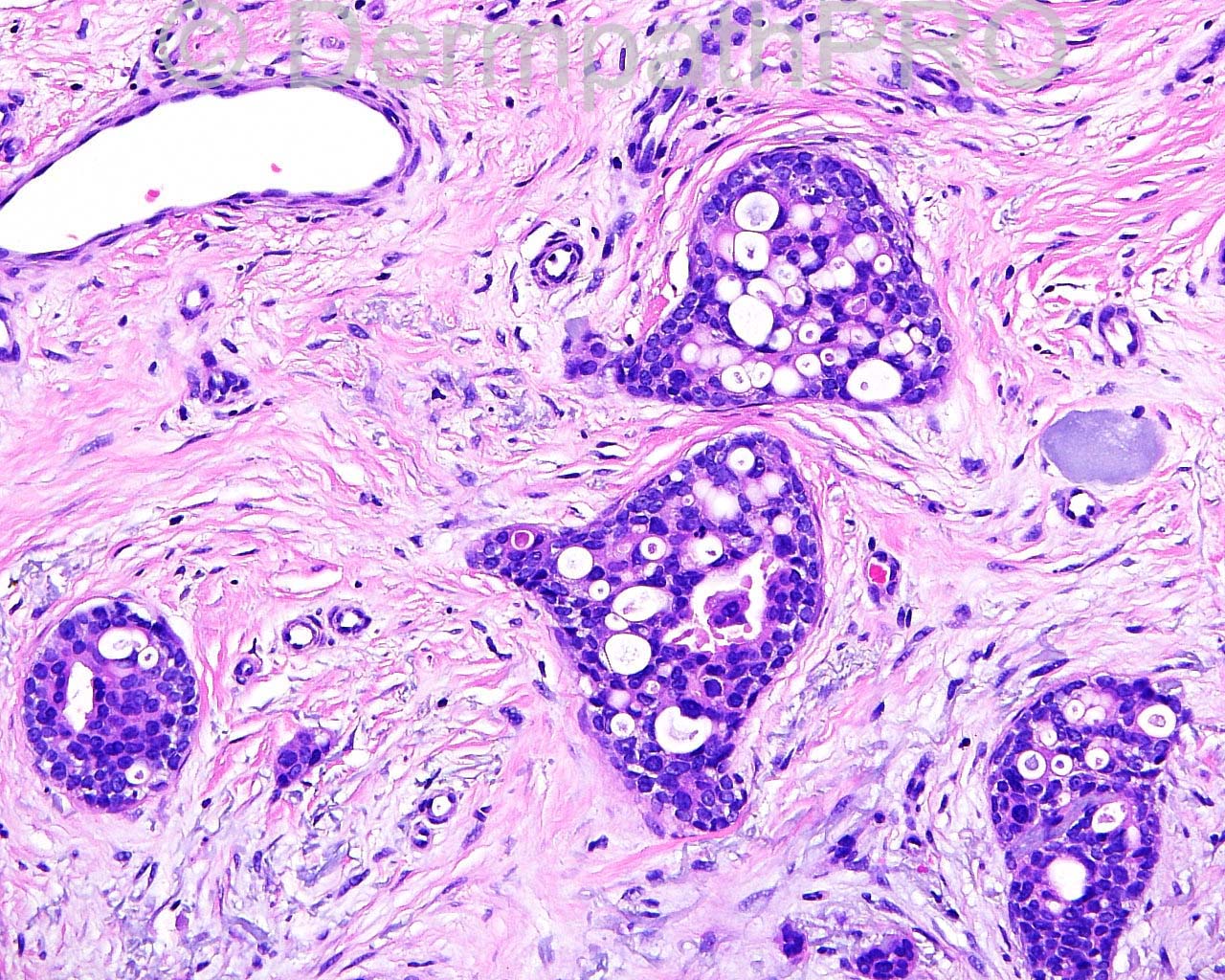
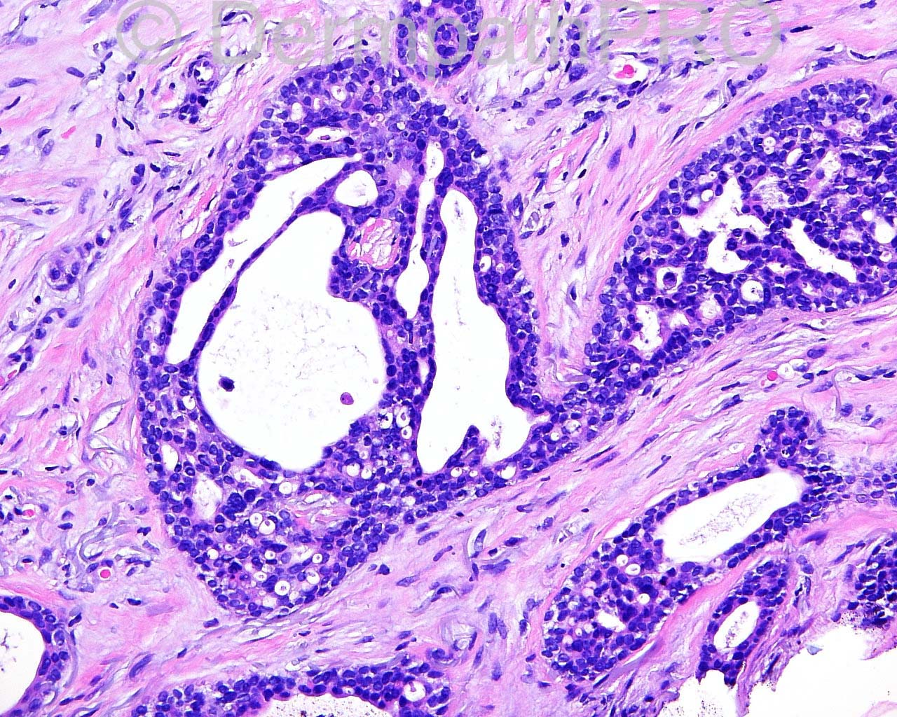
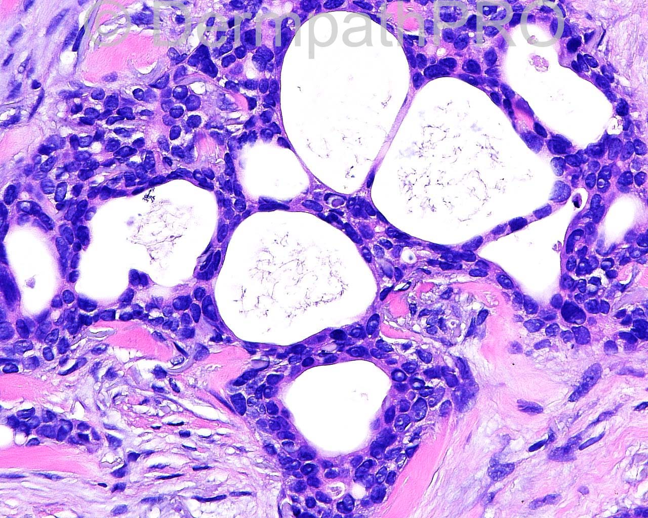
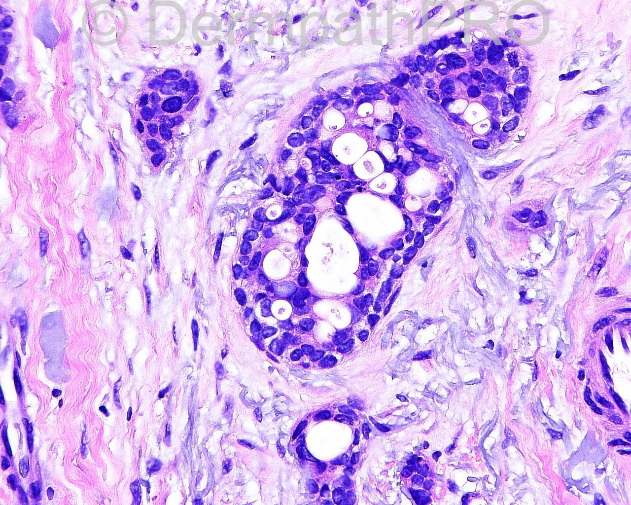
The patient is an 87-year-old woman with a biopsy of a lesion on the right distal lower leg.

Case posted by Dr. Mark Hurt


  Report Record

User Feedback


Robledo F. Rocha

Posted

Two malignant sweat gland tumors came to my mind, both rarely found on limbs: microcystic adnexal carcinoma and adenoid cystic carcinoma. I favor the former in virtue of keratocysts in the upper dermis and intracytoplasmic lumina. Also, metastatic adenocarcinoma should be considered.

Share this comment


Link to comment
share_externally

Dr. Mona Abdel-Halim

Posted

I think it is benign, papillary eccrine adenoma

Share this comment


Link to comment
share_externally

Dr. Richard Carr

Posted

I favoured a low grade ductal adenocarcinoma most likely a primary but should consider metastasis.

Share this comment


Link to comment
share_externally

Mark A. Hurt MD

Posted

This was an external consultation sent to me. The referring dermatopathologist provided the following information:

“[i]Clinically, it was rule out squamous cell carcinoma. This infiltrating epithelial tumor exhibits features for which I considered a sy[/i][i]ringomatous carcinoma (Ackerman[/i][i]), microcystic adnexal carcinoma or adenosquamous carcinoma. I must say I've not see many lesions like this"[/i]

Here is my diagnosis:

SKIN, RIGHT DISTAL LOWER LEG , BIOPSY :
[b]-- CRIBRIFORM APOCRINE CARCINOMA[/b]
[b]COMMENT: [/b]This is an unusual kind of carcinoma that has not been described in many case reports or series. One of the larger series is by Rütten, et al in 2009. Those lesions tended to occur in limbs, both upper and lower, with a slight female dominance. None of the patients studied had any local persistence or regrowth or evidence of metastasis in follow up from about two years to eighteen years. A number of the patients were lost to follow up in this series. I think that morphologically, this lesion is very similar to those that have been published, and also similar to the original series published by Requena et al in 1998. The differential is usually with adenoid cystic carcinoma, but I don't see the stereotyped pattern for adenoid cystic carcinoma, nor is there any evidence of neural involvement in this lesion, which is so common in adenoid cystic carcinoma. Regarding syringomatous carcinomas, those lesions tend to be uniform tubules rather than complex cribriform patterns. Microcytic carcinoma likewise tends to contain a gradient effect of tubules rather than cribriform patterns. Adenosquamous carcinoma does have some features of this current lesion, but it generally lacks the cribriform features.

[center]References:[/center]

Rütten A, Kutzner H, Mentzel T, Hantschke M, Eckert F, Angulo J, Rodríguez Peralto JL, Requena L. Primary cutaneous cribriform apocrine carcinoma: a clinicopathologic and immunohistochemical study of 26 cases of an under-recognized cutaneous adnexal neoplasm. J Am Acad Dermatol. 2009 Oct;61(4):644-51. doi: 10.1016/j.jaad.2009.03.032. PubMed PMID: 19751882.

Requena L, Kiryu H, Ackerman AB. Cribriform carcinoma. In: Requena L, Kiryu H, Ackerman AB, eds. [i]Neoplasms with apocrine differentiation.[/i] Philadelphia: Lippincott-Raven, Ardor Scribendi; 1998;879-905

Share this comment


Link to comment
share_externally

Mark A. Hurt MD

Posted

Here is my micro, to reveal the stains I used:

This lesion consists of aggregations and lobules of ductal structures with a prominent cribriform pattern. Some keloidal colonization is identified in the midst of the lesion. There is some connection to the surface in a few areas. The lesional cells contain fairly large and hyperchromatic nuclei with apocrine-type snouting at the apices of many of them. The lesion extends to the base of this shave. Cytologically, in addition to the apocrine differentiation, there is a considerable amount of nuclear variation with a few mitotic figures identified and some areas of apoptosis within these cribriform structures.

Immunohistochemically CEA monoclonal stains the lesion throughout the cytoplasm and rims the ducts especially well in many areas. There is 4+ staining with appropriate control. EMA marks the ducts and canaliculi in a 4+ membranous pattern with appropriate external control as well. CK7 is similar to EMA in lining small ductal structures in a membranous pattern of 4+ with appropriate control. CK20 is negative with appropriate 4+ cytoplasmic external control. CDX2 is negative with 4+ cytoplasmic external control. CK5/6, coupled with S100 protein, fails to show nerve within the substance of the lesion, thus I do not identify any neural involvement of this lesion. The background CK5/6 marker is a 4+ cytoplasmic staining, while the S100 protein marker is a cytoplasmic and nuclear marker of some dendritic cells and is 4+ as well. Smooth muscle actin shows a strong background staining of the interstitium but also stains very weakly the rimming around the periphery of these ducts. It is not obvious to me, however, that there is a myoepithelial layer within the substance of the ducts. p63 has strong nuclear staining in the periphery of the ductal structures and within some of the ductal structures but not all of them. In the cribriform areas there tends to be a paucity of staining, whereas the periphery is strong and 4+ nuclear. D2-40 also shows a strong membranous pattern and cytoplasmic pattern around the periphery of the ducts, at least about 50 percent of them. Estrogen receptor (clone 6F11), progesterone receptor (cone PGR16), and Her2neu (clone SP3) are all negative with appropriate external controls.

Share this comment


Link to comment
share_externally


Join the conversation

You can post now and register later. If you have an account, sign in now to post with your account.

Guest
Add a comment...

×   Pasted as rich text.   Paste as plain text instead

  Only 75 emoji are allowed.

×   Your link has been automatically embedded.   Display as a link instead

×   Your previous content has been restored.   Clear editor

×   You cannot paste images directly. Upload or insert images from URL.

Loading...

×
×
  • Create New...