Jump to content
In this section we have spot diagnoses posted on a daily basis since June 2010, now over 4000! You can review the archived cases and read the suggested diagnoses by users and the final comment by the contributors.
Case are uploaded each week day by 10 am UK time with the correct diagnosis will generally be posted at 8 pm UK time. Why not view the most recent spot diagnosis and proffer a diagnosis?

Case Number : Case 1127 - 17th October Posted By: Guest

Please read the clinical history and view the images by clicking on them before you proffer your diagnosis.
Submitted Date :
   (0 reviews)

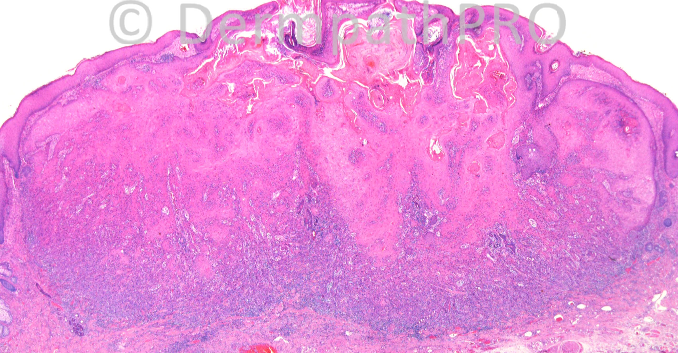
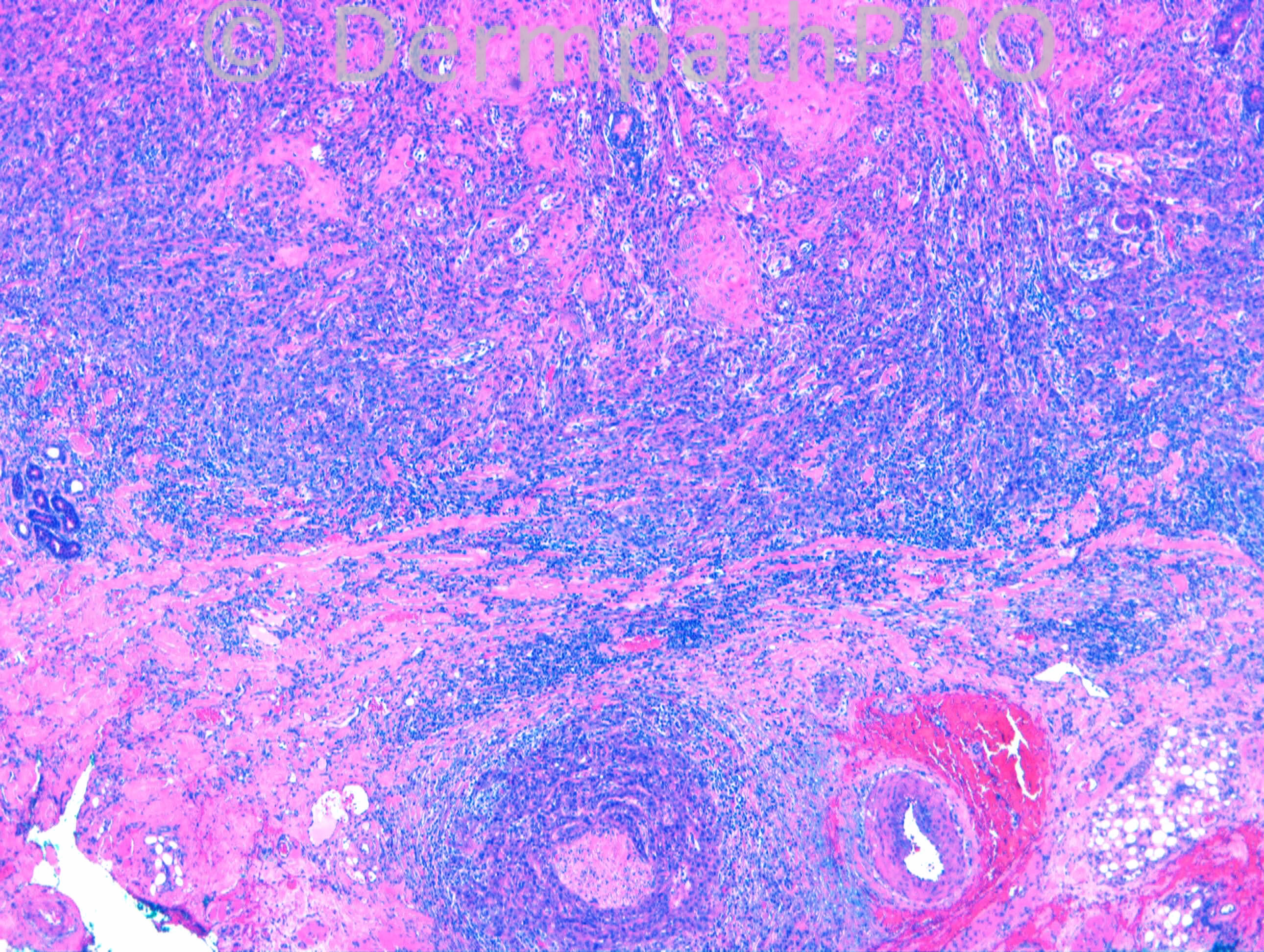
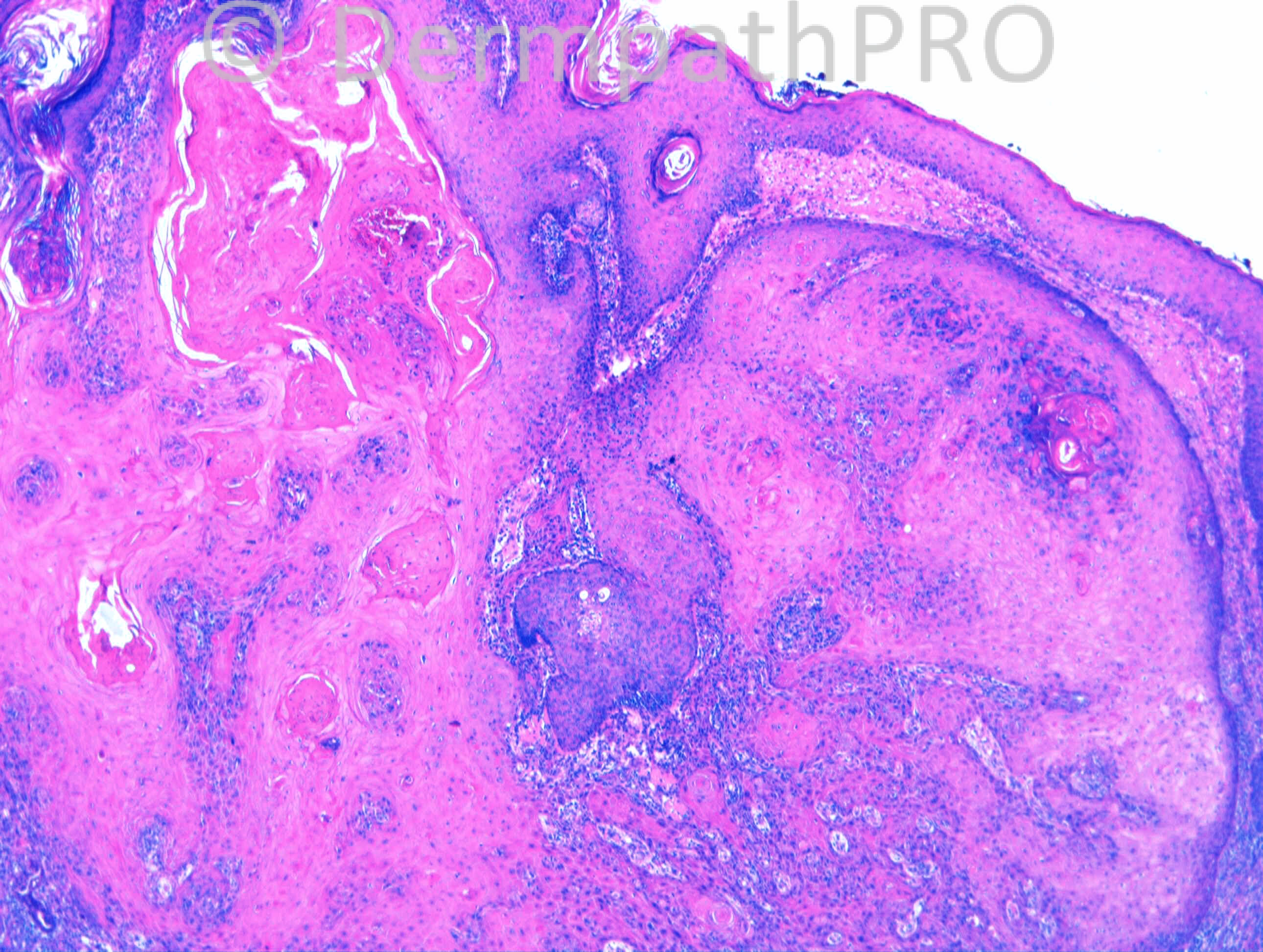
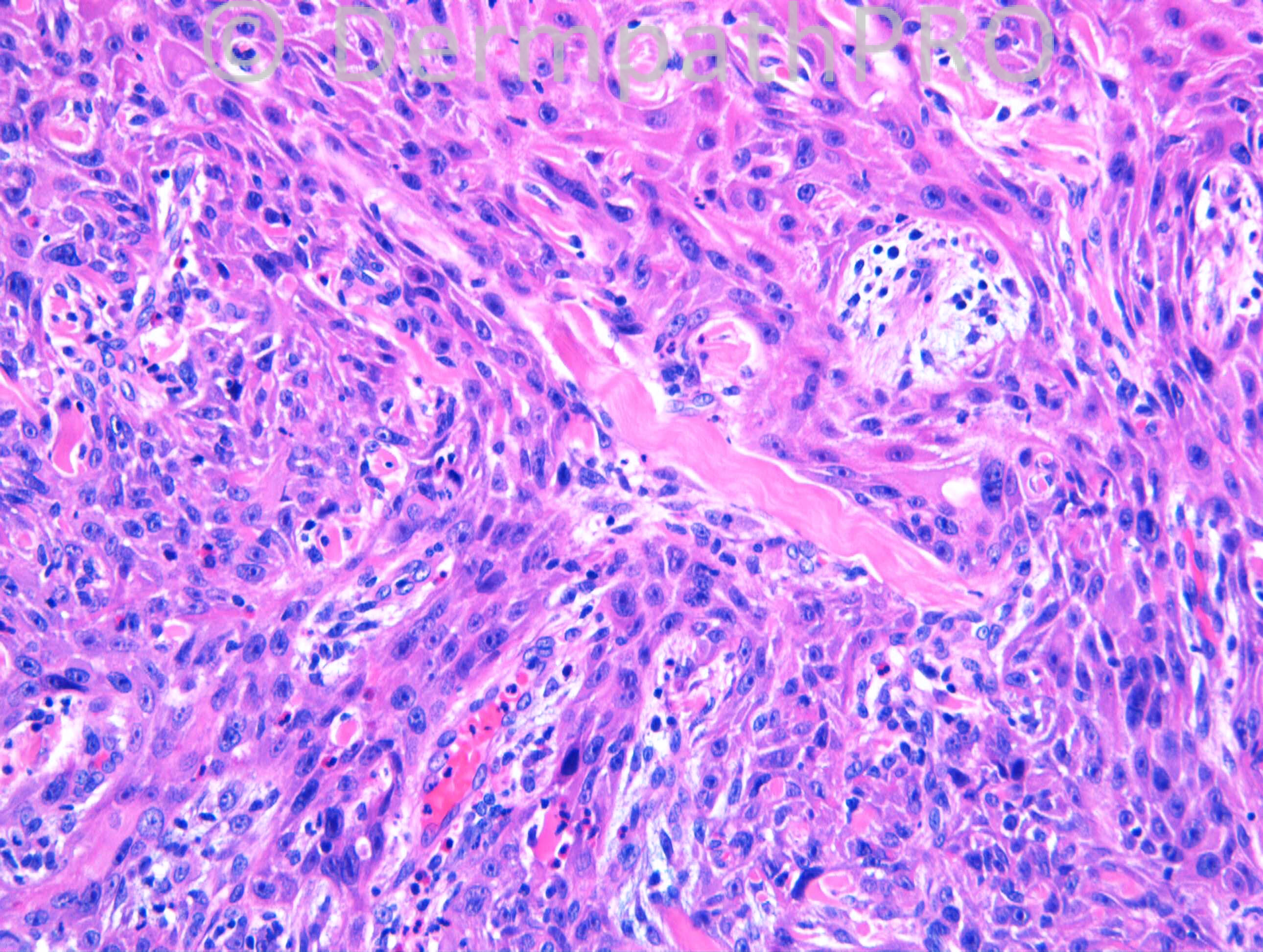
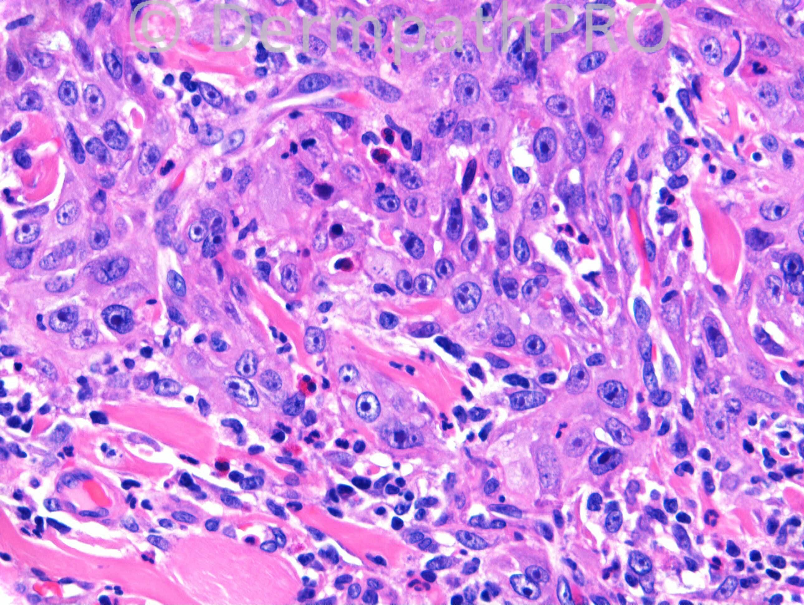
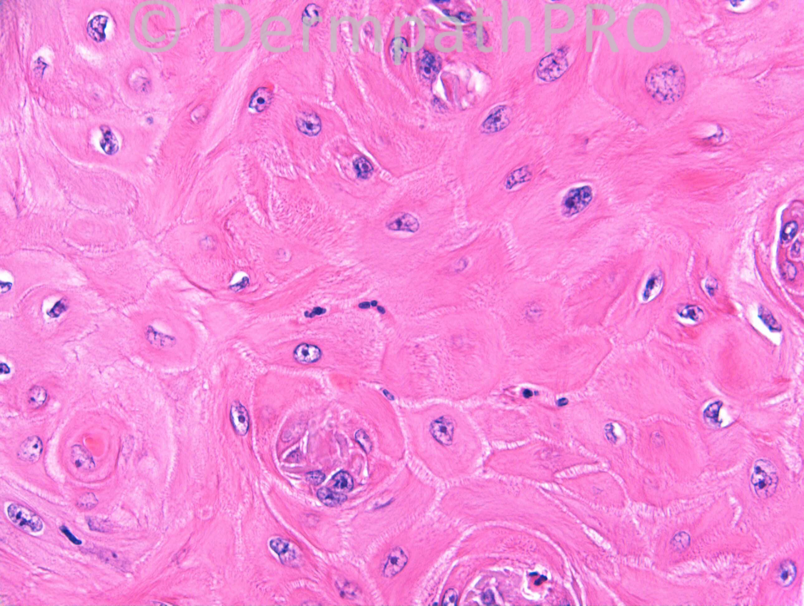
M60. Side of nose. 8/52 steady growth, keratotic centre. No sign of regression yet. ?SCC, ?KA

Case Posted by Dr Richard Carr

  Report Record

User Feedback


Dr. Mona Abdel-Halim

Posted

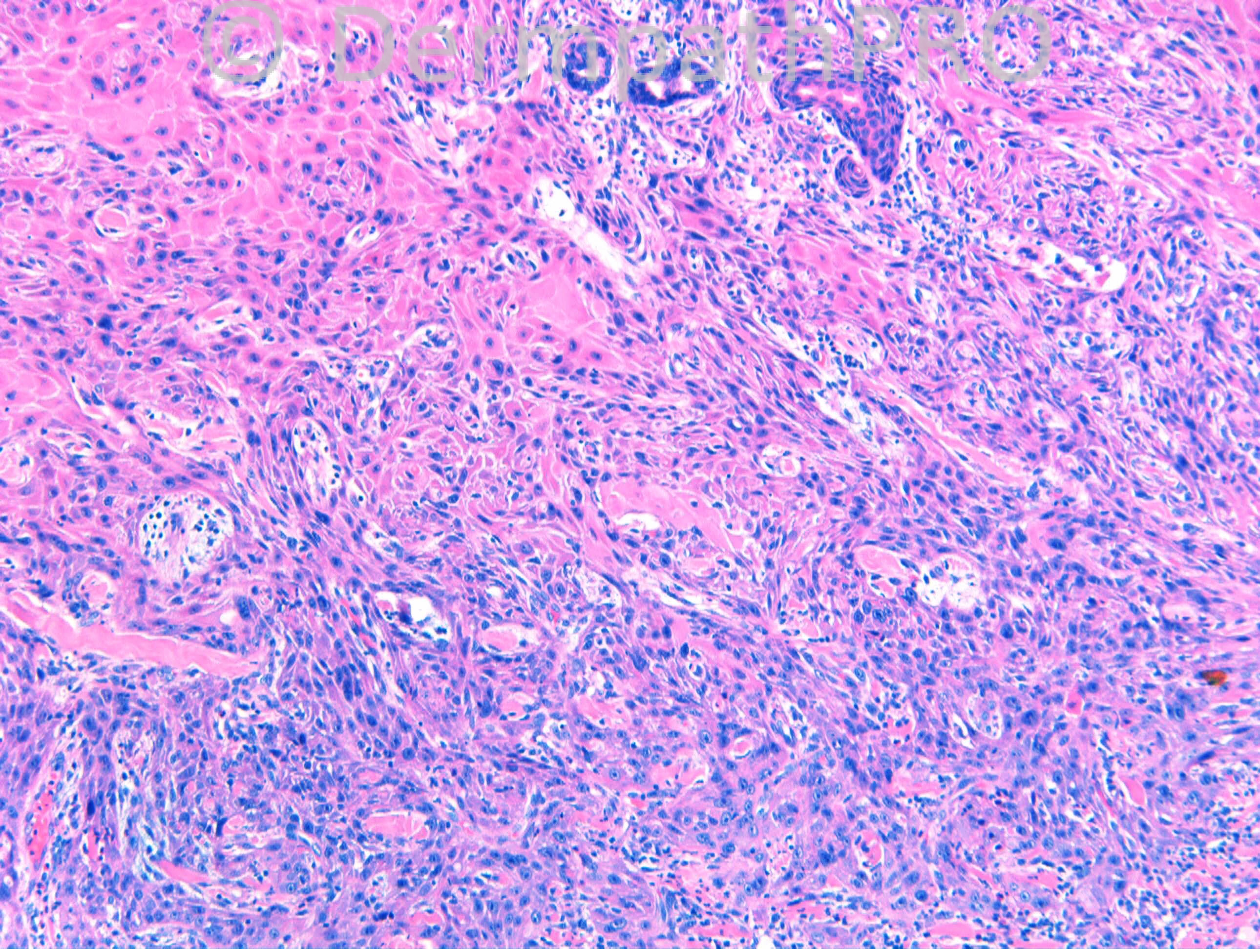
Although early proliferative KA may show infiltrative component with entrapment of collagen and elastic fibres and may show perineural invasion, I am still uncomfortable with KA. The lesion is not nicely rounded, the infiltrative component is very atypical with hyperchromatic and plemorphic nuclei and deeply extending in the dermis. Together with a steady growth, I am favouring follicular SCC.

Share this comment


Link to comment
share_externally

Dr. Richard Carr

Posted

I decided to post early as this case is clearly going to be controversial.
Although I may be in a minority of 1, my report read as follows:

A highly infitlrative / proliferative, symmetrical, squamoproliferative lesion in keeping with histologically early KA.
There is perinerual invasion. No vascular invasion.
Radial margin: 5mm
Deep margin: 0.3mm

Discussion:
Features that are typical of KA (in addition to the clinical information provided) are the symmetry, nice arciform / rounded profile to the base (excluding the perineural extension), the lesion stops at the level of the sweat gland coils, the highly infiltrative borders with full maturation in all areas, lack of a desmoplastic stroma and beautiful entrapment of collagen and elastic. The inflammatory reaction to the tumour is also replete with eosinophils and neutrophil microabscess. Other typical features are adenomatoid transformation of the preserved sweat gland coils and elastic entrapment within fully matured squamous epithelium in the upper areas (not illustrated at high power). In sum there are no features that are particularly worrisome for a follicular squamous cell carcinoma (in particular lack of, pushing borders at the base, acantholysis, follicular mucin, asymmetry, solid areas lacking full maturation). I do not use cellular atypia to diagnose KA in an otherwise typical lesion and believe that is not a helpful diagnostic criterion although I report some otherwise typical KA with atypia of uncertain / doubtful significance. I am happy to send a copy of our paper on follicular SCC (from Diagnostic Histopathology) to any one who has not requested it ([email="richard.carr@swft.nhs.uk"]richard.carr@swft.nhs.uk[/email]). I am pasting here from the paper:

"Roughly 15% of fSCC have an irregularly formed crateriform appearance and some keratoacanthoma-like features. The latter type of fSCC can be distinguished from KA (or KA with cellular atypia) by the presence of multiple features that added together rule out KA including lack of symmetry, focal ulceration or marked crusting, irregular often largely parakeratotic keratin plug, infiltration irregularly or deeply in to subcutis, absence of striking inflammatory regression reaction despite large size, irregular thick basaloid proliferative zones and prominent dyskeratosis20,22-26 (Fig. 7).
Generally incorporation of elastic and collagen in fSCC is uncommon, compared with KA, but can be seen occasionally in highly mitotically active and pleomorphic high grade areas that lack full maturation to distinctive tricholemmal epithelium throughout, the latter being an important diagnostic feature of KA.25,26"

Additional comments: Mark's point remains exceedingly important (although I cannot agree with his diagnosis on this case). I have seen several examples of fSCC that have well differentiated superficial tumour but poorly differentiated bases that certainly do not show the typical features of a KA seen, in my opinion, on this case. They tend to a more vertical growth down through subcutis and loose the symmetry and have solid areas that just push down through subcutis (rather than the characteristic and in my opinion very typical infiltrative pattern in early KA seen in this case). I have collected 6 cases of the latter tumours (fSCC) that metastasised to local nodes and they can be a real catch on biopsy!!! As a final point I do sit on the fence - depending upon the size of the biopsy - I think we should admit uncertainty in those cases and discuss the management most appropriate (akin to the situation of Spitzoid tumours of uncertain malignant potential). Given the close margin and perineural invasion in this case I would think it prudent to follow-up the patient closely but I personally would be neutral regarding the requirement for further excision or radiotherapy.

Share this comment


Link to comment
share_externally

Guest Tiberiu Tebeica

Posted

The length of dr. Carr's post reminds me why I hate the damn squamous proliferations :) Have a nice weekend everyone!

Share this comment


Link to comment
share_externally


Join the conversation

You can post now and register later. If you have an account, sign in now to post with your account.

Guest
Add a comment...

×   Pasted as rich text.   Paste as plain text instead

  Only 75 emoji are allowed.

×   Your link has been automatically embedded.   Display as a link instead

×   Your previous content has been restored.   Clear editor

×   You cannot paste images directly. Upload or insert images from URL.

Loading...

×
×
  • Create New...