Jump to content
In this section we have spot diagnoses posted on a daily basis since June 2010, now over 4000! You can review the archived cases and read the suggested diagnoses by users and the final comment by the contributors.
Case are uploaded each week day by 10 am UK time with the correct diagnosis will generally be posted at 8 pm UK time. Why not view the most recent spot diagnosis and proffer a diagnosis?

Case Number : Case 1129 - 21st October Posted By: Guest

Please read the clinical history and view the images by clicking on them before you proffer your diagnosis.
Submitted Date :
   (0 reviews)

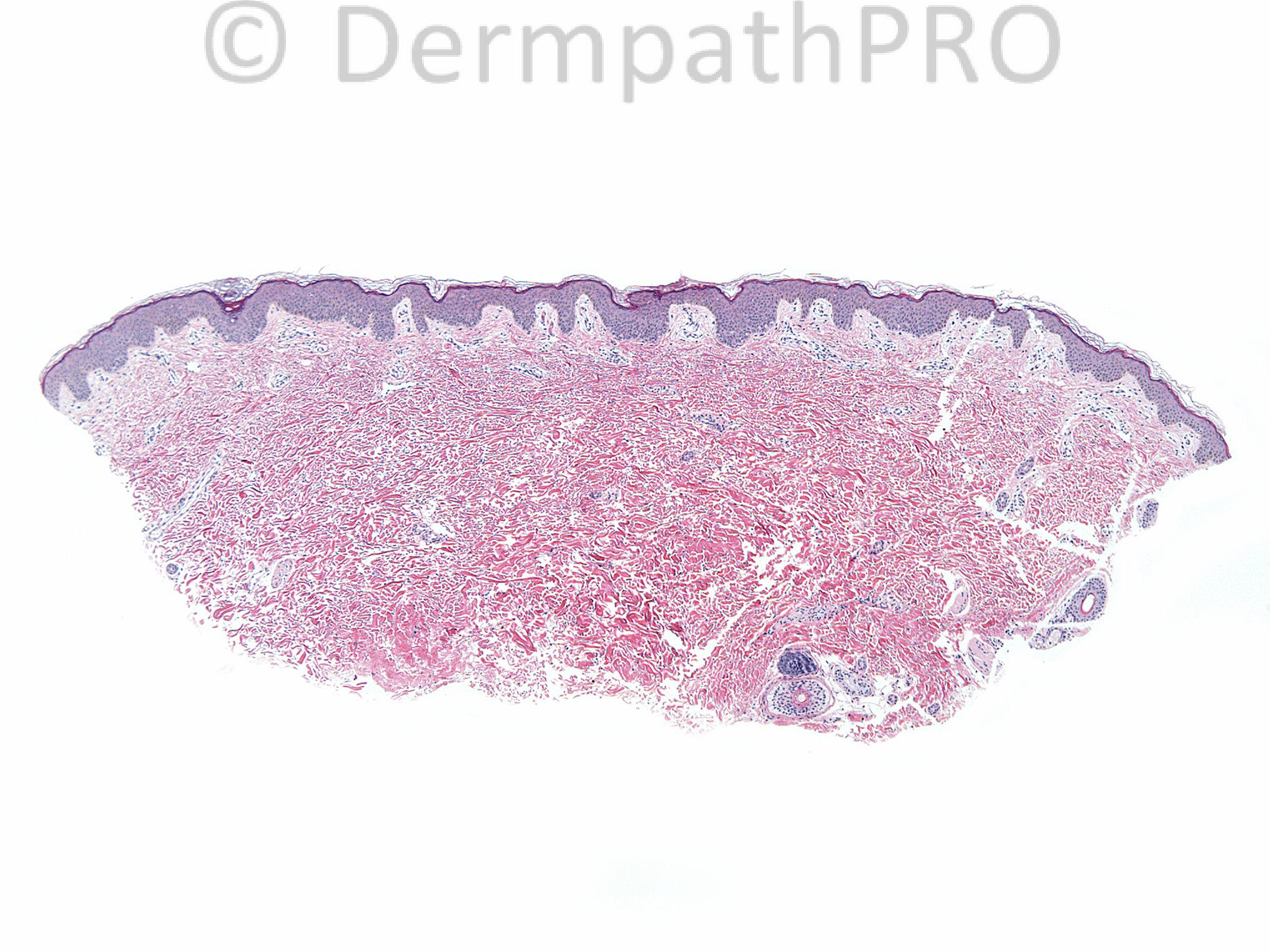
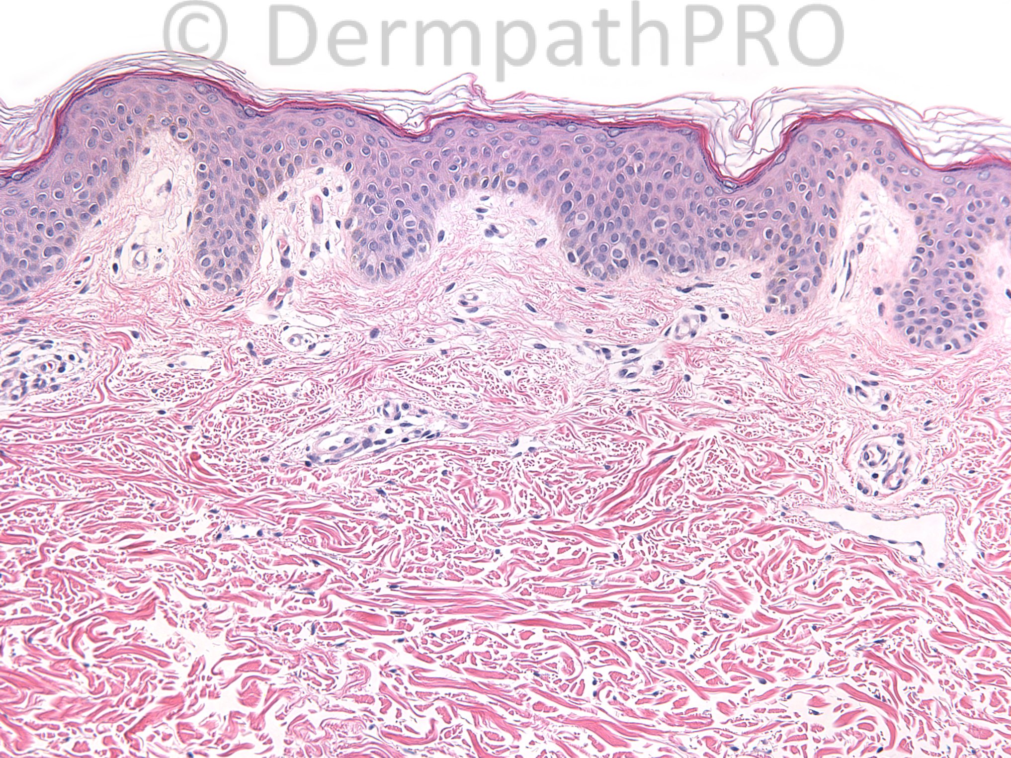
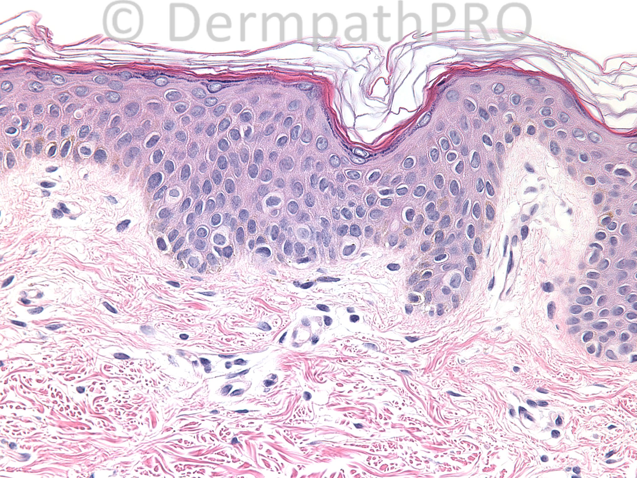
Three year old female with hypopigmented discrete macules (20-30) on suprapubic area since age 9 months, increasing in number and size.

Case posted by Dr.Uma Sundram.


  Report Record

User Feedback


Robledo F. Rocha

Posted

Non-inflammatory decrease in the epidermal pigment intensity and normal number of melanocytes, suggestive of progressive macular hypomelanosis. Ash leaf spots of tuberous sclerosis must be considered, too.

Share this comment


Link to comment
share_externally

Dr. Mona Abdel-Halim

Posted

Yes, very beautiful, very typical clear cell papulosis.. Typical site

Share this comment


Link to comment
share_externally

Clear cells among basal cells and supra basal consistent with clear cell papulosis. Very nice

Share this comment


Link to comment
share_externally

Guest Romualdo

Posted

Agree with Mona and Sadat: clear cell papulosis.

Share this comment


Link to comment
share_externally

Eman El-Nabarawy

Posted

What about nevus depigmentosus? Clear cell papulosis is a nice possibility but it presents with papules not macules as in our case!!

Share this comment


Link to comment
share_externally

Uma Sundram

Posted

[img]https://dermpathpro.com/uploads/spot_diagnosis_comment_img/CASE1129_SD_US_017_F4.jpeg[/img]

Share this comment


Link to comment
share_externally

Uma Sundram

Posted

[img]https://dermpathpro.com/uploads/spot_diagnosis_comment_img/CASE1129_SD_US_017_F5.jpeg[/img]

Share this comment


Link to comment
share_externally

Uma Sundram

Posted

We thought this was a great case of clear cell papulosis. The first immunohistochemical image is that of CK7 and the second of BRST-2. Increase in the presence of these types of clear cells, together with the clinical setting, led us to make the diagnosis of clear cell papulosis. We published this case report in Pediatric Dermatology and the reference has great clinical photos: here's the reference-
[url="http://www.ncbi.nlm.nih.gov/pubmed/22142549"]Clear-cell papulosis: a rare entity that may be misconstrued pathologically as normal skin.[/url]
Wysong A, [b]Sundram U[/b], [b]Benjamin L[/b].
Pediatr Dermatol. 2012 Mar-Apr;29(2):195-8.

Share this comment


Link to comment
share_externally


Join the conversation

You can post now and register later. If you have an account, sign in now to post with your account.

Guest
Add a comment...

×   Pasted as rich text.   Paste as plain text instead

  Only 75 emoji are allowed.

×   Your link has been automatically embedded.   Display as a link instead

×   Your previous content has been restored.   Clear editor

×   You cannot paste images directly. Upload or insert images from URL.

Loading...

×
×
  • Create New...